Article Text

Abstract
Acute ischaemic stroke is a major public health priority and will become increasingly relevant to neurologists of the future. The cornerstone of effective stroke care continues to be timely reperfusion treatment. This requires early recognition of symptoms by the public and first responders, triage to an appropriate stroke centre and efficient assessment and investigation by the attending stroke team. The aim of treatment is to achieve recanalisation and reperfusion of the ischaemic penumbra with intravenous thrombolysis and/or endovascular thrombectomy in appropriately selected patients. All patients should be admitted directly to an acute stroke unit for close monitoring for early neurological deterioration and prevention of secondary complications. Prompt investigation of the mechanism of stroke allows patients to start appropriate secondary preventative treatment. Future objectives include improving accessibility to endovascular thrombectomy, using advanced imaging to extend therapeutic windows and developing neuroprotective agents to prevent secondary neuronal damage.
- STROKE
- CEREBROVASCULAR DISEASE
- NEUROANATOMY
- NEUROEPIDEMIOLOGY
- NEURORADIOLOGY
- REHABILITATION
- DYSPHAGIA
This article is made freely available for use in accordance with BMJ's website terms and conditions for the duration of the COVID-19 pandemic or until otherwise determined by BMJ. You may use, download and print the article for any lawful, non-commercial purpose (including text and data mining) provided that all copyright notices and trade marks are retained.
https://bmj.com/coronavirus/usageStatistics from Altmetric.com
INTRODUCTION
Stroke is the fourth leading cause of death and the largest cause of adult neurological disability in the UK.1 2 The associated socioeconomic burden is huge; the aggregate cost of stroke, including long-term healthcare, rehabilitation and loss of employment, is estimated to be £25.6 billion per year.3 As such, it is one of the key diseases targeted by the National Health Service (NHS) Long Term Plan in England and Wales.4
In contrast to most other countries around the world, stroke medicine in the UK is not the sole preserve of neurologists; indeed, most stroke consultants in the NHS are geriatricians. While stroke medicine is indisputably multidisciplinary, appropriately trained neurologists are well placed to manage stroke and its mimics. In the UK, the new neurology training curriculum will produce consultants trained in stroke medicine, with the potential to expand the stroke workforce.4 Here, we review the diagnosis and management of acute ischaemic stroke and transient ischaemic attack (TIA) for the practising neurologist.
Service design
The introduction of intravenous thrombolysis with recombinant tissue-type plasminogen activator (rtPA, alteplase) to treat acute ischaemic stroke required a revolution in the organisation of stroke care. Recognition that ‘time is brain’ drove effective public and prehospital awareness campaigns, such as the ‘Face, Arm, Speech, Time’ (FAST) test5 and rapid prehospital triage to designated centres.
The organisation of stroke care depends upon local geography, but the implementation of dedicated acute stroke pathways varies widely in the UK. Comprehensive stroke centres provide all aspects of acute stroke care. Triage of patients eligible for endovascular thrombectomy directly to a comprehensive stroke centre (the ‘mothership’ model) may improve the likelihood of good outcome, even if other hospitals are closer. Primary stroke centres are usually smaller centres that initiate intravenous thrombolysis and transfer patients eligible for endovascular thrombectomy to a comprehensive stroke centre, the so-called ‘drip-and-ship’ model.6 Rural hospitals without a stroke team can be linked with stroke centres by telemedicine for thrombolysis calls.7 8 The key aspect of any stroke service model is that patients can access specialist expertise, neuroimaging and stroke unit care without delay.9
The distinction between TIA and stroke cannot be made while the patient remains symptomatic; therefore, all patients should be assessed rapidly. Patients with a completed TIA (symptom resolution within 24 hours) or minor, non-disabling, stroke require prompt mechanistic investigation and secondary preventative treatment, with expert review within 24 hours recommended for all suspected cases.10 Organisational models to achieve this commonly include rapid-access clinics (figure 1). The remainder of this article focuses on the assessment and treatment of acute disabling ischaemic stroke.
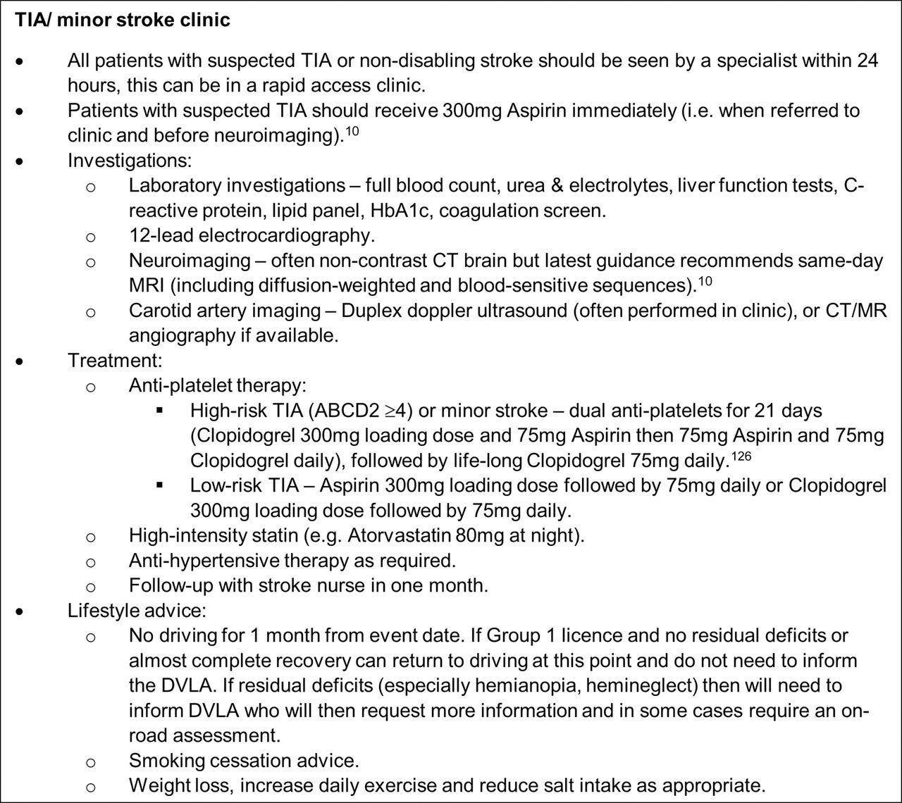
Eligibility, investigations, diagnosis and treatment in a rapid-access TIA clinic.
DVLA, Driver and Vehicle Licensing Agency; TIA, transient ischaemic attack.
Diagnosis
Initial history
As with all aspects of neurology, the history is crucial for diagnosis. However, in the setting of acute stroke, details need to be acquired efficiently and focused on answering a few key questions. Collateral history from witnesses or family members is essential as the nature of the deficit commonly prevents patients themselves from giving a reliable history.
‘When was the patient last seen to be well?’ Early determination of whether the patient is within the reperfusion therapy treatment window sets the pace of subsequent investigations and aids the triage of simultaneous referrals. Symptom onset should be documented as a clock time to avoid confusion. The time recorded for unwitnessed events or ‘wake-up’ strokes should be when the patient was definitely last well (rather than when found); the surrogate use of an activity can be useful, for example, waking to go to the toilet or successfully using a mobile phone.
‘How quickly did the symptoms develop?’ Stroke symptom onset is usually sudden, although notable exceptions include the stuttering nature of capsular warning syndrome, or prodromal symptoms of basilar artery occlusion. Fluctuating severity is common in the early hours after stroke, and initial improvement may be followed by deterioration, especially among those with intracranial vessel occlusion. More gradual evolution of symptoms may suggest alternative diagnoses.
‘Is there any significant past medical and drug history?’ A brief overview of the patient’s background, especially vascular risk factors, will influence the diagnostic decision process; these details can sometimes be obtained from electronic medical records before the patient’s arrival. Risk factors associated with ischaemic stroke include cigarette smoking, hypertension, hypercholesterolaemia, diabetes mellitus, cardiac or peripheral vascular disease, and drugs of abuse. A history of carotid stenosis or atrial fibrillation may suggest a cause.11 Reviewing the list of medication helps to screen for known relevant diagnoses, risk factors for stroke, and whether the patient is taking oral anticoagulation therapy as a potential contraindication to thrombolysis.
Stroke mimics account for at least 20–25% of acute presentations and many of them can be suspected from the history. In one study, the five most frequent stroke mimics were seizure, syncope, sepsis, migraine and brain tumours12; detailed reviews can be found in Practical Neurology.13 14
Posterior circulation strokes are misdiagnosed three times more often than anterior circulation strokes, as they frequently present with non-specific symptoms, including isolated ‘dizziness’ (vertigo or disequilibrium) or headache.15 Acute onset vertigo or disequilibrium with an additional posterior circulation symptom should necessitate further assessment.
Examination
An overview of the patient can be made immediately and should focus on the level of consciousness, head and/or gaze deviation, and laterality of purposeful movements. As in any emergency situation, an initial screen of the airway, breathing and circulation and vital signs will establish cardiovascular stability and suitability to go to scan.
Up to 80% of patients with acute ischaemic stroke have an elevated blood pressure (BP) (≥140 mmHg systolic),16 which spontaneously improves over the following week17 –19 and is associated with poorer outcomes in both ischaemic stroke and intracerebral haemorrhage.20 21 The cause of transient post-stroke hypertension is unknown, but potential mechanisms include disturbed cerebral autoregulation or non-stroke causes such as urinary retention or psychological stress.22 Pyrexia is also common and could reflect aspiration pneumonia, urinary tract infection or infective endocarditis.23
A focused, rather than extensive, neurological examination should be performed in order to identify the affected vascular territory and to quantify physical impairment using the National Institutes of Health Stroke Scale (NIHSS). Limitations to clinical examination in the hyperacute setting include the immaturity of physical signs (such as hypertonia or brisk reflexes) and the degree of patient cooperation. In agitated or dysphasic patients, there is a greater reliance on careful observation when assessing limb paresis, eye movements or visual fields.
The NIHSS is the most commonly used neurological deficit rating scale, with a maximum score of 42 (hypothetical due to several mutually exclusive items). Its advantages include an accredited training and certification system (http://www.nihstrokescale.org/), quick completion time (≤10 min24) and facilitation of communication between team members. It may be used to monitor deficit severity, to identify neurological deterioration and to select patients for reperfusion therapy. Its limitations include the underrepresentation of non-dominant hemisphere deficits,25 such as apraxia or anosognosia (which may be subtle but potentially significantly disabling), and low sensitivity for posterior circulation deficits.26
Quick recognition of common stroke syndromes increases diagnostic confidence and facilitates an efficient neurological examination. Despite limited use in the hyperacute setting, stroke syndromes often suggest the underlying cause. Large-vessel stroke syndromes (table 1) suggest an atheroembolic cause, whereas lacunar syndromes are classically associated with cerebral small-vessel disease. Lacunar syndromes include contralateral pure motor, pure sensory and sensorimotor impairment, the clumsy hand–dysarthria syndrome (which can also be cortical) and ataxic hemiparesis.
Large-vessel stroke syndromes (assumes left hemispheric dominance)
The three-step ‘HINTS’ (Head-Impulse-Nystagmus-Test-of-Skew) bedside examination is often used to assess patients presenting with acute vestibular syndromes and has a high sensitivity (100%) and specificity (96%) for detecting a central cause.30 31 As its positive predictive value is only 69%, an isolated abnormal head impulse test (suggesting unilateral peripheral vestibulopathy) should be interpreted with caution.32
Investigations
Pre-imaging
Rapid neuroimaging is essential for patients with acute stroke. The American Stroke Association guidelines advise that the only necessary prior investigation is a capillary blood glucose,33 which in practice is obtained by paramedics. An intravenous cannula is often required for contrast or perfusion imaging sequences, allowing a blood panel to be obtained simultaneously. This would usually include a screen for infection, renal function and, if the patient takes anticoagulants, a coagulation screen. Although many radiology departments require a recent renal function before giving contrast,34 recent studies have questioned the concept of contrast-induced nephropathy.35 36
Imaging
Stroke centres should establish protocols to eliminate delays to neuroimaging, for example, protocolled stroke imaging sequences and priority use of a designated scanner near to the emergency department.
Neuroimaging in the hyperacute acute stroke setting remains predominantly CT-based.37 A non-contrasted CT scan of head is quick, sensitive and cost-effective at ruling out intracranial haemorrhage, which is usually sufficient for making thrombolysis decisions.38 However, CT scanning has much lower sensitivity and specificity for acute ischaemia because the net tissue water content (and therefore visual change in parenchymal attenuation) changes over hours after the onset of ischaemia. Specificity is compromised by the high prevalence of existing ischaemic changes or old established infarcts. Signs of acute ischaemia on non-contrast CT include loss of grey–white matter differentiation (eg, at the insular ribbon), hemispheric sulcal effacement, loss of integrity of the lentiform nucleus or hyperdensity within an intracranial artery (the ‘dense artery sign’). Early ischaemic changes can be quantified to assess the extent of parenchymal damage using the 10-point Alberta Stroke Program Early CT Score (ASPECTS).39
Multimodal CT imaging comprises CT-perfusion and/or CT-angiography, as well as non-contrast CT, aiming to improve and broaden case selection for reperfusion therapy. Rapid multimodal CT can be performed in acute stroke care pathways. Stroke centres need clear protocols for efficient interpretation to prevent unnecessary delays in giving rtPA.40
CT-angiography of the cervicocranial and intracranial arteries should be performed urgently to detect intracranial large artery occlusion when endovascular thrombectomy is available. Intracranial large artery occlusion is a marker of poor prognosis in minor stroke and TIA41 and observational evidence suggests that patients with non-disabling symptoms due to intracranial large artery occlusion may benefit from thrombolysis,42 but a randomised trial is ongoing.43
CT-perfusion sequences can assess various aspects of cerebral perfusion (see discussion below), often with automated software, such as MIStar (Apollo Medical Imaging Technology) or Rapid Processing of Perfusion and Diffusion (RAPID) CT-perfusion (iSchemaView); these technologies ease interpretation by increasing inter-observer reproducibility and ensuring use of validated thresholds. A comprehensive review of CT-perfusion interpretation has recently been published in Practical Neurology.44
MRI has much greater sensitivity for ischaemia than CT, particularly in minor stroke where it can predict poor short- and long-term outcomes.45 Moreover, comparing different sequences offers an approximate indication of time since onset.46 Rapid stroke MRI protocols typically include diffusion-weighted imaging (DWI), time-of-flight MR-angiogram of the intracranial arteries, T2-fluid-attenuated inversion recovery (FLAIR) and a blood-sensitive sequence such as gradient-recalled echo or susceptibility-weighted imaging.47
Principles of acute stroke care
The main objective of acute ischaemic stroke treatment is to salvage ischaemic, but viable, brain tissue by recanalising occluded cerebral arteries and reperfusing the ischaemic penumbra.48 The penumbra is a region of electrically inexcitable, hypoperfused parenchyma surrounding the irreversibly damaged core49 that is temporarily supported by leptomeningeal collateral flow. Failure to recruit or maintain collaterals underlies the highly variable individual speed of evolution of the core; the mechanisms of collateral failure are currently poorly understood.50 Rapidly declining benefit from reperfusion therapies (‘time is brain’)51 reflects the average pathophysiological status of failure of collateral support over several hours. Some people, identified by imaging, maintain collaterals for longer periods, and later reperfusion is beneficial. ⇓Figure 2 offers a structured approach to acute stroke reperfusion; it is an overview of ‘best practice’ and should be used in conjunction with local protocols tailored to available services where necessary.

Process of acute ischaemic stroke reperfusion therapy: an overview of ‘best practice’.
Patients with severely elevated BP (≥185 mmHg systolic or ≥110 mmHg diastolic) are precluded from thrombolysis due to alteplase licensing restrictions; they may require intravenous antihypertensive therapy9 (eg, intravenous labetalol 5–10 mg or glyceryl trinitrate 50 mg in 50 mL starting at 1.5 mL/hour). However, the BP threshold is based on the original alteplase trial inclusion criteria52 and there is no evidence that reducing BP in this context helps clinically; indeed, recent data suggest a complex interaction between reperfusion status, BP and patient outcome, with one study suggesting that lowering BP before reperfusion treatment may be inappropriate.53
Acute reperfusion strategies
Intravenous thrombolysis
Tissue-type plasminogen activator (tPA) cleaves plasminogen on the surface of thrombi to form plasmin, a powerful endogenous fibrinolytic enzyme.54 Intravenous rtPA (alteplase) is proven and licenced to improve functional outcome in acute ischaemic stroke up to 4.5 hours after symptom onset.10 55 The treatment effect is heavily time-dependent: the number needed to treat for excellent functional outcome at 1.5 hours is five, compared with nine at 3.0–4.5 hours.56 The relative benefit of rtPA is not modified by baseline stroke severity or by age.56 57
UK guidelines recommend all patients with disabling symptoms should be considered for rtPA treatment within 3 hours of symptom onset, and up to 4.5 hours in those aged under 80. Patients presenting at 4.5–6 hours should be considered on an individual basis for treatment, recognising that the benefits are smaller than if treated earlier, but that the risks of a worse outcome, including death, are not increased.58 The UK performs poorly compared with other countries, both in the proportion of patients receiving rtPA (12% for the past 6 years59) and mean door-to-needle times (52 min last year in England and Wales60); considerable improvements in outcome are achievable if these could be bettered.
Informed consent is rarely possible and should not delay treatment. In one registry of nearly 2000 patients, a median door-to-needle time of only 20 min included a consent discussion of less than a minute61; however if unavailable, treatment should proceed in the patient’s best interests.
Currently, there is little evidence to support thrombolysis in patients with non-disabling ischaemic stroke.62 ⇓Table 2 shows the relative and absolute contraindications to rtPA. Symptomatic intracerebral haemorrhage is the most feared adverse effect of rtPA but haemorrhage associated with significant neurological deterioration occurs in only approximately 1.9% of treated patients.63 64 Radiological haemorrhagic transformation occurs due to reperfusion and is more common in people with larger infarcts (who therefore more severe baseline deficits). Neurological deterioration after rtPA infusion is common but usually reflects the initial ischaemic injury; in one recent case series, only 1 of 511 patients deteriorated during the rtPA infusion due to intracerebral haemorrhage. Most deterioration related to intracerebral haemorrhage occurred after the complete rtPA infusion, and deterioration was four times more likely to be due to initial ischaemia rather than to intracerebral haemorrhage.65 Deteriorating patients need urgent repeat neuroimaging to clarify the cause and rtPA infusion is usually suspended pending imaging.
American Heart Association/American Stroke Association absolute and relative contraindications to treatment of acute ischaemic stroke with alteplase
Targets for maintaining homeostasis in acute ischaemic stroke patients
Orolingual angioedema is a recognised complication of rtPA; while most cases are mild and self-limiting, severe attacks requiring airway management can occur in up to 1% of treated patients; people taking ACE inhibitors or those with insular ischaemia are at increased risk.69 Stroke centres should develop local protocols with the anaesthetic department for assessing and urgently managing angioedema. Although management in this setting is not evidence based, treatment should be consistent with that of other drug reactions (figure 3).

Suggested treatment algorithm for rtPA-associated angioedema.
rtPA, recombinant tissue-type plasminogen activator.
Endovascular thrombectomy
Despite the overall benefit of rtPA, the subgroup of patients with large proximal intracranial vessel occlusion (large artery occlusion; carotid, proximal middle cerebral arteries) have low rates of recanalisation with thrombolysis and only a 25% chance of a good outcome.70 71 Endovascular thrombectomy in addition to best medical therapy has been proven in nine randomised trials as superior to best medical therapy alone (including intravenous rtPA in the majority of patients) for patients with anterior circulation large artery occlusion.72 –80 The number needed to treat to achieve a reduction of one or more points on modified Rankin Scale (mRS) is 2.6.81 A detailed guide has recently been published in Practical Neurology.82
Unfortunately, the UK has been slow to provide this service; current endovascular thrombectomy rates are 5.5 per 1000 ischaemic strokes in the UK, versus 50 in the US and Western Europe.59 Parts of the UK, notably Scotland, have no access to thrombectomy at all. In England and Wales, the NHS Long Term Plan aims for a 10-fold increase by 2022, in part by expanding endovascular thrombectomy training to specialities other than interventional neuroradiology.4
Guidelines from the UK’s National Institute of Health and Care Excellence (NICE) recommend endovascular thrombectomy for patients with disabling acute ischaemic stroke (arbitrarily defined as NIHSS ≥6) due to imaging-proven anterior circulation large-vessel occlusion up to 6 hours, and posterior circulation (basilar or posterior cerebral artery) large-vessel occlusion up to 24 hours after symptom onset.10 Patients with lower NIHSS but functionally disabling symptoms may also be considered due to the high risk of deterioration associated with large-vessel occlusion.83
As with rtPA, the benefit of endovascular thrombectomy is highly time-dependent.84 However, several clinical trials showed favourable outcome of endovascular thrombectomy versus medical management in anterior circulation large-vessel occlusion beyond 6 hours, although based on small numbers of patients.73 76 85 Two trials have extended the therapeutic window even further: up to 16 hours in DEFUSE 386 and 24 hours in DAWN87 with CT perfusion or DWI-perfusion imaging with clinical mismatch. These trials demonstrated that imaging can select patients with large artery occlusion whose good collateral supply makes them likely to benefit from endovascular thrombectomy.
The optimal mode of anaesthesia during endovascular thrombectomy has yet to be determined; retrospective data suggested that general anaesthesia may be harmful (although potentially biased by patient selection),88 whereas single-centre randomised trials have shown neutral or beneficial effects.89 Multicentre randomised trials are ongoing.82
The complication rates of endovascular thrombectomy are in keeping with other emergency procedures and serious adverse events are rare.90 Although adverse events occur in approximately 15% of patients (including vasospasm, arterial perforation or dissection, device misplacement, symptomatic intracerebral haemorrhage or embolisation to new or target vessel territory), clinical outcome is not affected overall; the number needed to treat of 2.6 includes these complications.91
Acute stroke unit and early complications
Guidelines recommend that everyone with acute ischaemic stroke is admitted directly to an acute stroke unit.9 Stroke unit care has an number needed to treat of 17 to avoid death or disability, a benefit that is sustained over time without lengthening hospital stays.92 93 Key features of the acute stroke unit include stroke-specific multidisciplinary care (physiotherapy, speech and language therapy, occupational therapy) and high nursing ratios.94 95 However, for the past 5 years, only 58% of patients in England and Wales were admitted to an acute stroke unit within 4 hours.60
Key functions of an acute stroke unit are the prevention of secondary brain insults by maintaining physiological homeostasis (table 3) and monitoring of neurological status.96 The patient should also undergo bedside cardiac telemetry if atrial fibrillation has not been confirmed.
Neurological deterioration should prompt urgent repeat neuroimaging; early neurological complications include recurrent ischaemia, cerebral oedema or haemorrhagic transformation. Repeat brain imaging around 24 hours following rtPA administration is widely undertaken to inform on intracerebral haemorrhage incidence as a quality of care metric, and visualisation of an infarct may provide prognostic and mechanistically relevant information, but the role for routine repeat imaging is debatable. Once haemorrhagic complications have been excluded at 24 hours, antiplatelet therapy should start, most often 300 mg aspirin daily for 2 weeks followed by lifelong clopidogrel monotherapy.
Patients with large volume hemispheric infarcts from acute occlusion of the proximal middle cerebral artery or internal carotid artery are particularly vulnerable to ‘malignant’ cerebral oedema, with a mortality rate of up to 78%.97 Decompressive hemicraniectomy increases the chance of survival (number needed to treat of 2), but patients are often left with significant disability (mRS 4–5 at 1 year in 43% with decompressive hemicraniectomy vs 17% with medical management)98; however, the great majority rate their quality of life as being satisfactory despite disability.99 Updated NICE guidance10 has removed the upper age limit for consideration of decompressive hemicraniectomy, in line with trial evidence. The current eligibility criteria are as follows:
Surgery may be performed 48 hours from stroke onset
Clinical deficits that suggest middle cerebral artery infarction with NIHSS >15
Decreased level of consciousness (≥1 on level of consciousness on NIHSS)
Infarction of ≥50% of middle cerebral artery territory as seen on CT scanning or infarct volume >145 cm3 on DWI
The high incidence of dysphagia after stroke is a risk factor for aspiration pneumonia and is associated with increased mortality and disability.100 Guidelines recommend that patients receive a bedside swallowing assessment and appropriate adaptation of oral intake to prevent aspiration.10 Although there are no randomised studies to determine whether screening methods improve outcomes,101 observational data suggest that delayed assessment is associated with a higher risk of aspiration pneumonia.102 Prophylactic antibiotics have not proven effective.103
Non-ambulatory patients with ischaemic stroke are at high risk of deep vein thrombosis.104 Prophylaxis with low-molecular-weight heparin is not recommended due to the risk of haemorrhagic transformation,9 although some studies have shown no significant additional risk.105 Intermittent pneumatic compression devices are effective (compared to compression stockings) at reducing the risk of deep vein thrombosis and are recommended for all non-ambulatory stroke patients.9 106
Future directions
There is a wealth of active clinical research in stroke medicine, driven by the significant public health implications of this common and socioeconomically impactful disease. A particular priority in the UK is to improve systems that reduce onset-to-needle times, increase access to endovascular thrombectomy and admission rates to acute stroke units. Audits, including the Sentinel Stroke National Audit Programme (SSNAP), measure the processes and structure of stroke care and use these data to drive improvements.
Mobile stroke units with in-built CT scanners and telemedicine links with stroke centres are associated with earlier thrombolytic delivery and improved clinical outcome in urban settings but are resource intensive and their optimal deployment depends on accurate prehospital triage.107
Alternatives to alteplase that are more fibrin-specific may be safer, more effective and may increase the therapeutic window. However, desmoteplase did not improve functional outcome compared with placebo in acute ischaemic patients 3–9 hours after symptom onset.108 Although tenecteplase has not proven superior to alteplase in minor ischaemic stroke patients109 (a trial in patients with non-disabling symptoms due to large-vessel occlusion is ongoing43), it doubled recanalisation rates in pre-endovascular treatment of strokes from large-vessel occlusion with improved functional outcome.110 In addition, the single bolus administration of tenecteplase may be advantageous for drip-and-ship thrombectomy service pathways.
Ongoing trials are investigating the efficacy of endovascular thrombectomy in patient subgroups, including basilar artery occlusion (BASICS111), low NIHSS (MOSTE112 and ENDOLOW113), or low ASPECTS score (TESLA,114 TENSION115 and IN EXTREMIS112). The optimal prehospital service pathway is another unanswered question, and mothership and drip-and-ship models are also being compared in a multicentre trial.116
Multiple preclinical and clinical studies to prevent secondary neuronal injury following ischaemic stroke have been unsuccessful, and to date there are no evidence-based neuroprotective agents.117 Although the neuroprotectant nerinetide did not improve outcomes in endovascular-treated patients compared with placebo in one recent randomised trial, secondary subgroup analyses suggest further investigation may be warranted in patients not treated with alteplase.118 Translational studies of neuroprotective therapies may be aided by novel tissue banking of thrombi extracted by endovascular thrombectomy.119 The CHARM trial aims to assess whether glibenclamide (BIIB093) improves functional outcome in patients with malignant brain oedema.120
CONCLUSION
Stroke medicine is a varied and rapidly developing field that provides the opportunity to offer life-changing treatments to patients affected by the leading cause of neurological disability. Stroke care will have increasing relevance for neurologists of the future and as a specialty we have a lot to offer, in particular with diagnostic expertise. Equally, we may need to develop our skills further, for example, through managing acutely unwell patients with general medical problems on the acute stroke unit or by learning how to perform mechanical thrombectomy.
Key points
Stroke is a public health priority and prompt specialist intervention significantly reduces the burden of death and disability.
We need effective systems in place for prehospital recognition and appropriate triage of suspected acute stroke.
Patients with non-disabling stroke or TIA should be assessed within 24 hours.
Reperfusion with intravenous thrombolysis and/or endovascular thrombectomy are highly effective therapies, but time-dependent.
All patients should be treated in an acute stroke unit, for monitoring to detect and act on physiological insults including brain oedema, and for investigating promptly to allow initiation of mechanism-appropriate secondary preventative treatments.
Further Reading
REFERENCES
Footnotes
Contributors RH drafted the manuscript. Other authors revised the manuscript.
Acknowledgement The authors would like to thank Mr Philip Baker for his assistance in designing the figures.
Funding The authors have not declared a specific grant for this research from any funding agency in the public, commercial or not-for-profit sectors.
Competing interests None declared.
Patient consent for publication Not required.
Provenance and peer review Commissioned externally reviewed by John Fink, Christchurch, New Zealand and Michael O’Sullivan, Brisbane, Australia.
Other content recommended for you
- Management of acute ischemic stroke
- Advances in mechanical thrombectomy for acute ischaemic stroke from large vessel occlusions
- Relationship between reperfusion and intracranial hemorrhage after thrombectomy
- Follow-up neutrophil-lymphocyte ratio after stroke thrombectomy is an independent biomarker of clinical outcome
- Thrombectomy for acute ischemic stroke in the elderly: a ‘real world’ experience
- Predictors and impact of hemorrhagic transformations after endovascular thrombectomy in patients with acute large vessel occlusions
- Artificial intelligence to diagnose ischemic stroke and identify large vessel occlusions: a systematic review
- Endovascular thrombectomy beyond 12 hours of stroke onset: a stroke network’s experience of late intervention
- ASPECTS decay during inter-facility transfer in patients with large vessel occlusion strokes
- Interaction between time, ASPECTS, and clinical mismatch