
Main menu

  • Home
  • Content
    • Latest
    • Ahead of print
    • Archive
  • Info for
    • Authors
    • Reviewers
    • Subscribers
    • Institutions
    • Advertisers
  • About Us
    • About Us
    • Editorial Office
    • Editorial Board
  • More
    • Alerts
    • Feedback
    • Folders
    • Help
  • Other Publications
    • Saudi Medical Journal

User menu

  • My alerts
  • Log in

Search

  • Advanced search
Neurosciences Journal
  • Other Publications
    • Saudi Medical Journal
  • My alerts
  • Log in
Neurosciences Journal

Advanced Search

  • Home
  • Content
    • Latest
    • Ahead of print
    • Archive
  • Info for
    • Authors
    • Reviewers
    • Subscribers
    • Institutions
    • Advertisers
  • About Us
    • About Us
    • Editorial Office
    • Editorial Board
  • More
    • Alerts
    • Feedback
    • Folders
    • Help
  • Follow psmmc on Twitter
  • Visit psmmc on Facebook
  • RSS
OtherClinical Note
Open Access

Value of diffusion-weighted imaging in hypoglycemic encephalopathy

Fuldem Y. Dönmez, Serdar Arslan, Ahmed M. Agildere and Ayse Ecevit
Neurosciences Journal July 2015, 20 (3) 296-297; DOI: https://doi.org/10.17712/nsj.2015.3.20140696
Fuldem Y. Dönmez
From the Departments of Radiology (Dönmez, Arslan, Agildere), and Pediatrics (Ecevit), Faculty of Medicine, Baskent University, Ankara, Turkey
MD
  • Find this author on Google Scholar
  • Find this author on PubMed
  • Search for this author on this site
  • For correspondence: [email protected]
Serdar Arslan
From the Departments of Radiology (Dönmez, Arslan, Agildere), and Pediatrics (Ecevit), Faculty of Medicine, Baskent University, Ankara, Turkey
MD
  • Find this author on Google Scholar
  • Find this author on PubMed
  • Search for this author on this site
Ahmed M. Agildere
From the Departments of Radiology (Dönmez, Arslan, Agildere), and Pediatrics (Ecevit), Faculty of Medicine, Baskent University, Ankara, Turkey
MD
  • Find this author on Google Scholar
  • Find this author on PubMed
  • Search for this author on this site
Ayse Ecevit
From the Departments of Radiology (Dönmez, Arslan, Agildere), and Pediatrics (Ecevit), Faculty of Medicine, Baskent University, Ankara, Turkey
MD
  • Find this author on Google Scholar
  • Find this author on PubMed
  • Search for this author on this site
  • Article
  • Figures & Data
  • eLetters
  • Info & Metrics
  • References
  • PDF
Loading

Hypoglycemia is the most common metabolic disorder that may cause neonatal encephalopathy and result in permanent brain injury.1 Prompt diagnosis is important as urgent treatment may change the progression of the disease and the future prognosis. Conventional MRI is useful to demonstrate the signal changes that are prominent on the occipital lobes; however, MRI may not show the pathology in the very early period. Diffusion-weighted-imaging (DWI) should be obtained prior to other sequences to show any early signal changes that are not visible on MRI.

We report a 3-day-old baby who was admitted to the emergency room with the complaint of poor feeding and cyanosis. There was no problem perinatally, Apgar score was 10 on the first and fifth minutes, and the physical examination was normal at birth and when discharged from the hospital. At the time of admission, he had generalized seizures. The laboratory tests showed severe hypoglycemia, blood glucose level was 21 mg/dl (normal value 50-80 mg/dl). He was given 2cc/kg 10% dextrose, control blood glucose was 78 mg/dl and saturation was 95%. Fifteen minutes later, the blood glucose decreased again (32 mg/dl) and 100 cc/kg 10% dextrose was injected. Despite these injections, control glucose levels were 42, 47, and 57 mg/dl, and during the acute treatment, seizures repeated. Antiepileptic drugs were started (Phenobarbital 20mg/kg infusion and 5 mg/kg maintenance dose) and dextrose was continued. An MRI of the brain was performed after the cessation of the seizures. The conventional MRI was normal except for suspicious faint signal increase on the left occipital lobe. Fluid-attenuated inversion recovery (FLAIR) sequence showed no signal changes (Figure 1A). However, diffusion weighted MRI showed significant perirolandic and right occipital diffusion restriction, which was hyperintense on DWI and hypointense on apparent diffusion coefficient map (Figure 1B). The left occipital lobe was normal. An MR angiography was also normal ruling out any vascular occlusion. During follow-up, he did not have any more seizures and serum blood glucose was within normal range. Ten days later he was discharged with per oral Phenobarbital (2 × 9 mg) and Diazoxide (3 × 20 mg). An informed consent was obtained from his parents.

Figure 1
  • Download figure
  • Open in new tab
  • Download powerpoint
Figure 1

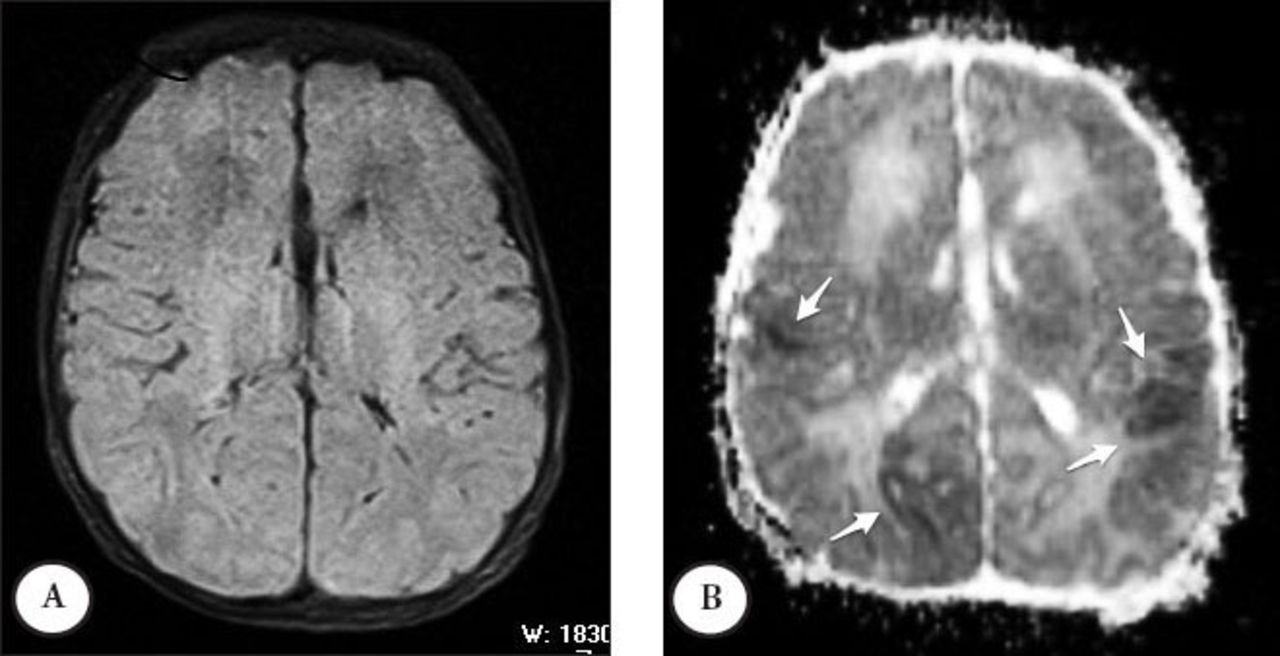
Axial fluid-attenuated inversion-recovery image A) does not show any abnormal signal. Apparent coefficient map (ADC) shows B) restriction of diffusion on the right insula, posterior parietal, and left parietal lobes.

Neonatal hypoglycemia is associated with corticospinal tract injury and adverse motor and cognitive outcomes.2 Infants who are small for gestational age, the smaller of the twins, babies of toxemic mothers, hypocalcemic, and polycythemic patients are at risk for hypoglycemia; however, there may not be an underlying cause at all times.3 Although it is not easy to distinguish hypoxic ischemic encephalopathy and hypoglycemic encephalopathy, various injury patterns in hypoglycemia are described based on MRI findings. Wong et al4 showed that selective posterior white matter and pulvinar edema were most predictive of clinical hypoglycemia, and no injury or a watershed pattern of injury was seen more often in severe hypoglycemia. The conventional MRI is good at showing increased signal on FLAIR or T2-weighted imaging; however, it may not be sensitive in the very early period. The DWI is also important as well as conventional techniques. Our case did not show any abnormal signal intensity on T2-weighted and FLAIR imaging on the regions that showed restriction of diffusion on the DWI. The DWI is not only necessary to distinguish vasogenic edema versus cytotoxic edema, but also leads to the diagnosis itself in case of normal conventional MRI findings.

In conclusion, in neonates who are suspicious for hypoglycemic encephalopathy and have poor sedation, priority should be given to DWI before other sequences and the protocol should be organized accordingly.

  • Received November 5, 2014.
  • Accepted May 8, 2015.
  • Copyright: © Neurosciences

Neurosciences is an Open Access journal and articles published are distributed under the terms of the Creative Commons Attribution-NonCommercial License (CC BY-NC). Readers may copy, distribute, and display the work for non-commercial purposes with the proper citation of the original work.

References

  1. ↵
    1. Gataullina S,
    2. De Lonlay P,
    3. Dellatolas G,
    4. Valayannapoulos V,
    5. Napuri S,
    6. Damaj L,
    7. et al.
    (2013) Topography of brain damage in metabolic hypoglycaemia is determined by age at which hypoglycaemia occurred. Dev Med Child Neurol 55, 162–166.
  2. ↵
    1. Tam EW,
    2. Haeusslein LA,
    3. Bonifacio SL,
    4. Glass HC,
    5. Rogers EE,
    6. Jeremy RJ,
    7. et al.
    (2012) Hypoglycemia is associated with increased risk for brain injury and adverse neurodevelopmental outcome in neonates at risk for encephalopathy. J Pediatr 161, 88–93.
  3. ↵
    1. Harris DL,
    2. Weston PJ,
    3. Harding JE
    (2012) Incidence of neonatal hypoglycemia in babies identified as at risk. J Pediatr 161, 787–791.
  4. ↵
    1. Wong DS,
    2. Poskitt KJ,
    3. Chau V,
    4. Miller SP,
    5. Roland E,
    6. Hill A,
    7. et al.
    (2013) Brain injury patterns in hypoglycemia in neonatal encephalopathy. AJNR Am J Neuroradiol 34, 1456–1461.
PreviousNext
Back to top

In this issue

Neurosciences Journal: 20 (3)
Neurosciences Journal
Vol. 20, Issue 3
1 Jul 2015
  • Table of Contents
  • Cover (PDF)
  • Index by author
Print
Download PDF
Email Article

Thank you for your interest in spreading the word on Neurosciences Journal.

NOTE: We only request your email address so that the person you are recommending the page to knows that you wanted them to see it, and that it is not junk mail. We do not capture any email address.

Enter multiple addresses on separate lines or separate them with commas.
Value of diffusion-weighted imaging in hypoglycemic encephalopathy
(Your Name) has sent you a message from Neurosciences Journal
(Your Name) thought you would like to see the Neurosciences Journal web site.
Citation Tools
Value of diffusion-weighted imaging in hypoglycemic encephalopathy
Fuldem Y. Dönmez, Serdar Arslan, Ahmed M. Agildere, Ayse Ecevit
Neurosciences Journal Jul 2015, 20 (3) 296-297; DOI: 10.17712/nsj.2015.3.20140696

Citation Manager Formats

  • BibTeX
  • Bookends
  • EasyBib
  • EndNote (tagged)
  • EndNote 8 (xml)
  • Medlars
  • Mendeley
  • Papers
  • RefWorks Tagged
  • Ref Manager
  • RIS
  • Zotero
Share
Value of diffusion-weighted imaging in hypoglycemic encephalopathy
Fuldem Y. Dönmez, Serdar Arslan, Ahmed M. Agildere, Ayse Ecevit
Neurosciences Journal Jul 2015, 20 (3) 296-297; DOI: 10.17712/nsj.2015.3.20140696
Twitter logo Facebook logo Mendeley logo
  • Tweet Widget
  • Facebook Like
  • Google Plus One
Bookmark this article

Jump to section

  • Article
    • References
  • Figures & Data
  • eLetters
  • References
  • Info & Metrics
  • PDF

Related Articles

  • No related articles found.
  • Google Scholar

Cited By...

  • No citing articles found.
  • Google Scholar

More in this TOC Section

  • How to make a decision on the choice of therapy for multiple sclerosis with sarcoidosis comorbidity?
  • Mycoplasma pneumonia and stroke: causation, association, or coincidence?
  • Posterior reversible encephalopathy syndrome overshadowing COVID-19: An Abstruse scenario or a conspicuous association?
Show more Clinical Note

Similar Articles

Navigate

  • home

More Information

  • Help

Additional journals

  • All Topics

Other Services

  • About

© 2025 Neurosciences Journal Neurosciences is copyright under the Berne Convention and the International Copyright Convention. All rights reserved. Neurosciences is an Open Access journal and articles published are distributed under the terms of the Creative Commons Attribution-NonCommercial License (CC BY-NC). Readers may copy, distribute, and display the work for non-commercial purposes with the proper citation of the original work. Electronic ISSN 1658-3183. Print ISSN 1319-6138.

Powered by HighWire