
Main menu

  • Home
  • Content
    • Latest
    • Ahead of print
    • Archive
  • Info for
    • Authors
    • Reviewers
    • Subscribers
    • Institutions
    • Advertisers
  • About Us
    • About Us
    • Editorial Office
    • Editorial Board
  • More
    • Alerts
    • Feedback
    • Folders
    • Help
  • Other Publications
    • Saudi Medical Journal

User menu

  • My alerts
  • Log in

Search

  • Advanced search
Neurosciences Journal
  • Other Publications
    • Saudi Medical Journal
  • My alerts
  • Log in
Neurosciences Journal

Advanced Search

  • Home
  • Content
    • Latest
    • Ahead of print
    • Archive
  • Info for
    • Authors
    • Reviewers
    • Subscribers
    • Institutions
    • Advertisers
  • About Us
    • About Us
    • Editorial Office
    • Editorial Board
  • More
    • Alerts
    • Feedback
    • Folders
    • Help
  • Follow psmmc on Twitter
  • Visit psmmc on Facebook
  • RSS
Review ArticleReview Article
Open Access

A Closer Look at Angiitis of the central nervous system

Cheng Wan and Hua Su
Neurosciences Journal October 2017, 22 (4) 247-254; DOI: https://doi.org/10.17712/nsj.2017.4.20170052
Cheng Wan
From the Department of Nephrology, Union Hospital, Tongji Medical College, Huazhong University of Science and Technology, Wuhan, China
MD, PhD
  • Find this author on Google Scholar
  • Find this author on PubMed
  • Search for this author on this site
Hua Su
From the Department of Nephrology, Union Hospital, Tongji Medical College, Huazhong University of Science and Technology, Wuhan, China
MD, PhD
  • Find this author on Google Scholar
  • Find this author on PubMed
  • Search for this author on this site
  • For correspondence: [email protected]
  • Article
  • Figures & Data
  • eLetters
  • Info & Metrics
  • References
  • PDF
Loading

Article Figures & Data

Figures

  • Tables
  • Figure 1
    • Download figure
    • Open in new tab
    • Download powerpoint
    Figure 1

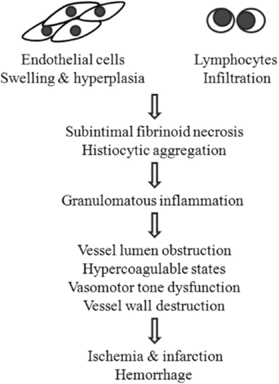
    Pathogenesis of PACNS From the point of pathology, the natural processes of PACNS were suggested to follow 3 steps: 1) endothelial cell swelling and hyperplasia, and lymphocytic infiltration; 2) subintimal fibrinoid necrosis of small vessels and histiocytic infiltration; and 3) marked granulomatous inflammation. From the point of pathophysiology, ischemia and infarction or hemorrhage were considered to be results of the inflammation process, which leads to the obstruction of the vessel lumen, hypercoagulable states, vasomotor tone dysfunction, and destruction of the vascular wall.

Tables

  • Figures
    • View popup
    Table 1

    Common clinical manifestations of PACNS.

    Adult PACNS*
     Headache
     Cognitive dysfunction
     Hemiparesis
     Consistent neurologic deficit or stroke
     Visual symptoms
     Transient ischemic attack
     Aphasia
     Seizures
     Ataxia
     Intracranial hemorrhage
    Large-medium vessel cPACNS†
     Focal neurologic deficits
      1. Acute hemiparesis
      2. Hemisensory loss
      3. Fine motor skill loss
      4. Hemifacial weakness
     Headaches
     Seizures
     Diffuse neurologic deficit
    Small-vessel cPACNS
     Any neurologic or psychiatric symptoms
    • ↵* PACNS - primary angiitis of the central nervous system,

    • ↵† cPACNS - children primary angiitis of the central nervous system

    • View popup
    Table 2

    Differential diagnosis of PACNS.

    Secondary central nervous system vasculitis
     Systemic vasculitides: Takayasu arteritis, giant cell arteritis, polyarteritis nodosa, Kawasaki disease, granulomatosis with polyangiitis, eosinophilic granulomatosis with polyangiitis, Behçet disease, etc.
     Vasculitis associated with systemic diseases: lupus vasculitis, rheumatoid vasculitis, Gougerot Sjögren’s syndrome, etc.
     Infections: viral (e.g., herpes zoster, HIV1), bacterial (e.g., tuberculosis, syphilis), fungal (e.g., aspergillosis, cryptococcus), mycoplasmal, etc
     Cancer-associated: Hodgkin and non-Hodgkin lymphoma, leukemia, etc.
    Nonvasculitic autoimmune and inflammatory brain diseases
     Neuromyelitis optica, N-Methyl-D-aspartate receptor–mediated encephalitis, Susac syndrome, optic neuritis, multiple sclerosis, acute demyelinating encephalomyelitis, Rasmussen encephalitis.
    Non-inflammatory vasculopathies
     RCVS:2 Call-Fleming syndrome, postpartum angiopathy, migrainous vasospasm, drug-induced arteritis, BACNS3.
     Others such as fibromuscular dysplasia, Moyamoya disease, intracranial dissection, radiation vasculopathy, etc.
    Miscellaneous
     thromboembolic disease, bacterial endocarditis, anti-phospholipid syndrome and other hypercoagulable states, cardiac myxoma embolism, cholesterol atheroembolism, hemoglobin disorders, etc.
    • HIV - human immunodeficiency virus, RCVS - reversible cerebral vasoconstriction syndrome, BACNS - benign angiitis of the central nervous system

PreviousNext
Back to top

In this issue

Neurosciences Journal: 22 (4)
Neurosciences Journal
Vol. 22, Issue 4
1 Oct 2017
  • Table of Contents
  • Cover (PDF)
  • Index by author
Print
Download PDF
Email Article

Thank you for your interest in spreading the word on Neurosciences Journal.

NOTE: We only request your email address so that the person you are recommending the page to knows that you wanted them to see it, and that it is not junk mail. We do not capture any email address.

Enter multiple addresses on separate lines or separate them with commas.
A Closer Look at Angiitis of the central nervous system
(Your Name) has sent you a message from Neurosciences Journal
(Your Name) thought you would like to see the Neurosciences Journal web site.
Citation Tools
A Closer Look at Angiitis of the central nervous system
Cheng Wan, Hua Su
Neurosciences Journal Oct 2017, 22 (4) 247-254; DOI: 10.17712/nsj.2017.4.20170052

Citation Manager Formats

  • BibTeX
  • Bookends
  • EasyBib
  • EndNote (tagged)
  • EndNote 8 (xml)
  • Medlars
  • Mendeley
  • Papers
  • RefWorks Tagged
  • Ref Manager
  • RIS
  • Zotero
Share
A Closer Look at Angiitis of the central nervous system
Cheng Wan, Hua Su
Neurosciences Journal Oct 2017, 22 (4) 247-254; DOI: 10.17712/nsj.2017.4.20170052
Twitter logo Facebook logo Mendeley logo
  • Tweet Widget
  • Facebook Like
  • Google Plus One
Bookmark this article

Jump to section

  • Article
    • Abstract
    • Footnotes
    • References
  • Figures & Data
  • eLetters
  • References
  • Info & Metrics
  • PDF

Related Articles

  • No related articles found.
  • PubMed
  • Google Scholar

Cited By...

  • No citing articles found.
  • Google Scholar

More in this TOC Section

  • Structural and functional changes in the hippocampus induced by environmental exposures
  • Tumefactive demyelinating lesions: A literature review of recent findings
  • Epilepsia partialis continua: A review
Show more Review Article

Similar Articles

Navigate

  • home

More Information

  • Help

Additional journals

  • All Topics

Other Services

  • About

© 2025 Neurosciences Journal Neurosciences is copyright under the Berne Convention and the International Copyright Convention. All rights reserved. Neurosciences is an Open Access journal and articles published are distributed under the terms of the Creative Commons Attribution-NonCommercial License (CC BY-NC). Readers may copy, distribute, and display the work for non-commercial purposes with the proper citation of the original work. Electronic ISSN 1658-3183. Print ISSN 1319-6138.

Powered by HighWire