
Main menu

  • Home
  • Content
    • Latest
    • Ahead of print
    • Archive
  • Info for
    • Authors
    • Reviewers
    • Subscribers
    • Institutions
    • Advertisers
  • About Us
    • About Us
    • Editorial Office
    • Editorial Board
  • More
    • Alerts
    • Feedback
    • Folders
    • Help
  • Other Publications
    • Saudi Medical Journal

User menu

  • My alerts
  • Log in

Search

  • Advanced search
Neurosciences Journal
  • Other Publications
    • Saudi Medical Journal
  • My alerts
  • Log in
Neurosciences Journal

Advanced Search

  • Home
  • Content
    • Latest
    • Ahead of print
    • Archive
  • Info for
    • Authors
    • Reviewers
    • Subscribers
    • Institutions
    • Advertisers
  • About Us
    • About Us
    • Editorial Office
    • Editorial Board
  • More
    • Alerts
    • Feedback
    • Folders
    • Help
  • Follow psmmc on Twitter
  • Visit psmmc on Facebook
  • RSS
OtherClinical Image
Open Access

Neuroregression, coarse features, and oligosaccharides in urines

Ahmed Y. BoAli, Kalthoum Tlili-Graiess, Amal M. AlHashem and Brahim M. Tabarki
Neurosciences Journal October 2017, 22 (4) 325-327; DOI: https://doi.org/10.17712/nsj.2017.4.20170225
Ahmed Y. BoAli
From the Divisions of Pediatric Neurology (BoAli, Tabarki), and Genetics (AlHashem), Department of Pediatrics, and the Division of Neuroradiology (Tlili), Department of Radiology, Prince Sultan Military Medical City, Riyadh, Kingdom of Saudi Arabia
MD
  • Find this author on Google Scholar
  • Find this author on PubMed
  • Search for this author on this site
Kalthoum Tlili-Graiess
From the Divisions of Pediatric Neurology (BoAli, Tabarki), and Genetics (AlHashem), Department of Pediatrics, and the Division of Neuroradiology (Tlili), Department of Radiology, Prince Sultan Military Medical City, Riyadh, Kingdom of Saudi Arabia
MD
  • Find this author on Google Scholar
  • Find this author on PubMed
  • Search for this author on this site
Amal M. AlHashem
From the Divisions of Pediatric Neurology (BoAli, Tabarki), and Genetics (AlHashem), Department of Pediatrics, and the Division of Neuroradiology (Tlili), Department of Radiology, Prince Sultan Military Medical City, Riyadh, Kingdom of Saudi Arabia
MD
  • Find this author on Google Scholar
  • Find this author on PubMed
  • Search for this author on this site
Brahim M. Tabarki
From the Divisions of Pediatric Neurology (BoAli, Tabarki), and Genetics (AlHashem), Department of Pediatrics, and the Division of Neuroradiology (Tlili), Department of Radiology, Prince Sultan Military Medical City, Riyadh, Kingdom of Saudi Arabia
MD
  • Find this author on Google Scholar
  • Find this author on PubMed
  • Search for this author on this site
  • For correspondence: [email protected]
  • Article
  • Figures & Data
  • eLetters
  • Info & Metrics
  • References
  • PDF
Loading

Clinical Presentation

A 5-year-old boy, the product of a consanguineous marriage, presented with neuroregression; and spasticity since the age of one year. There were no seizures. His sister, aged 3 years had similar features. Examination revealed impaired cognition, generalized spasticity, brisk deep tendon reflexes, and bilateral extensor plantar responses. The head circumference was normal, and there was mild facial coarsening. There were no organomegaly, or angiokeratomas, and the ophthalmological examination was unremarkable. A brain MRI/MR spectroscopy was performed and is shown in (Figures 1 and 2).

Figure 1
  • Download figure
  • Open in new tab
  • Download powerpoint
Figure 1

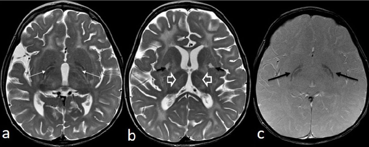
A brain MRI/MR axial (a and b) T2-weighted MR images show severe hypomyelination with a low signal of the globus pallidus within the medial and lateral segments separated by the hyperintense medial medullary laminae (white long arrows). The internal medullary laminae of the thalami are hyperintense (open arrows). A high signal intensity alteration is seen in both the putamina (short black arrows). The gradient echo weighted image (c) clearly shows the low signal of the globus pallidus (long black arrows)

Figure 2
  • Download figure
  • Open in new tab
  • Download powerpoint
Figure 2

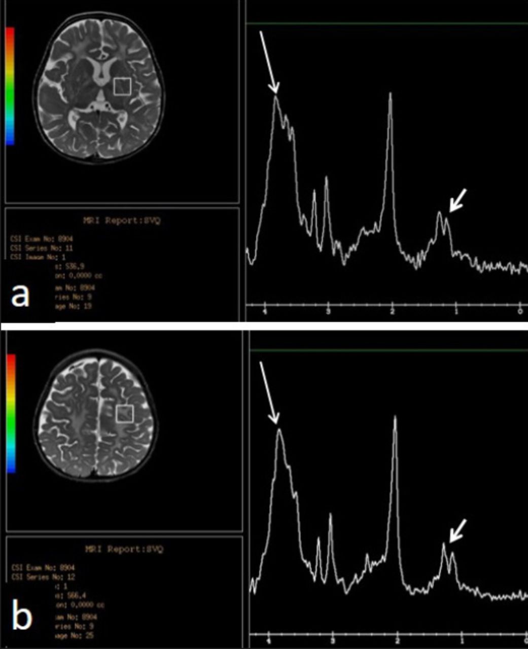
Single voxel MR spectroscopy (TE 35 ms) in the left basal ganglia (a) and the anterior centrum semiovale (b) demonstrates an abnormal broad and high peak at approximately 3.5-3.9 ppm composed of multiple peaks, the highest at 3.8 compatible with mannose (long arrow) and a second peak at 1.2 ppm (short arrow) representing fucose (this peak can easily be confused with the lactate peak, usually at 1.3 ppm).

Questions:

  1. What are the findings inferred from Figures 1 and 2

  2. What is the most likely diagnosis?

  3. How is the diagnosis confirmed?

Answers & Discussion

  1. The brain MRI revealed abnormalities within the periventricular and subcortical white matter, the globus pallidus, and the putamina. The MRS showed a peak at the 3.8-3.9 ppm

  2. Based on the clinical presentation and the neuroimaging findings, the most likely diagnosis is fucosidosis.

  3. Confirmatory diagnosis of fucosidosis requires positive test for urine oligosaccharides, absent α-L-fucosidase activity in the plasma and leukocytes, and genetic study. In our patient, the urine was positive for oligosaccharides, and the genetic study showed a mutation in the FUCA1 gene (c.1123del). Fucosidosis is an autosomal recessive lysosomal storage disease caused by defective alpha -L- fucosidase, with accumulation of fucose in the tissues. Clinical features include progressive regression of milestones, spasticity, coarse facial features, angiokeratoma, and dysostosis multiplex.1-3 Fucosidosis has been classified into 2 major types. Type 1 is characterized by an early onset, rapid and severe psychomotor regression, and death within the first decade of life. Type 2 is characterized by milder neurological phenotype, the development of angiokeratoma, and longer survival.1-2 Neuroimaging plays an important role in the diagnosis of fucosidosis. The MRI of the brain shows prominent and progressive changes in white matter signal intensity, including periventricular, lobar, and subcortical supratentorial areas, internal and external capsules, internal medullary laminae of the thalami, putamina, and both hypothalami. The globi pallidi and substantia nigra have high signal intensity on T1 and low signal intensity on T2 and FLAIR. The MR sprectroscopy shows a broad and high “hump” corresponding to multiple peaks from approximately 3.5-3.9 ppm, presumed to be the result of accumulation of carbohydrate containing macromolecules. The highest peak at 3.8 is compatible with mannose and a doublet at 1.2 ppm, which invertsat long echotime, suggests fucose peak. The combination of imaging and MR spectroscopy findings is probably specific.1-2 Supportive diagnosis of fucosidosis also includes dysostosis multiplex; and high amounts of oligosaccharides in the urine.1-3 The treatment of fucosidosis is mainly supportive. Hematopoietic transplant or umbilical cord blood transplantation could reduce the severity and retard the progression of clinical neurological deterioration.3

Footnotes

  • Notice: Authors are encouraged to submit clinical images for possible publication in the Journal. These may be in any field of Clinical Neurosciences, and should approximately follow the format used here. Please address any submissions to the Assistant Editor, Neurosciences Journal, Prince Sultan Military Medical City, PO Box 7897, Riyadh 11159, Kingdom of Saudi Arabia. E-mail: malaskar{at}psmmc.med.sa

  • Copyright: © Neurosciences

Neurosciences is an Open Access journal and articles published are distributed under the terms of the Creative Commons Attribution-NonCommercial License (CC BY-NC). Readers may copy, distribute, and display the work for non-commercial purposes with the proper citation of the original work.

References

  1. ↵
    1. Ediz SS,
    2. Aralasmak A,
    3. Yilmaz TF,
    4. Toprak H,
    5. Yesil G,
    6. Alkan A
    (2016) MRI and MRS findings in fucosidosis;a rare lysosomal storage disease. Brain Dev 38, 435–438.
  2. ↵
    1. Malatt C,
    2. Koning JL,
    3. Naheedy J
    (2015) Skeletal and brain abnormalities in fucosidosis, a rare lysosomal storage disorder. J Radiol Case Rep 9, 30–38.
  3. ↵
    1. Jiang M,
    2. Liu S,
    3. Jiang H,
    4. Lin Y,
    5. Shao Y,
    6. Hu H,
    7. et al.
    (2017) Brain abnormalities in fucosidosis: transplantation or supportive therapy? Metab Brain Dis 32, 317–320.
PreviousNext
Back to top

In this issue

Neurosciences Journal: 22 (4)
Neurosciences Journal
Vol. 22, Issue 4
1 Oct 2017
  • Table of Contents
  • Cover (PDF)
  • Index by author
Print
Download PDF
Email Article

Thank you for your interest in spreading the word on Neurosciences Journal.

NOTE: We only request your email address so that the person you are recommending the page to knows that you wanted them to see it, and that it is not junk mail. We do not capture any email address.

Enter multiple addresses on separate lines or separate them with commas.
Neuroregression, coarse features, and oligosaccharides in urines
(Your Name) has sent you a message from Neurosciences Journal
(Your Name) thought you would like to see the Neurosciences Journal web site.
Citation Tools
Neuroregression, coarse features, and oligosaccharides in urines
Ahmed Y. BoAli, Kalthoum Tlili-Graiess, Amal M. AlHashem, Brahim M. Tabarki
Neurosciences Journal Oct 2017, 22 (4) 325-327; DOI: 10.17712/nsj.2017.4.20170225

Citation Manager Formats

  • BibTeX
  • Bookends
  • EasyBib
  • EndNote (tagged)
  • EndNote 8 (xml)
  • Medlars
  • Mendeley
  • Papers
  • RefWorks Tagged
  • Ref Manager
  • RIS
  • Zotero
Share
Neuroregression, coarse features, and oligosaccharides in urines
Ahmed Y. BoAli, Kalthoum Tlili-Graiess, Amal M. AlHashem, Brahim M. Tabarki
Neurosciences Journal Oct 2017, 22 (4) 325-327; DOI: 10.17712/nsj.2017.4.20170225
Twitter logo Facebook logo Mendeley logo
  • Tweet Widget
  • Facebook Like
  • Google Plus One
Bookmark this article

Jump to section

  • Article
    • Clinical Presentation
    • Questions:
    • Answers & Discussion
    • Footnotes
    • References
  • Figures & Data
  • eLetters
  • References
  • Info & Metrics
  • PDF

Related Articles

  • No related articles found.
  • Google Scholar

Cited By...

  • No citing articles found.
  • Google Scholar

More in this TOC Section

  • Multiple cranial nerve enhancement in a case of neuronal ceroid lipofuscinosis type 8
  • Unilateral Lennox–Gastaut syndrome associated with Klippel–Trénaunay syndrome
  • A child with leukemia and behavioral changes
Show more Clinical Image

Similar Articles

Navigate

  • home

More Information

  • Help

Additional journals

  • All Topics

Other Services

  • About

© 2025 Neurosciences Journal Neurosciences is copyright under the Berne Convention and the International Copyright Convention. All rights reserved. Neurosciences is an Open Access journal and articles published are distributed under the terms of the Creative Commons Attribution-NonCommercial License (CC BY-NC). Readers may copy, distribute, and display the work for non-commercial purposes with the proper citation of the original work. Electronic ISSN 1658-3183. Print ISSN 1319-6138.

Powered by HighWire