


Pneumocephalus is the collection of air within the cranial cavity. It was first described by Thomas Du in 1866 in an autopsy report of a trauma patient. In 1914, Wolff was the first to use the term pneumocephalus. Published studies and reviews indicated that trauma and surgery are the most common causes of pneumocephalus accounting for 75-90% of cases. In addition, neoplasms account for 13% and infections account for 9% of cases. Other causes include lumbar puncture, angiography, Valsalva’s maneuver, subarachnoid-pleural fistula, congenital skull defects, basilar skull fractures, sinus fractures, osteoma, central venous catheterization, epidural steroid injections, and epidural anesthesia. Spontaneous or idiopathic pneumocephalus was only found in 0.6% of the cases.1,2 In this article, we aim to report a case of post-dural puncture headache and pneumocephalus complicating epidural analgesia using the loss of resistance to air technique. Post-epidural puncture headache was successfully treated with an autologous blood patch. However, headache persisted, and another cause was investigated further. This patient was found to have both post-dural puncture headache and pneumocephalus.
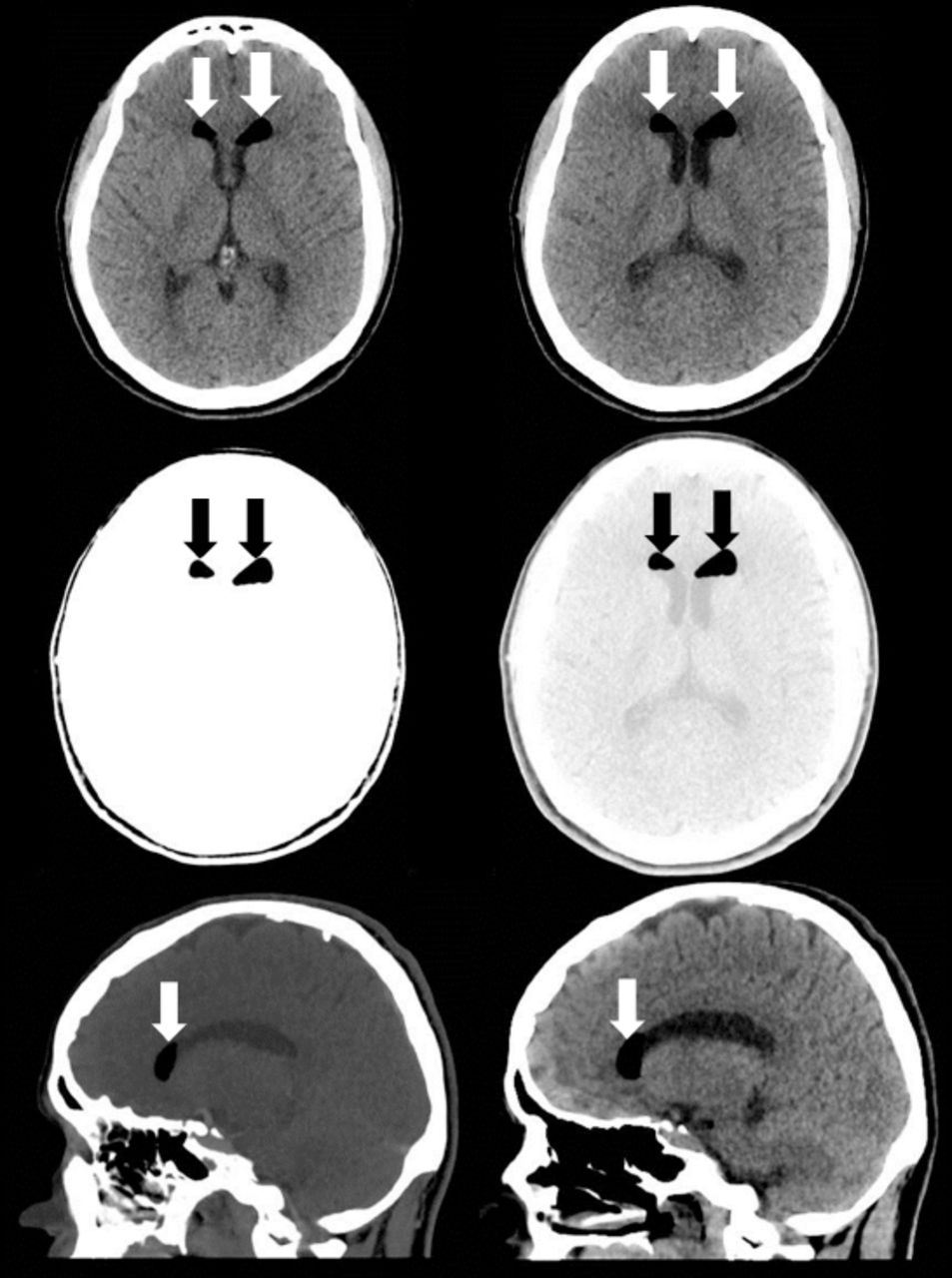
A 36-year-old female delivered a healthy male infant 24 hours prior to presentation through a spontaneous vaginal delivery. She received an epidural analgesia using Tuohy needle 18 gauge for labor pain. Immediately after the epidural injection, she developed a severe headache which was constant and throbbing in nature located in both frontal and occipital regions with radiation to the neck. Due to labor pain and being occupied by giving birth, she did not complain about this headache except after delivery. The pain was worse with sitting or standing up and relieved by lying flat. The pain was not similar to her usual migraine headaches, which started at the age of 16. There were no associated fever, photophobia, neck stiffens, sensory symptoms, or weakness. There was no nasal discharge, epistaxis, anosmia, facial pain, or ear discharge. Her past surgical history was remarkable for previous varicose vein surgery, which was carried out 3 years ago under local anesthesia. During this admission, there was no history of cannulation of major arteries or veins in the neck. She had 2 previous pregnancies that were uneventful. On examination, her vital signs were normal. She was in severe pain, and she liked to stay in a supine position. Her neurological examination showed a normal higher mental functions, cranial nerve examination, motor, sensory and coordination systems. Her gait was not assessed, and her fundus was normal. There were no signs of meningeal irritation. Her basic blood work including chemistry was normal. The patient was treated conservatively using intravenous hydration, non-steroidal anti-inflammatory drugs, and caffeine. Twenty-four hours later, she continued to be symptomatic, and an epidural autologous blood patch (22 cc) was carried out by the same anesthesia consultant who performed the initial epidural anesthesia with some improvement in her symptoms. The patient started to walk with disappearance of the postural headache despite having a constant headache in the frontal region of her head that had no precipitating or aggravating factors. Cerebrospinal fluid (CSF) analysis was normal including opening pressure, physical appearance, and analysis. Computed tomography (CT) of the brain with CT venography was carried out to rule out secondary headache disorders showed pockets of air within the lateral ventricles, diagnostic of pneumocephalus (Figure 1). The patient gradually improved and discharged home 24 hours later. On a visit to the clinic 2 weeks later, she was free of pain with a normal clinical examination.

CT of the brain showing pockets of air within the lateral ventricles, diagnostic of pneumocephalus.
The pathophysiology of pneumocephalus involves several mechanisms based on the cause or underlying condition. Several mechanisms may work simultaneously in the same patient. Pneumocephalus may be due to an excessive leak of the CSF causing intracranial hypotension. As a result, air is drawn into the cranial cavity similar to the entry of air into an inverted bottle of water as the fluid escapes. This mechanism is considered the most common during neurosurgical and anesthesia procedures. The second mechanism may involve ball-valve effect with air being forced through the area of intracranial dural defect from sudden changes in nasopharyngeal pressure such as during coughing or sneezing. Other mechanisms of pneumocephalus include the admission of air into the cranium from penetrating skull injuries and intracranial infection from gas-forming organisms resulting in intracerebral gas collection.4 Pneumocephalus is usually a benign and self-limiting condition with the air being spontaneously absorbed without producing symptoms. However, if the air accumulates in the brain or the cranium, elevated intracranial pressure and neurological deficit may occur possibly due to a mass effect. This condition is called tension pneumocephalus, which may result in tonsillar herniation syndrome.1,2
The technique used in epidural analgesia is called the loss of resistance to air technique. This technique is based on the perception of loss of resistance as the advancing needle pacing thorough the ligamentum flavum into the epidural space during compression of the plunger of the syringe. This technique is associated with more complications if air is used to identify the epidural space instead of normal saline. Complications associated with this technique may include paraesthesia, air venous embolism, epidural hematoma due to blood vessel lesions, neurological complications, accidental puncture of the dura mater, epilepsy, total subarachnoid block, and pneumoencephalus.3
The occurrence of pneumocephalus after epidural analgesia is rare with only a few cases reported in the literature. It develops due to inadvertent dural puncture and injection of air into the subarachnoid or subdural space with migration to the cranial cavity. Headache is the most common symptom of pneumocephalus due to the fast brain motion as a result of air injection and meningeal irritation. Other symptoms vary depending on the distribution and amounts of air in the intracranial cavity and may include elevated intracranial pressure, seizures, vomiting, and unstable vital signs.1,2
Post-dural puncture headache is the most common complication of unintentional dural puncture with an epidural needle. Pneumocephalus with the subsequent headache can be differentiated from post-dural puncture headache by differences in clinical presentation. Post-dural puncture headache usually occurs within one to 4 days of the inadvertent dural puncture and continues for approximately 4 days and is characterized by a reduction of the headache in the supine position. The headache in pneumocephalus usually occurs immediately following the procedure and continues to deteriorate in spite of any postural changes and persists even in the supine position.4
Diagnosis of pneumocephalus is confirmed by a brain CT, which has the ability to detect as little as 0.5 ml of intracranial air. Air appears as dark black in the CT that is darker than CSF and will have a different distribution pattern depending on the localization.1,2
Pneumocephalus resolves spontaneously with conservative management. In symptomatic cases of pneumocephalus, treatment with 100% oxygen in the supine position has been shown to increase the rate of reabsorption. The mechanism behind that is the reabsorption of intracranial air through intensifying the diffusion concentration gradient for nitrogen between the air collection and the surrounding cerebral tissue. Nitrous oxide may lead to the expansion of pneumocephalus and should be avoided. In addition to oxygen, it is important to provide aggressive hydration, caffeine, or analgesics. Tension pneumocephalus is considered a neurosurgical emergency and must be evacuated similar to an intracranial hematoma.2
The epidural blood patch is a procedure in which the autologous blood is injected into the epidural space in order to close the hole in the dura mater that was created by the inadvertent dural puncture. It is commonly performed for post-dural puncture headache. Cases of pneumocephalus following an epidural blood patch have been reported previously in the literature. Pneumocephalus after the epidural blood patch procedure is thought to occur as a result of the movement of air bubbles from the epidural space to subdural and subarachnoid spaces through the defect that was formed during the inadvertent dural puncture.5
In conclusion, we report a case of adverse events of epidural analgesia using the loss of resistance to air technique. We believe that this technique should be studied further in both human and animal models in regards to complications, particularly pneumocephalus. At least, the patient should be informed about this potential complication if this technique is used. Post-partum patients who have persisted headache after blood patch should be investigated further to explore other potential causes.
Footnotes
Disclosure. Authors have no conflict of interests and the work was not supported or funded by any drug company.
- Received October 19, 2017.
- Accepted March 14, 2018.
- Copyright: © Neurosciences
Neurosciences is an Open Access journal and articles published are distributed under the terms of the Creative Commons Attribution-NonCommercial License (CC BY-NC). Readers may copy, distribute, and display the work for non-commercial purposes with the proper citation of the original work.
In this issue


Related Articles
Cited By...
- No citing articles found.