


Article Figures & Data
Figures
- Figure 1
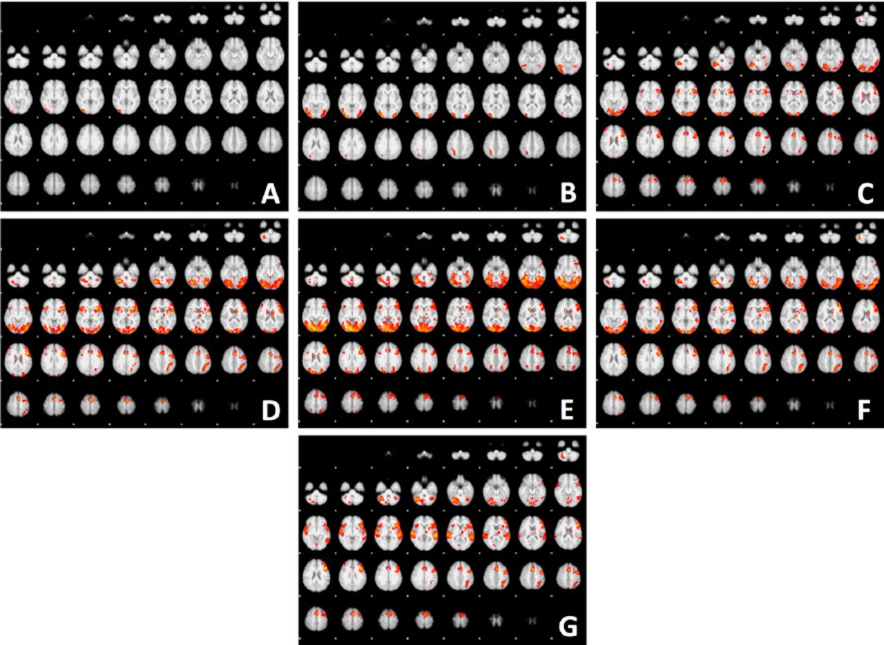
Statistical grand average map of fMRI response to A) Words Rhyme (RH), B) Symbols Rhyme (RH), C) Semantic Category Generation (SCG) task, D) Silent Word Generation (SWG) task, E) Picture Verb Generation (VGp) task, to F) Verb Generation words (VGw) task, G) Verb Generation audio (VGa) task overlaid on 36 axial slices of the MNI152_T1_2mm standard image included in FSL. The (Red-yellow) color shows fMRI signal level (Z-scores) above the 0.05 significance threshold.

Tables
- Table 1
Summary of all 6 tasks; Picture Verb Generation (VGp), Semantic Category Generations (SCG), Silent Word Generation (SWG), Verb Generation words (VGw), Verb Generation audio (VGa), and Rhyming (RH).
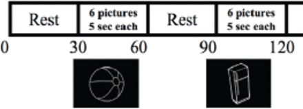
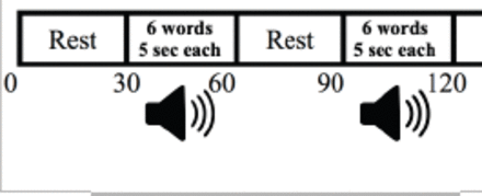
# Task Task description Duration Images •Alternating 4-ON/5-OFF blocks with 30 s per block. •In each ON block; 6 black/white images were presented. 1 VGp •Participant was asked to think of as much as he/she can of (action words) from each image that showed during the scan. And to relax when the crosshair appeared on the screen, with his/her eyes kept open. 270 sec 
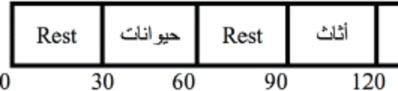
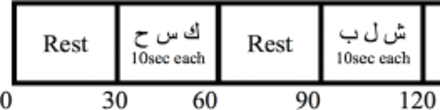
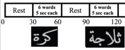
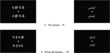
•Alternating 4-ON/5-OFF blocks with 30 s per block. •It consisted of different categories displayed during ON blocks. 2 SCG •The participant was asked to think as much as he/she can about names in the same category displayed (animals, furniture, fruit & vegetable, cloths). 270 sec •Alternating 4-ON/5-OFF blocks with 30 s per block. •Three Arabic letters were displayed in each ON block 3 SWG •Participant was asked to think about words starting with the letter displayed, and to do nothing at rest with keeping his/her eyes open. 270 sec •Alternating 4-ON/5-OFF blocks with 30 s per block. •In each ON block; six nouns were presented. 4 VGw •Participant instructed to think of a verb that is associated with the noun that showed during the scan. For example, the word “ball” might generate the verb “hit”. And to relax when the crosshair appeared on the screen, with his/her eyes kept open. 270 sec •It is similar to VGw, although stimuli were presented in different order through the headphones. 5 VGa •Patients were instructed to think of the response, but not to vocalize it. 270 sec •This paradigm started and ended with 12 sec rest moment. •Consisted of 16 blocks, 8 blocks of pair words and 8 blocks of pair symbols interleaved to each other. 6 RH •Total of 64 pairs of word and 64 pairs of symbols, where pairs remained for 3 sec. 408 sec •Participant was asked to press the button on the response pad when non-rhyme. VGp - Verb Generation - Picture Format, SCG - Semantic Category Generations, SWG - Silent Word Generation, VGw - Verb Generation words, VGa - Verb Generation audio, RH - rhyming, sec - second
- Table 2
Lateralization of brain activity has been assessed for each participant by calculating the laterality index (LI) across these region of interest (ROI).
IFG (POP) Inferior Frontal Gyrus (Pars Opercularis) IFG (PTR) Inferior Frontal Gyrus (Pars Triangularis) AG Angular Gyrus SMG Supramarginal Gyrus MTG Middle Temporal Gyrus STG Superior Temporal Gyrus - Table 3
Details of activated cerebral areas in 24 volunteers (mean values) during the Verb Generation audio (VGa) task.
Cluster Index Voxels P -log10(P) Z MAX Z-MAX X (mm) Z-MAX Y (mm) Z-MAX Z (mm) Region Name 1 13898 0.000 80.5 7.3 -44 18 26 L Inferior Frontal Gyrus (pars opercularis) 6.68 -66 -18 10 L Superior Temporal Gyrus 6.65 -34 20 2 L Insular Cortex 6.45 -44 26 22 L Inferior Frontal Gyrus (pars triangularis) 2 3116 0.000 28.8 6.54 -2 14 48 L Paracingulate Gyrus 6.11 -4 6 58 L Supplementary Motor Cortex 5.94 2 20 48 R Paracingulate Gyrus 5.62 -10 4 64 L Superior Frontal Gyrus 5.57 -12 14 38 L Paracingulate Gyrus 3 1777 0.000 19.3 6.32 62 -14 6 R Planum Temporale 5.29 40 22 -2 R Frontal Operculum Cortex 5.21 62 4 -2 R Superior Temporal Gyrus 5.19 64 -8 4 R Planum Temporale 4.9 30 28 4 R Frontal Orbital Cortex 4.87 60 4 -10 R Superior Temporal Gyrus 4 967 0.000 12.4 5.87 -28 -66 48 L Lateral Occipital cortex (superior division) 5.64 -32 -58 44 L Superior Parietal Lobule 4.92 -40 -50 48 4.29 -30 -74 42 L Lateral Occipital Cortex 4.28 -32 -70 40 4.05 -30 -46 40 L Superior Parietal Lobule 5 317 0.000 5.13 4.68 -28 -22 -2 L Left Putamen 4.6 -10 -28 -6 Brain-Stem 6 180 0.001 3.06 3.81 18 4 16 R Caudate 3.74 18 10 4 R Putamen 3.57 18 12 16 R Caudate 7 170 0.001 2.89 4.34 46 44 30 R Frontal Pole 4.06 38 50 30 3.83 42 36 30 3.75 38 36 22 8 137 0.005 2.31 4.13 2 -60 -12 Cerebellum (R V) 4.04 -2 -54 -14 Cerebellum (L I-IV) 3.96 2 -52 -6 Cerebellum (R I-IV) 3.76 0 -50 -14 Cerebellum (L I-IV) 3.31 -8 -52 -14 Cerebellum (L V) 9 121 0.010 2.01 4.07 8 -24 -12 Brain-Stem 3.81 6 -30 -4 3.43 10 -32 -6 10 89 0.043 1.37 3.63 14 -78 14 R Intracalcarine Cortex 3.52 10 -80 10 - Table 4
Details of activated cerebral areas in 24 volunteers (mean values) during the Silent Word Generation (SWG) task.
Cluster Index Voxels P -log10(P) Z MAX Z-MAX X (mm) Z-MAX Y (mm) Z-MAX Z (mm) Region Names 1 13118 0.000 81.8 6.57 -44 -68 -20 L Occipital Fusiform Gyrus 6.4 -40 -78 -2 L Lateral Occipital Cortex 6.33 26 -88 14 R Lateral Occipital Cortex 2 8645 0.000 61.6 7.05 -4 4 60 L Supplementary Motor Cortex 6.72 -44 20 24 L Inferior Frontal Gyrus (pars opercularis) 6.63 -32 20 2 L Insular Cortex 6.52 -8 18 34 L Cingulate Gyrus 6.28 -2 14 48 L Para-Cingulate Gyrus 6.18 -28 24 2 L Insular Cortex 3 1519 0.000 18.4 6.16 -40 -42 46 L Superior Parietal Lobule 5.33 -28 -64 36 L Lateral Occipital Cortex 5.32 -28 -64 50 5.2 -28 -56 50 L Superior Parietal Lobule 4.98 -26 -60 44 L Lateral Occipital Cortex 4.95 -34 -44 38 L Supramarginal Gyrus 4 845 0.000 12 5.72 34 18 2 R Insular cortex 5.49 30 28 0 R Frontal Orbital Cortex 5 46 20 -4 R Frontal Operculum Cortex 4.86 52 16 2 R Inferior Frontal Gyrus (pars opercularis) 4.74 40 28 2 R Frontal Operculum Cortex 4.3 46 18 8 R Inferior Frontal Gyrus (pars opercularis) 5 382 0.000 6.53 5.14 22 8 6 R Putamen 4.29 16 -2 22 R Caudate 4.23 18 10 18 4.15 26 4 0 R Putamen 4.13 12 6 8 R Caudate 6 242 0.000 4.43 4.49 44 32 18 R Middle Frontal Gyrus 4 42 38 22 R Frontal Pole 3.89 42 40 34 3.55 40 48 28 7 224 0.000 4.14 3.98 2 -52 -12 Cerebellum (R I-IV) 3.89 2 -46 -8 3.84 8 -40 -22 3.83 2 -40 -20 3.83 -2 -48 -20 Cerebellum (L I-IV) 8 108 0.009 2.01 4.69 42 12 28 R Inferior Frontal Gyrus (pars opercularis) 9 98 0.016 1.8 3.47 34 -28 -26 Temporal Fusiform Cortex - Table 5
Details of activated cerebral areas in 24 volunteers (mean values) during the Verb Generation words (VGw) task.
Cluster Index Voxels P -log10(P) Z MAX Z-MAX X (mm) Z-MAX Y (mm) Z-MAX Z (mm) Region Name 1 7813 0.000 57.7 6.27 -42 18 24 L Inferior Frontal Gyrus (pars opercularis) 6.1 -50 20 26 6.04 -6 6 58 L Supplementary Motor Cortex 5.73 -44 8 26 L Inferior Frontal Gyrus (pars opercularis) 5.72 -2 16 48 L Para-cingulate Gyrus 5.67 -50 18 16 L Inferior Frontal Gyrus (pars opercularis) 2 5527 0.000 45.6 6.37 34 -60 -26 Cerebellum (R VI) 6.21 40 -64 -28 Cerebellum (R Crus) 6.12 18 -96 0 R Occipital Pole 6.08 20 -98 8 6.04 16 -86 -6 R Lingual Gyrus 5.99 26 -96 8 R Occipital Pole 3 5172 0.000 43.5 6.29 -18 -94 6 L Occipital Pole 6.14 -16 -98 0 6.14 -16 -100 8 6.02 -50 -66 -12 L Lateral Occipital Cortex 5.97 -18 -90 -12 L Occipital Fusiform Gyrus 5.94 -34 -36 -24 L Temporal Fusiform Cortex 4 1318 0.000 16.7 5 -30 -48 38 L Supramarginal Gyrus (posterior division) 4.88 -28 -70 52 L Lateral Occipital Cortex 4.87 -26 -60 44 4.82 -28 -64 48 4.72 -34 -56 46 L Superior Parietal Lobule 4.66 -24 -66 44 L Lateral Occipital Cortex 5 257 0.000 4.69 4.36 40 18 4 R Frontal Operculum Cortex 4.29 30 24 4 R Insular Cortex 3.67 46 18 10 R Inferior Frontal Gyrus (pars opercularis) 3.28 36 32 -2 R Frontal Orbital Cortex - Table 6
Details of activated cerebral areas in 24 volunteers (mean values) during the Picture Verb Generation (VGp) task.
Cluster Index Voxels P -log10(P) Z MAX Z-MAX X (mm) Z-MAX Y (mm) Z-MAX Z (mm) Region Name 1 24524 0.000 111 7.02 8 -84 -4 R Lingual Gyrus 6.98 16 -96 14 R Occipital Pole 6.96 8 -90 -4 6.79 12 -82 -10 R Lingual Gyrus 6.77 30 -52 -18 R Temporal Occipital Fusiform Cortex 6.66 -14 -100 8 R Occipital Pole 2 7798 0.000 50.9 6.11 -46 20 26 L Middle Frontal Gyrus 5.39 -2 14 48 L Para-Cingulate Gyrus 5.31 -30 28 4 L Insular Cortex 5.27 -42 10 28 L Inferior Frontal Gyrus (pars opercularis) 5.25 -38 26 -4 L Frontal Orbital Cortex 3 351 0.000 5.3 4.98 30 28 -2 R Frontal Orbital Cortex 4.72 36 20 0 R Insular Cortex 4.02 40 28 -6 R Frontal Orbital Cortex 3.84 44 16 0 R Frontal Operculum Cortex 3.22 48 26 0 R Inferior Frontal Gyrus (pars triangularis) 4 317 0.000 4.86 4.86 -18 0 18 L Caudate 4.22 -18 12 12 4.18 -18 8 6 L Putamen 4.08 -14 10 8 L Caudate 5 162 0.002 2.64 6.43 -22 -30 0 L Thalamus 5.02 -6 -30 -2 Brain-Stem 3.78 -16 -32 -8 L Para-hippocampal Gyrus 6 159 0.003 2.59 4.62 40 6 30 R Precentral Gyrus 7 145 0.004 2.36 4.31 18 2 18 R Caudate 4.08 18 12 18 3.72 20 12 6 R Putamen 8 90 0.043 1.37 5.81 22 -28 2 R Thalamus 5.54 24 -28 8 - Table 7
Details of activated cerebral areas in 24 volunteers (mean values) during the Semantic Category Generation (SCG) task.
Cluster Index Voxels P -log10(P) Z MAX Z-MAX X (mm) Z-MAX Y (mm) Z-MAX Z (mm) Region Name 1 6583 0.000 45 6.73 18 -94 4 R Occipital Pole 6.01 -16 -98 12 5.91 -14 -98 2 L Occipital Pole 5.75 -16 -98 6 2 3513 0.000 29.2 6.43 -34 24 4 L Frontal Operculum Cortex 5.86 -18 -8 22 L Caudate 5.48 -40 20 24 5.42 -46 22 24 L Middle Frontal Gyrus 5.3 -46 18 28 5.16 -48 38 16 L Frontal Pole 3 2602 0.000 23.6 5.83 -4 14 48 L Para-Cingulate Gyrus 5.8 -6 6 60 5.69 -2 4 60 L Supplementary Motor Cortex 5.23 -2 22 44 L Para-Cingulate Gyrus 5.23 4 8 60 R Supplementary Motor Cortex 5.13 4 4 62 9 511 0.000 7.22 4.93 -26 -62 44 4.13 -28 -70 44 L Lateral Occipital cortex (superior division) 4.04 -28 -70 52 5 468 0.000 6.62 5.28 32 20 2 R Insular cortex 4.97 30 28 0 R Frontal Orbital Cortex 6 216 0.000 3.45 4.8 18 6 18 4.79 18 -2 24 R Caudate 4.7 18 -8 24 3.61 24 6 6 R Putamen 7 181 0.001 2.92 4.02 42 48 24 R Frontal Pole 3.64 48 48 20
In this issue


Jump to section
Related Articles
Cited By...
- No citing articles found.