
Main menu

  • Home
  • Content
    • Latest
    • Ahead of print
    • Archive
  • Info for
    • Authors
    • Reviewers
    • Subscribers
    • Institutions
    • Advertisers
  • About Us
    • About Us
    • Editorial Office
    • Editorial Board
  • More
    • Alerts
    • Feedback
    • Folders
    • Help
  • Other Publications
    • Saudi Medical Journal

User menu

  • My alerts
  • Log in

Search

  • Advanced search
Neurosciences Journal
  • Other Publications
    • Saudi Medical Journal
  • My alerts
  • Log in
Neurosciences Journal

Advanced Search

  • Home
  • Content
    • Latest
    • Ahead of print
    • Archive
  • Info for
    • Authors
    • Reviewers
    • Subscribers
    • Institutions
    • Advertisers
  • About Us
    • About Us
    • Editorial Office
    • Editorial Board
  • More
    • Alerts
    • Feedback
    • Folders
    • Help
  • Follow psmmc on Twitter
  • Visit psmmc on Facebook
  • RSS
Review ArticleReview Article
Open Access

Tumefactive demyelinating lesions: A literature review of recent findings

Yaser M. Al Malik
Neurosciences Journal July 2024, 29 (3) 153-160; DOI: https://doi.org/10.17712/nsj.2024.3.20230111
Yaser M. Al Malik
From the College of Medicine, King Saud bin Abdulaziz University for Health Sciences (KSAU-HS), from King Abdullah International Medical Research Center, and from the Divison of Neurology, King Abdulaziz Medical City, Ministry of the National Guard - Health Affairs, Riyadh, Kingdom of Saudi Arabia
MBBS, FRCPC
  • Find this author on Google Scholar
  • Find this author on PubMed
  • Search for this author on this site
  • ORCID record for Yaser M. Al Malik
  • For correspondence: [email protected]
  • Article
  • Figures & Data
  • eLetters
  • Info & Metrics
  • References
  • PDF
Loading

Article Figures & Data

Figures

  • Tables
  • Figure 1
    • Download figure
    • Open in new tab
    • Download powerpoint
    Figure 1

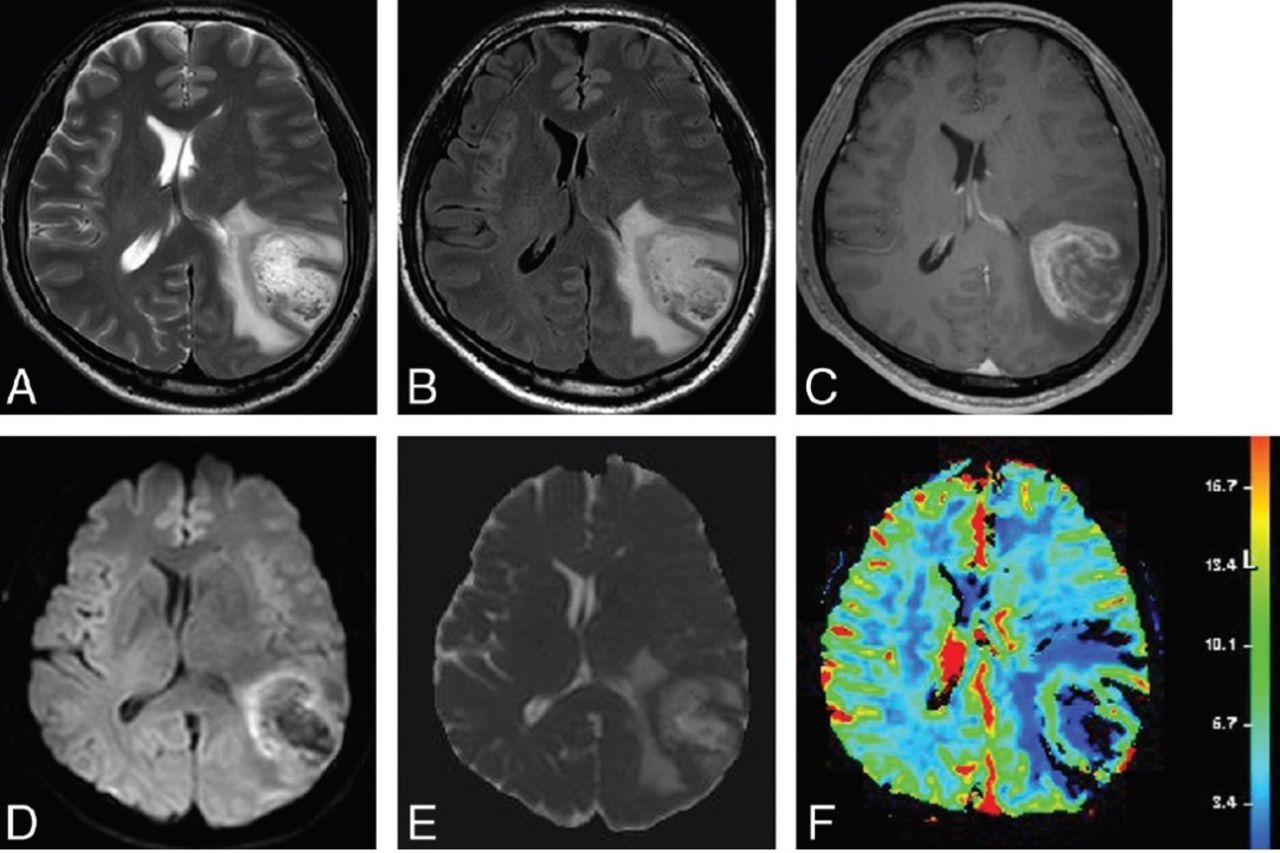
    - Magnetic resonance images from a male patient with biopsy-confirmed TDL. A) Axial T2-weighted and (B) axial FLAIR images show a well-defined hyperintense lesion in the left cerebral hemisphere with mild perilesional edema. (C) Contrast-enhanced axial T1WI shows open ring enhancement. (D) Axial DWI and (E) the corresponding ADC map reveal high ADC in the lesion and peripheral restricted diffusion. (F) DSC demonstrates low cerebral blood volume. Adapted from Suh et al.9

  • Figure 2
    • Download figure
    • Open in new tab
    • Download powerpoint
    Figure 2

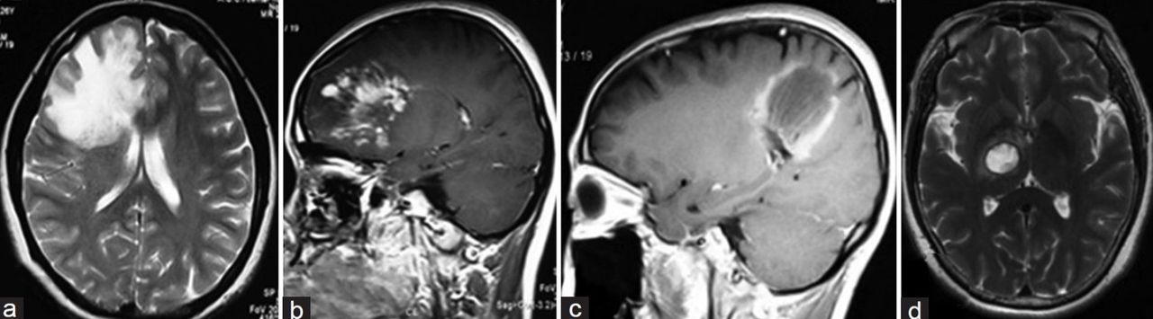
    - Three cases of TDLs showing the three different types of morphology on MRI: (a) Axial T2-weighted image showing diffuse, infiltrating lesion with indistinct margins. (b) Sagittal T1-weighted image with contrast highlighting infiltrating lesion heterogeneous enhancement. (c) Sagittal T1-weighted image with contrast showing ring-shaped enhancement. (d) Axial T2-weighted image demonstrating megacystic lesion with distinct margins.5

Tables

  • Figures
    • View popup
    Table 1

    - Common differential diagnoses of TDL and their main distinguishing laboratory and imaging features.

    Para-clinical featureTDLMSPCNSLHGG
    MRI T1WIHypointense Open/closed-ringGadolinium enhancing and non-enhancing lesions present2Uniform, contrast-enhancing in 98.9%, often contacting subarachnoid space Solitary in 50-81%3Heterogeneous enhancement4
    Gadolinium enhancementMultiple plaquesMay have necrotic areas4
    Distinct borders1
    MRI T2WIFLAIR: hyper-intense, ≥2 cm lesion (round, infiltrative or cystic)≥1 Ovoid lesion (>3 mm in size), in ≥2 sites of CNS5Usually hypointenseMostly hyper-intense
    Hypointense rimPerivenular2Mass effect, perilesional edema4Mass effect, edema4
    No/mild mass effect and/or edema1Central vein sign6
    Advanced imagingDSC/DWI: heterogeneous ADC values4Reduced amyloid PET activity in black hole areas in T1-weighted MR images9DSC/DWI: Homogenous and lower ADC values than TDL4DSC/DWI: higher ADC and CVB values than PCNSL and TDL4
    DSC/ASL perfusion: low CVB1,7Higher CVB than TDLs4,7
    FDG/MET-PET: high uptake10
    FDG/MET-PET: high uptake10
    FDG/MET-PET: No/low uptake8
    CSF OCBsPositive in 30%11 – 80%12Positive in 90%2Negative in 90%11Negative11
    Cell count42+-+++-
    (< 50/mL)
    MBP42-/+++-/++-/+-
    CSF biomarkers42IL-6 -/+IL-6 -sIL-2R -/+++sIL-2R -
    IL-10 -/++IL-10 -
    IL-6 -/++IL-6 -

    MRI T1WI: Magnetic resonance imaging T1 weighted image, MRI T2WI: Magnetic resonance imaging T2 weighted image, CSF OCBs: Cerebrospinal fluid Oligoclonal bands, TDL: Tumefactive demyelinating lesion, MS: Multiple Sclerosis, PCNSL: Primary Central Nervous system lymphoma, ADC: Apparent diffusion coefficient, CVB: Cerebral blood volume, DSC: Dynamic susceptibility contrast, DWI: Diffusion-weighted imaging, MBP: Myelin basic protein, FLAIR: Fluid attenuated inversion recovery, PET: Positron emission tomography, FDG: Fludeoxyglucose, MET: Metalized Polyethylene Terephthalate, IL: Interleukin

    PreviousNext
    Back to top

    In this issue

    Neurosciences Journal: 29 (3)
    Neurosciences Journal
    Vol. 29, Issue 3
    1 Jul 2024
    • Table of Contents
    • Cover (PDF)
    • Index by author
    Print
    Download PDF
    Email Article

    Thank you for your interest in spreading the word on Neurosciences Journal.

    NOTE: We only request your email address so that the person you are recommending the page to knows that you wanted them to see it, and that it is not junk mail. We do not capture any email address.

    Enter multiple addresses on separate lines or separate them with commas.
    Tumefactive demyelinating lesions: A literature review of recent findings
    (Your Name) has sent you a message from Neurosciences Journal
    (Your Name) thought you would like to see the Neurosciences Journal web site.
    Citation Tools
    Tumefactive demyelinating lesions: A literature review of recent findings
    Yaser M. Al Malik
    Neurosciences Journal Jul 2024, 29 (3) 153-160; DOI: 10.17712/nsj.2024.3.20230111

    Citation Manager Formats

    • BibTeX
    • Bookends
    • EasyBib
    • EndNote (tagged)
    • EndNote 8 (xml)
    • Medlars
    • Mendeley
    • Papers
    • RefWorks Tagged
    • Ref Manager
    • RIS
    • Zotero
    Share
    Tumefactive demyelinating lesions: A literature review of recent findings
    Yaser M. Al Malik
    Neurosciences Journal Jul 2024, 29 (3) 153-160; DOI: 10.17712/nsj.2024.3.20230111
    Twitter logo Facebook logo Mendeley logo
    • Tweet Widget
    • Facebook Like
    • Google Plus One
    Bookmark this article

    Jump to section

    • Article
      • Abstract
      • Acknowledgments
      • Footnotes
      • References
    • Figures & Data
    • eLetters
    • References
    • Info & Metrics
    • PDF

    Related Articles

    • No related articles found.
    • PubMed
    • Google Scholar

    Cited By...

    • No citing articles found.
    • Google Scholar

    More in this TOC Section

    • Structural and functional changes in the hippocampus induced by environmental exposures
    • Epilepsia partialis continua: A review
    Show more Review Article

    Similar Articles

    Navigate

    • home

    More Information

    • Help

    Additional journals

    • All Topics

    Other Services

    • About

    © 2025 Neurosciences Journal Neurosciences is copyright under the Berne Convention and the International Copyright Convention. All rights reserved. Neurosciences is an Open Access journal and articles published are distributed under the terms of the Creative Commons Attribution-NonCommercial License (CC BY-NC). Readers may copy, distribute, and display the work for non-commercial purposes with the proper citation of the original work. Electronic ISSN 1658-3183. Print ISSN 1319-6138.

    Powered by HighWire