
Main menu

  • Home
  • Content
    • Latest
    • Ahead of print
    • Archive
  • Info for
    • Authors
    • Reviewers
    • Subscribers
    • Institutions
    • Advertisers
  • About Us
    • About Us
    • Editorial Office
    • Editorial Board
  • More
    • Alerts
    • Feedback
    • Folders
    • Help
  • Other Publications
    • Saudi Medical Journal

User menu

  • My alerts
  • Log in

Search

  • Advanced search
Neurosciences Journal
  • Other Publications
    • Saudi Medical Journal
  • My alerts
  • Log in
Neurosciences Journal

Advanced Search

  • Home
  • Content
    • Latest
    • Ahead of print
    • Archive
  • Info for
    • Authors
    • Reviewers
    • Subscribers
    • Institutions
    • Advertisers
  • About Us
    • About Us
    • Editorial Office
    • Editorial Board
  • More
    • Alerts
    • Feedback
    • Folders
    • Help
  • Follow psmmc on Twitter
  • Visit psmmc on Facebook
  • RSS
Clinical ImageClinical Image
Open Access

Multiple cranial nerve enhancement in a case of neuronal ceroid lipofuscinosis type 8

Hanin Alsini, Reem Zakzouk, Khaled Hundallah and Brahim Tabarki
Neurosciences Journal January 2022, 27 (1) 50-51; DOI: https://doi.org/10.17712/nsj.2022.1.20210107
Hanin Alsini
From the Division of Paediatrics Neurology (Alsini, Hundallah, Tabarki), Department of Paediatrics; and the Division of Neuroradiology (Zakzouk), Department of Radiology, Prince Sultan Military Medical City, Riyadh, Kingdom of Saudi Arabia. Address correspondence and reprint request to: Dr. Hanin Alsini, Division of Neurology, Department of Paediatrics, Prince Sultan Military Medical, Riyadh, Kingdom of Saudi Arabia. E-mail: Orcid ID:
MD
  • Find this author on Google Scholar
  • Find this author on PubMed
  • Search for this author on this site
  • For correspondence: [email protected]
Reem Zakzouk
From the Division of Paediatrics Neurology (Alsini, Hundallah, Tabarki), Department of Paediatrics; and the Division of Neuroradiology (Zakzouk), Department of Radiology, Prince Sultan Military Medical City, Riyadh, Kingdom of Saudi Arabia. Address correspondence and reprint request to: Dr. Hanin Alsini, Division of Neurology, Department of Paediatrics, Prince Sultan Military Medical, Riyadh, Kingdom of Saudi Arabia. E-mail: Orcid ID:
MD
  • Find this author on Google Scholar
  • Find this author on PubMed
  • Search for this author on this site
  • For correspondence: [email protected]
Khaled Hundallah
From the Division of Paediatrics Neurology (Alsini, Hundallah, Tabarki), Department of Paediatrics; and the Division of Neuroradiology (Zakzouk), Department of Radiology, Prince Sultan Military Medical City, Riyadh, Kingdom of Saudi Arabia. Address correspondence and reprint request to: Dr. Hanin Alsini, Division of Neurology, Department of Paediatrics, Prince Sultan Military Medical, Riyadh, Kingdom of Saudi Arabia. E-mail: Orcid ID:
MD
  • Find this author on Google Scholar
  • Find this author on PubMed
  • Search for this author on this site
  • For correspondence: [email protected]
Brahim Tabarki
From the Division of Paediatrics Neurology (Alsini, Hundallah, Tabarki), Department of Paediatrics; and the Division of Neuroradiology (Zakzouk), Department of Radiology, Prince Sultan Military Medical City, Riyadh, Kingdom of Saudi Arabia. Address correspondence and reprint request to: Dr. Hanin Alsini, Division of Neurology, Department of Paediatrics, Prince Sultan Military Medical, Riyadh, Kingdom of Saudi Arabia. E-mail: Orcid ID:
MD
  • Find this author on Google Scholar
  • Find this author on PubMed
  • Search for this author on this site
  • For correspondence: [email protected]
  • Article
  • Figures & Data
  • eLetters
  • Info & Metrics
  • References
  • PDF
Loading

A 4 year-old boy, born of consanguineous parents, with a normal birth, and developmental history. He was referred to our clinic with history of ataxia, and exhibited no signs of seizures, visual impairment, or cognitive decline. Physical examination revealed an ataxic gait, and normal cranial nerves. Ophthalmological examination of both eyes revealed healthy optic discs. On the electroretinogram, the ABC wave was present of deformed shape and small amplitude, suggestive of left side visual pathway (retinal) abnormality. Electroencephalogram showed no epileptiform discharges. T2-weighted brain magnetic resonance imaging (MRI) revealed multiple areas of white-matter hyper-intensity, as well as thalamic hypo-intensity (Figure1). Post-contrast axial T1 weighted imaging (Figure 2) showed smooth enhancement of the cranial nerves, and cauda equina. Whole-exome sequencing revealed a homozygous pathogenic variant in the CLN8 gene (c.709G>A, p.(Gly237Arg).

Figure 1
  • Download figure
  • Open in new tab
  • Download powerpoint
Figure 1

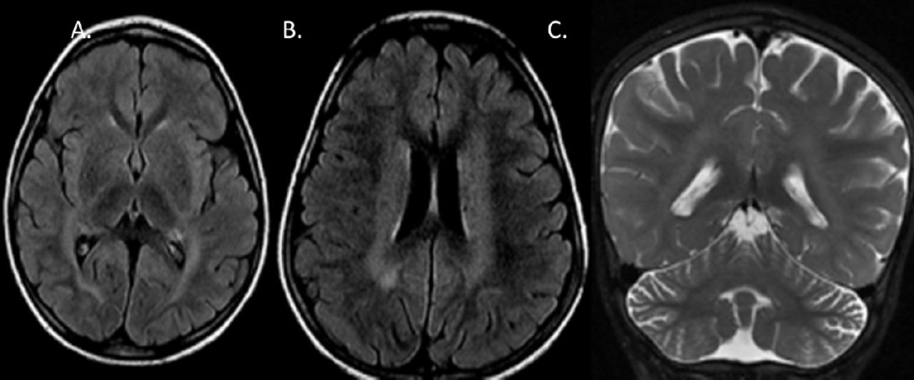
- Brain MRI. A) Axial FLAIR imaging shows hypointense thalami and hyperintense insular/sub-insular region and posterior limb of the internal capsule, B) hyper-intense periventricular and deep white matter. C) Coronal T2-weighted MR imaging shows mild cerebellar volume loss

Figure 2
  • Download figure
  • Open in new tab
  • Download powerpoint
Figure 2

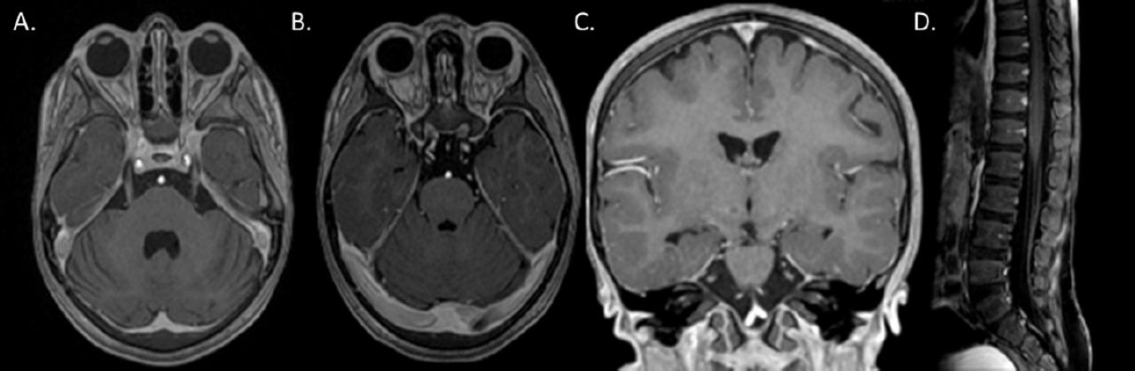
- Brain MRI. A, B) Axial T1-weighted MR with contrast shows striking enhancement of the trigeminal and abducens nerves bilaterally. C) Coronal T1 C+ MPRAGE MR reformat shows enhancement of facial and trigeminal nerves. D) Sagittal T1 C+ Fat suppressed MR of the lumbar spine shows nerve root enhancement

The MRI appearance of abnormal cranial nerve enhancement in MRI can be seen in various diseases ranging from common infectious, demyelinating, neoplastic, and autoimmune diseases, to rare neurodegenerative diseases.1 Neuronal ceroid lipofuscinoses (NCL) are the most common neurodegenerative disorders of childhood that involve the accumulation of lysosomal storage material. Although imaging findings of CLN8 has not been extensively described, reports have shown similar features compared to other CLN subtypes with presence of T2/FLAIR hyperintensity in the periventricular and deep white matter, subinsular regions, and posterior limb of internal capsule; along with T2 hypointense thalami, cerebral and cerebellar atrophy. Cranial nerve enhancement in NCL however has not been described in literature to the best of our knowledge.2 Cranial nerve enhancement in CLN8 suggests secondary autoimmunity, and inflammatory response in the central nervous system, as described in the literature in a mouse model of NCL.3 The reason for cranial nerve enhancement in our case in unclear, and may suggest an inflammatory process, as has been described in a mouse model of NCL.

  • Received August 22, 2021.
  • Accepted December 1, 2021.
  • Copyright: © Neurosciences

Neurosciences is an Open Access journal and articles published are distributed under the terms of the Creative Commons Attribution-NonCommercial License (CC BY-NC). Readers may copy, distribute, and display the work for non-commercial purposes with the proper citation of the original work.

References

  1. 1.↵
    1. Saremi F,
    2. Helmy M,
    3. Farzin S,
    4. Zee CS,
    5. Go JL.
    MRI of cranial nerve enhancement. Am J Roentgenol 2005; 185: 1487–1497.
    OpenUrlCrossRefPubMed
  2. 2.↵
    1. Biswas A,
    2. Krishnan P,
    3. Amirabadi A,
    4. Blaser S,
    5. Mercimek-Andrews S,
    6. Shroff M.
    Expanding the Neuroimaging Phenotype of Neuronal Ceroid Lipofuscinoses. AJNR Am J Neuroradiol 2020; 41: 1930–1936.
    OpenUrlAbstract/FREE Full Text
  3. 3.↵
    1. Xiong J,
    2. Kielian T.
    Microglia in juvenile neuronal ceroid lipofuscinosis are primed toward a pro-inflammatory phenotype. J Neurochem 2013; 127: 245–258.
    OpenUrlCrossRefPubMed
PreviousNext
Back to top

In this issue

Neurosciences Journal: 27 (1)
Neurosciences Journal
Vol. 27, Issue 1
1 Jan 2022
  • Table of Contents
  • Cover (PDF)
  • Index by author
Print
Download PDF
Email Article

Thank you for your interest in spreading the word on Neurosciences Journal.

NOTE: We only request your email address so that the person you are recommending the page to knows that you wanted them to see it, and that it is not junk mail. We do not capture any email address.

Enter multiple addresses on separate lines or separate them with commas.
Multiple cranial nerve enhancement in a case of neuronal ceroid lipofuscinosis type 8
(Your Name) has sent you a message from Neurosciences Journal
(Your Name) thought you would like to see the Neurosciences Journal web site.
Citation Tools
Multiple cranial nerve enhancement in a case of neuronal ceroid lipofuscinosis type 8
Hanin Alsini, Reem Zakzouk, Khaled Hundallah, Brahim Tabarki
Neurosciences Journal Jan 2022, 27 (1) 50-51; DOI: 10.17712/nsj.2022.1.20210107

Citation Manager Formats

  • BibTeX
  • Bookends
  • EasyBib
  • EndNote (tagged)
  • EndNote 8 (xml)
  • Medlars
  • Mendeley
  • Papers
  • RefWorks Tagged
  • Ref Manager
  • RIS
  • Zotero
Share
Multiple cranial nerve enhancement in a case of neuronal ceroid lipofuscinosis type 8
Hanin Alsini, Reem Zakzouk, Khaled Hundallah, Brahim Tabarki
Neurosciences Journal Jan 2022, 27 (1) 50-51; DOI: 10.17712/nsj.2022.1.20210107
Twitter logo Facebook logo Mendeley logo
  • Tweet Widget
  • Facebook Like
  • Google Plus One
Bookmark this article

Jump to section

  • Article
    • References
  • Figures & Data
  • eLetters
  • References
  • Info & Metrics
  • PDF

Related Articles

  • No related articles found.
  • PubMed
  • Google Scholar

Cited By...

  • No citing articles found.
  • Google Scholar

More in this TOC Section

  • Unilateral Lennox–Gastaut syndrome associated with Klippel–Trénaunay syndrome
  • A child with leukemia and behavioral changes
Show more Clinical Image

Similar Articles

Navigate

  • home

More Information

  • Help

Additional journals

  • All Topics

Other Services

  • About

© 2025 Neurosciences Journal Neurosciences is copyright under the Berne Convention and the International Copyright Convention. All rights reserved. Neurosciences is an Open Access journal and articles published are distributed under the terms of the Creative Commons Attribution-NonCommercial License (CC BY-NC). Readers may copy, distribute, and display the work for non-commercial purposes with the proper citation of the original work. Electronic ISSN 1658-3183. Print ISSN 1319-6138.

Powered by HighWire