
Main menu

  • Home
  • Content
    • Latest
    • Ahead of print
    • Archive
  • Info for
    • Authors
    • Reviewers
    • Subscribers
    • Institutions
    • Advertisers
  • About Us
    • About Us
    • Editorial Office
    • Editorial Board
  • More
    • Alerts
    • Feedback
    • Folders
    • Help
  • Other Publications
    • Saudi Medical Journal

User menu

  • My alerts
  • Log in

Search

  • Advanced search
Neurosciences Journal
  • Other Publications
    • Saudi Medical Journal
  • My alerts
  • Log in
Neurosciences Journal

Advanced Search

  • Home
  • Content
    • Latest
    • Ahead of print
    • Archive
  • Info for
    • Authors
    • Reviewers
    • Subscribers
    • Institutions
    • Advertisers
  • About Us
    • About Us
    • Editorial Office
    • Editorial Board
  • More
    • Alerts
    • Feedback
    • Folders
    • Help
  • Follow psmmc on Twitter
  • Visit psmmc on Facebook
  • RSS
OtherClinical Image
Open Access

Developmental regression and irritability

Naif M. AlAnazy, Saad A. Al-Shahwan, Zeeshan Azmat and Brahim Tabarki
Neurosciences Journal July 2014, 19 (3) 240-241;
Naif M. AlAnazy
MD
  • Find this author on Google Scholar
  • Find this author on PubMed
  • Search for this author on this site
Saad A. Al-Shahwan
MD
  • Find this author on Google Scholar
  • Find this author on PubMed
  • Search for this author on this site
Zeeshan Azmat
MD
  • Find this author on Google Scholar
  • Find this author on PubMed
  • Search for this author on this site
Brahim Tabarki
MD
  • Find this author on Google Scholar
  • Find this author on PubMed
  • Search for this author on this site
  • For correspondence: [email protected]
  • Article
  • Figures & Data
  • eLetters
  • Info & Metrics
  • References
  • PDF
Loading

Clinical Presentation

A 6-month-old girl was admitted with a 2-month history of irritability, unexplained intermittent fever, developmental regression, and myoclonic seizures. She is the product of first-degree consanguinity. The family history was remarkable for one sister who died at the age of 2 years with a similar neurological condition. Neurological examination showed excessive irritability, hypertonicity of the 4 limbs, hyperreflexia, and bilateral extensor plantar reflex. Systematic examination was unremarkable. A brain MRI was performed and is shown in Figures 1 & 2.

Figure 1
  • Download figure
  • Open in new tab
  • Download powerpoint
Figure 1

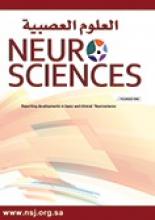
Axial T2-weighted patient brain MRI.

Figure 2
  • Download figure
  • Open in new tab
  • Download powerpoint
Figure 2

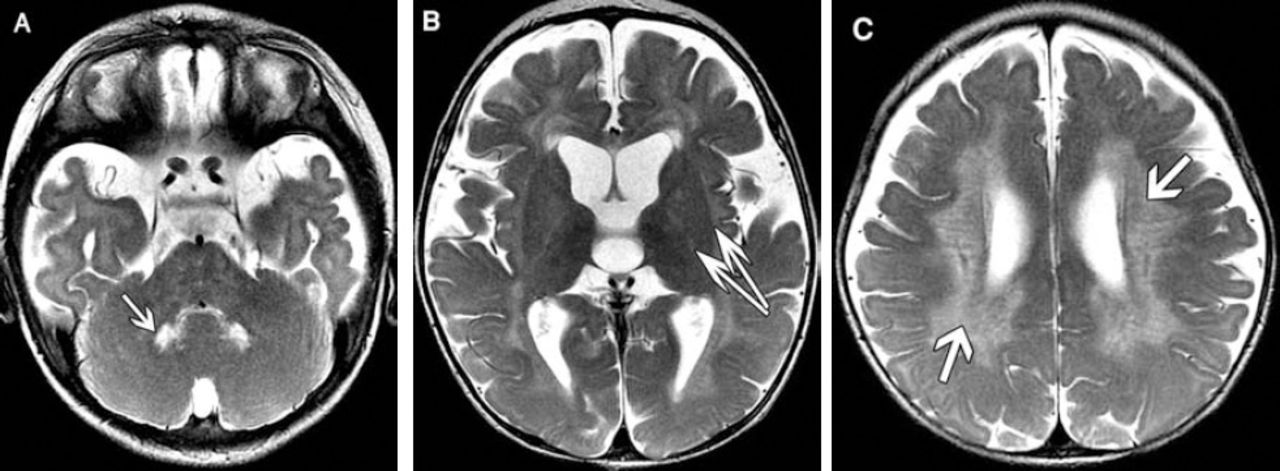
Axial and coronal T2-weighted patient brain MRI.

Questions

  1. What findings are seen in Figures 1 & 2?

  2. What is the most likely diagnosis?

  3. How is the diagnosis confirmed?

Answers & Discussion

  1. MRI findings. Figure 1: Axial T2-weighted images show A) hyperintense signal in deep cerebellar nuclei (arrow) bilaterally, B) in the bilateral posterior limb of the internal and external capsules, and C) bilateral symmetrical confluent hyperintense signals in the centrum semiovale and periventricular region sparing subcortical U fibers. Tubular linear areas of hypointensity are noted within the demyelinating bright areas extending from the periventricular region toward the cortex giving a tigroid pattern (stripe sign). Figure 2: Axial and coronal T2-weighted images (A & B) shows marked thickening of the optic nerves (arrow) and chiasm.

  2. Diagnosis. Based on the clinical presentation and neuroimaging findings, the most likely diagnosis is leukodystrophy type Krabbe disease.

  3. Confirmatory diagnosis of Krabbe disease requires enzymatic assay: b-galactocerebrosidase activity in peripheral blood leukocytes or skin fibroblasts. In our patient b-galactocerebrosidase enzyme activity was deficient. Genetic study showed mutations in the galactocerebrosidase gene.

Krabbe disease is an autosomal recessive lysosomal storage disease caused by mutations in the galactocerebrosidase gene leading to deficiency of the enzyme b-galactocerebrosidase, a key enzyme in the catabolism of myelin-enriched sphingolipids. Accumulation of globoid cells results in optic nerve enlargement.1 Clinically, the disease manifests early in infancy in 70-80% of patients. Infants usually present with spastic quadriparesis, tonic spasms, extreme irritability, inconsolable crying, progressive optic atrophy, peripheral neuropathy, and death usually due to severe respiratory infection at around the second year of life. The juvenile and adult forms are characterized by insidious visual compromise, cognitive deterioration, and gait disorders.1

The MRI abnormalities differed among phenotypes.2 Early infantile patients had a predominance of increased intensity in the dentate/cerebellar white matter as well as changes in the deep cerebral white matter. Later onset patients did not demonstrate involvement in the dentate/cerebellar white matter, but had extensive involvement of the deep cerebral white matter, parieto-occipital region, and posterior corpus callosum. Late infantile patients exhibited a mixed pattern. Adolescent/adult patients demonstrated isolated corticospinal tract involvement. The brain MRI may also show enlargement of the optic nerve and optic chiasma. Spinal MRI with contrast shows lumbosacral nerve root contrast enhancement. The differential diagnosis of optic nerve enlargement includes glioma, nerve sheath meningioma, dural ectasia, and neurofibromatosis type I.3

The diagnosis of Krabbe disease is challenging because of the large number of leukodystrophies, as well as the similarities between their clinical and imaging findings. Specific neuroimaging findings may help to establish this diagnosis. The detection of optic nerve enlargement, or lumbosacral nerve root contrast enhancement not found in other types of leukodystrophies, is an auxiliary tool in the differential diagnosis of KD.

Treatment of Krabbe disease is mainly symptomatic. At present, the only clinical treatment is hematopoietic cell transplantation. It is beneficial if performed before the onset of symptoms, but its efficacy in correcting the severe neurological disease is variable. Gene therapy is a promising alternative.1

Footnotes

  • Notice: Authors are encouraged to submit clinical images for possible publication in the Journal. These may be in any field of Clinical Neurosciences, and should approximately follow the format used here. Please address any submissions to the Assistant Editor, Neurosciences Journal, Prince Sultan Military Medical City, PO Box 7897, Riyadh 11159, Kingdom of Saudi Arabia. E-mail: sdouglas{at}psmmc.med.sa

  • Copyright: © Neurosciences

Neurosciences is an Open Access journal and articles published are distributed under the terms of the Creative Commons Attribution-NonCommercial License (CC BY-NC). Readers may copy, distribute, and display the work for non-commercial purposes with the proper citation of the original work.

References

  1. ↵
    1. Pastores GM
    (2009) Krabbe disease: an overview. Int J Clin Pharmacol Ther 47, Suppl 1, S75–S81.
  2. ↵
    1. Abdelhalim AN,
    2. Alberico RA,
    3. Barczykowski AL,
    4. Duffner PK
    (2014) Patterns of magnetic resonance imaging abnormalities in symptomatic patients with Krabbe disease correspond to phenotype. Pediatr Neurol 50, 127–134.
  3. ↵
    1. Patel B,
    2. Gimi B,
    3. Vachha B,
    4. Agadi S,
    5. Koral K
    (2008) Optic nerve and chiasm enlargement in a case of infantile Krabbe disease: quantitative comparison with 26 age-matched controls. Pediatr Radiol 38, 697–699.
PreviousNext
Back to top

In this issue

Neurosciences Journal: 19 (3)
Neurosciences Journal
Vol. 19, Issue 3
1 Jul 2014
  • Table of Contents
  • Cover (PDF)
  • Index by author
Print
Download PDF
Email Article

Thank you for your interest in spreading the word on Neurosciences Journal.

NOTE: We only request your email address so that the person you are recommending the page to knows that you wanted them to see it, and that it is not junk mail. We do not capture any email address.

Enter multiple addresses on separate lines or separate them with commas.
Developmental regression and irritability
(Your Name) has sent you a message from Neurosciences Journal
(Your Name) thought you would like to see the Neurosciences Journal web site.
Citation Tools
Developmental regression and irritability
Naif M. AlAnazy, Saad A. Al-Shahwan, Zeeshan Azmat, Brahim Tabarki
Neurosciences Journal Jul 2014, 19 (3) 240-241;

Citation Manager Formats

  • BibTeX
  • Bookends
  • EasyBib
  • EndNote (tagged)
  • EndNote 8 (xml)
  • Medlars
  • Mendeley
  • Papers
  • RefWorks Tagged
  • Ref Manager
  • RIS
  • Zotero
Share
Developmental regression and irritability
Naif M. AlAnazy, Saad A. Al-Shahwan, Zeeshan Azmat, Brahim Tabarki
Neurosciences Journal Jul 2014, 19 (3) 240-241;
Twitter logo Facebook logo Mendeley logo
  • Tweet Widget
  • Facebook Like
  • Google Plus One
Bookmark this article

Jump to section

  • Article
    • Clinical Presentation
    • Questions
    • Answers & Discussion
    • Footnotes
    • References
  • Figures & Data
  • eLetters
  • References
  • Info & Metrics
  • PDF

Related Articles

  • No related articles found.
  • Google Scholar

Cited By...

  • No citing articles found.
  • Google Scholar

More in this TOC Section

  • Multiple cranial nerve enhancement in a case of neuronal ceroid lipofuscinosis type 8
  • Unilateral Lennox–Gastaut syndrome associated with Klippel–Trénaunay syndrome
  • A child with leukemia and behavioral changes
Show more Clinical Image

Similar Articles

Navigate

  • home

More Information

  • Help

Additional journals

  • All Topics

Other Services

  • About

© 2025 Neurosciences Journal Neurosciences is copyright under the Berne Convention and the International Copyright Convention. All rights reserved. Neurosciences is an Open Access journal and articles published are distributed under the terms of the Creative Commons Attribution-NonCommercial License (CC BY-NC). Readers may copy, distribute, and display the work for non-commercial purposes with the proper citation of the original work. Electronic ISSN 1658-3183. Print ISSN 1319-6138.

Powered by HighWire