
Main menu

  • Home
  • Content
    • Latest
    • Ahead of print
    • Archive
  • Info for
    • Authors
    • Reviewers
    • Subscribers
    • Institutions
    • Advertisers
  • About Us
    • About Us
    • Editorial Office
    • Editorial Board
  • More
    • Alerts
    • Feedback
    • Folders
    • Help
  • Other Publications
    • Saudi Medical Journal

User menu

  • My alerts
  • Log in

Search

  • Advanced search
Neurosciences Journal
  • Other Publications
    • Saudi Medical Journal
  • My alerts
  • Log in
Neurosciences Journal

Advanced Search

  • Home
  • Content
    • Latest
    • Ahead of print
    • Archive
  • Info for
    • Authors
    • Reviewers
    • Subscribers
    • Institutions
    • Advertisers
  • About Us
    • About Us
    • Editorial Office
    • Editorial Board
  • More
    • Alerts
    • Feedback
    • Folders
    • Help
  • Follow psmmc on Twitter
  • Visit psmmc on Facebook
  • RSS
Review ArticleReview Article
Open Access

Focal brainstem gliomas

Advances in intra-operative management

Abdulrahman J. Sabbagh and Ahmed M. Alaqeel
Neurosciences Journal April 2015, 20 (2) 98-106; DOI: https://doi.org/10.17712/nsj.2015.2.20140621
Abdulrahman J. Sabbagh
From the Departments of Pediatric Neurosurgery (Sabbagh), National Neuroscience Institute, King Fahd Medical City, the Faculty of Medicine (Sabbagh), King Saud University for Health Sciences, the Division of Neurosurgery (Alaqeel), Department of Surgery, College of Medicine, King Saud University, Riyadh, Kingdom of Saudi Arabia, and the Department of Clinical Neurosciences (Alaqeel), Division of Neurosurgery, University of Calgary, Alberta, Canada
MBBS, FRCSC
  • Find this author on Google Scholar
  • Find this author on PubMed
  • Search for this author on this site
  • For correspondence: [email protected]
Ahmed M. Alaqeel
From the Departments of Pediatric Neurosurgery (Sabbagh), National Neuroscience Institute, King Fahd Medical City, the Faculty of Medicine (Sabbagh), King Saud University for Health Sciences, the Division of Neurosurgery (Alaqeel), Department of Surgery, College of Medicine, King Saud University, Riyadh, Kingdom of Saudi Arabia, and the Department of Clinical Neurosciences (Alaqeel), Division of Neurosurgery, University of Calgary, Alberta, Canada
MD
  • Find this author on Google Scholar
  • Find this author on PubMed
  • Search for this author on this site
  • Article
  • Figures & Data
  • eLetters
  • Info & Metrics
  • References
  • PDF
Loading

Article Figures & Data

Figures

  • Figure 1
    • Download figure
    • Open in new tab
    • Download powerpoint
    Figure 1

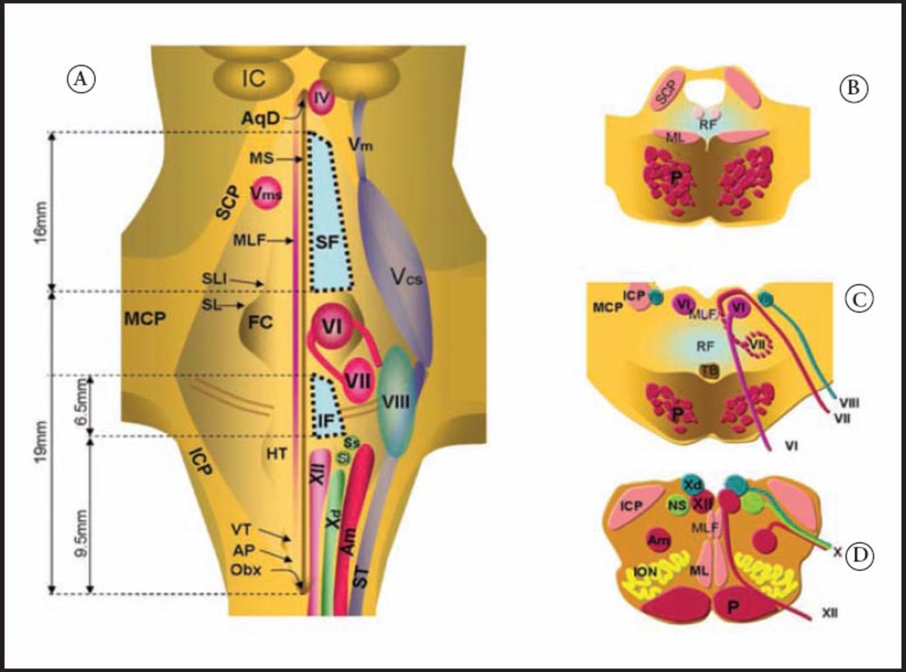
    *Illustration showing the A) dorsal view of the fourth ventricular floor. This illustration shows the supra-facial (SF) and infra-facial (IF) triangles. The column of illustrations on the right show corresponding axial sections through B) upper, C) mid, and D) lower pons. AqD - aqueduct of Sylvius, N - nucleus, IC - inferior colliculus, MS - median sulcus, Vm - mesencephalic N of the 5th cranial nerve (V), Vcs - chief (sensory) N of V, Vms - motor (mastication) N of V, MLF - medial longitudinal fascicle, FC - facial colliculus, IV - trochlear N, CTT - central tegmental tract, SL - sulcus limitans, SLI - sulcus limitans incisure, HT - hypoglossal triangle, SM - striae medullaris, SCP, MCP, SCP - superior, middle and inferior cerebellar peduncle, VT - vagal triangle, AP - area postrema, Obx - Obex, VI, Abducent N - VII, facial N. and fiber tracks and nerve, VIII - vestibular N. and nerve, XII - hypoglossal N. and nerve, Xd - dorsal vagal N, Am - N ambiguus of 9th and 10th cranial nerves with parasympathetics on its medial border, Ss & Si - superior and inferior salivatory NN., ST - spinal trigeminal tract, STT - spinothalamic tract, ML - medial lemniscus, ION - inferior olivary N., P - Pyramid, TB - trapezoid body, Pn TPF - pontine NN. and transverse pontine fibers, SF - Suprafacial triangle, IF - infrafacial triangle. *Figure 58.4 found on page 201 in Sabbagh AJ, Albanyan AA, Alyamany MA, Bunyan R, Abdelmoity AT, Soualmi LB. Case 58. In: Nader R, Sabbagh A, editors. Neurosurgery Case Review. NY (USA): Thieme; 2010 (reprinted with permission).

  • Figure 2
    • Download figure
    • Open in new tab
    • Download powerpoint
    Figure 2

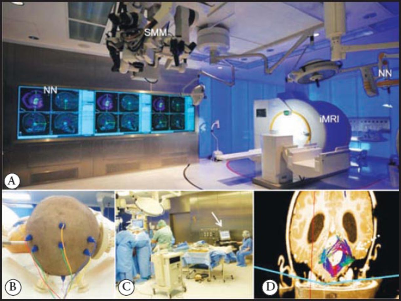
    Some of the available technologies A) intraoperative MRI suite where state of the art neuronavigation and intraoperative monitoring and imaging are performed at the National Neurosciences Institute, King Fahad Medical City, Riyadh, Saudi Arabia. B) patient’s head in prone position connected through corkscrew iOM electrodes shown inside the MRI coil. C) Surgery starts after connecting the patient, iOM staff and equipment (arrow). D) Tractography imaging showing tracts displaced by tumor (dotted arrow). iMRI - intraoperative MRI device, NN - neuronavigation equipment and screen, ceiling mounted microscope, iOM - intraoperative neurophysiologic monitoring

  • Figure 3
    • Download figure
    • Open in new tab
    • Download powerpoint
    Figure 3

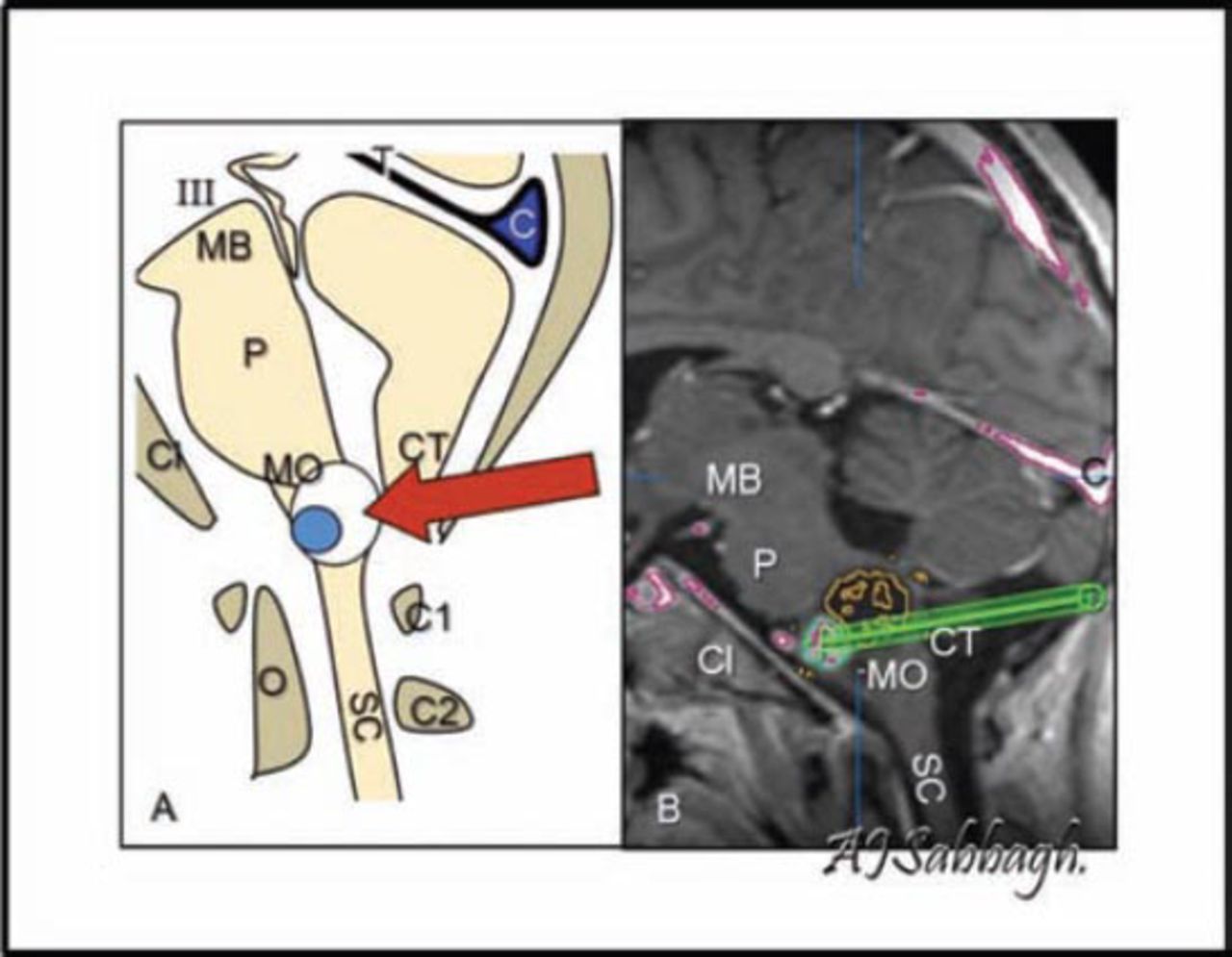
    Illustration depicting the posterior fossa, showing the trajectory to a medulla oblongata tumor (arrow) by artist rendering A) and neuronavigation screenshot B). The illustration shows the position of the neuronavigation probe (green rod), the third nerve (III), midbrain (MB), pons (P), medulla oblongata (MO), spinal cord (SC) clivus (CL), cerebellar tonsils (CT), odontoid process, tentorium (T), confluence of sinuses (C), cervical vertebrae one (C1), cervical vertebrae 2 (C2), odontoid (O)

  • Figure 4
    • Download figure
    • Open in new tab
    • Download powerpoint
    Figure 4

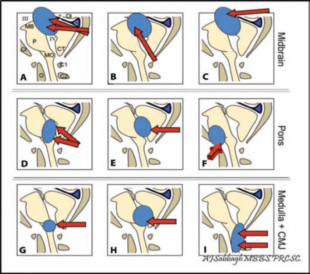
    Shows the diagrammatic illustration of some example approaches to the brainstem depending on region and location. Tectal region approaches (A, B, and C): A) supracerebellar-infratentorial, B) subvermial trans-forth ventricular, and C) occipital transtentorial approaches. Pontine region approaches (D, E, and F): D) telovelotonsillar, E) trans/subvermian, and F) transpetrosal (for anterolateral lesions) approaches. Cervico -medullary region approaches (G, H, and I): G) subvermian, H) Telovelotonsillar, and I) cervical approaches. The illustration shows the third nerve (III), midbrain (MB), pons (P), medulla oblongata (MO), spinal cord (SC) clivus (CL), cerebellar tonsils (CT), odontoid process, tentorium (T), and confluence of sinuses (C).

PreviousNext
Back to top

In this issue

Neurosciences Journal: 20 (2)
Neurosciences Journal
Vol. 20, Issue 2
1 Apr 2015
  • Table of Contents
  • Cover (PDF)
  • Index by author
Print
Download PDF
Email Article

Thank you for your interest in spreading the word on Neurosciences Journal.

NOTE: We only request your email address so that the person you are recommending the page to knows that you wanted them to see it, and that it is not junk mail. We do not capture any email address.

Enter multiple addresses on separate lines or separate them with commas.
Focal brainstem gliomas
(Your Name) has sent you a message from Neurosciences Journal
(Your Name) thought you would like to see the Neurosciences Journal web site.
Citation Tools
Focal brainstem gliomas
Abdulrahman J. Sabbagh, Ahmed M. Alaqeel
Neurosciences Journal Apr 2015, 20 (2) 98-106; DOI: 10.17712/nsj.2015.2.20140621

Citation Manager Formats

  • BibTeX
  • Bookends
  • EasyBib
  • EndNote (tagged)
  • EndNote 8 (xml)
  • Medlars
  • Mendeley
  • Papers
  • RefWorks Tagged
  • Ref Manager
  • RIS
  • Zotero
Share
Focal brainstem gliomas
Abdulrahman J. Sabbagh, Ahmed M. Alaqeel
Neurosciences Journal Apr 2015, 20 (2) 98-106; DOI: 10.17712/nsj.2015.2.20140621
Twitter logo Facebook logo Mendeley logo
  • Tweet Widget
  • Facebook Like
  • Google Plus One
Bookmark this article

Jump to section

  • Article
    • Abstract
    • Acknowledgment
    • Footnotes
    • References
  • Figures & Data
  • eLetters
  • References
  • Info & Metrics
  • PDF

Related Articles

  • No related articles found.
  • PubMed
  • Google Scholar

Cited By...

  • No citing articles found.
  • Google Scholar

More in this TOC Section

  • Structural and functional changes in the hippocampus induced by environmental exposures
  • Tumefactive demyelinating lesions: A literature review of recent findings
  • Epilepsia partialis continua: A review
Show more Review Article

Similar Articles

Navigate

  • home

More Information

  • Help

Additional journals

  • All Topics

Other Services

  • About

© 2025 Neurosciences Journal Neurosciences is copyright under the Berne Convention and the International Copyright Convention. All rights reserved. Neurosciences is an Open Access journal and articles published are distributed under the terms of the Creative Commons Attribution-NonCommercial License (CC BY-NC). Readers may copy, distribute, and display the work for non-commercial purposes with the proper citation of the original work. Electronic ISSN 1658-3183. Print ISSN 1319-6138.

Powered by HighWire