
Main menu

  • Home
  • Content
    • Latest
    • Ahead of print
    • Archive
  • Info for
    • Authors
    • Reviewers
    • Subscribers
    • Institutions
    • Advertisers
  • About Us
    • About Us
    • Editorial Office
    • Editorial Board
  • More
    • Alerts
    • Feedback
    • Folders
    • Help
  • Other Publications
    • Saudi Medical Journal

User menu

  • My alerts
  • Log in

Search

  • Advanced search
Neurosciences Journal
  • Other Publications
    • Saudi Medical Journal
  • My alerts
  • Log in
Neurosciences Journal

Advanced Search

  • Home
  • Content
    • Latest
    • Ahead of print
    • Archive
  • Info for
    • Authors
    • Reviewers
    • Subscribers
    • Institutions
    • Advertisers
  • About Us
    • About Us
    • Editorial Office
    • Editorial Board
  • More
    • Alerts
    • Feedback
    • Folders
    • Help
  • Follow psmmc on Twitter
  • Visit psmmc on Facebook
  • RSS
OtherClinical Image
Open Access

An adult with focal neurological deficits and inter-arm blood pressure difference

Ramachandiran Nandhagopal
Neurosciences Journal October 2015, 20 (4) 402-403; DOI: https://doi.org/10.17712/nsj.2015.4.20150042
Ramachandiran Nandhagopal
From the Neurology Unit, Department of Medicine, College of Medicine and Health Sciences, Al-Khod, Muscat, Oman
MBBS, DM
  • Find this author on Google Scholar
  • Find this author on PubMed
  • Search for this author on this site
  • For correspondence: [email protected]
  • Article
  • Figures & Data
  • eLetters
  • Info & Metrics
  • References
  • PDF
Loading

Clinical Presentation

A 62-year-old man, with no vascular risk factors or prior history of myocardial infarction, presented to the emergency department after experiencing a sudden collapse while driving. Neurological consultation was sought for a Glasgow coma scale of 9/15 and transient acute right hemiparesis 6 hours after the onset of his initial symptom. He scored 13 points on National Institute of Health stroke scale (maximum possible score of 42). His brachial systolic blood pressure was unequal: 90 mm Hg in the right arm and 120 mm Hg in the left arm. Imaging scans of brain (Figure A & B) and blood vessels (Figure C) were obtained.

Questions

  1. What findings are observed in Figure A & B?

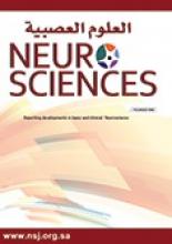
  2. What does the vascular imaging scan show (Figure C)?

  3. What is the diagnosis?

Answers

  1. Cranial diffusion weighted MRI (Figures A & B) and corresponding apparent diffusion coefficient map (insets in answer Figures A & B) revealed multiple, acute infarctions (arrows) in the right frontal and parietal areas, and left basal ganglia.

  2. Computed tomographic angiography showed the dissection of ascending, arch, and descending aorta (white arrows pointing to the true lumen) extending into the brachiocephalic trunk and left common carotid artery (arrowheads) (Figure C)

  3. Type A aortic dissection, causing bilateral carotid dissection, accounted for bilateral, multifocal acute infarctions in both cerebral hemispheres.

Discussion

Aortic dissection that involves the ascending aorta is termed type A aortic dissection and is a rare cause of acute ischemic stroke.1 Bedside monitoring of pulses and blood pressure in both arms could aid in suspecting aortic dissection in stroke patients with altered mental status (especially in those who could not provide history of chest or abdominal pain). An inter-arm pressure difference of more than 20 mm Hg is abnormal and suggests an underlying vascular disease such as aortic dissection or subclavian stenosis.2 If aortic dissection is suspected or confirmed, thrombolytic therapy should be avoided in the case of hyperacute presentation (within 4.5 hours of ischemic stroke onset). This patient presented beyond the thrombolytic time window and the extension of the dissection into bilateral cervical carotid arteries accounted for multiple infarctions in the bilateral carotid distribution (Figure 1). He was considered unsuitable for surgical (repair such as segmental resection and interposition of Dacron graft) or endovascular intervention, as there was an extensive dissection involving the ascending, arch and descending aorta along with complications/poor prognostic factors such as preoperative bilateral ischemic stroke and renal dysfunction as a result of ischemic nephropathy. He was managed conservatively and died of a fatal cardiac arrest 7 days later. Beta-blockers are utilized for treating high blood pressure in aortic dissection with a target systolic blood pressure of 100-120 mm Hg and heart rate of 60-80/minute.3

Figure 1
  • Download figure
  • Open in new tab
  • Download powerpoint
Figure 1

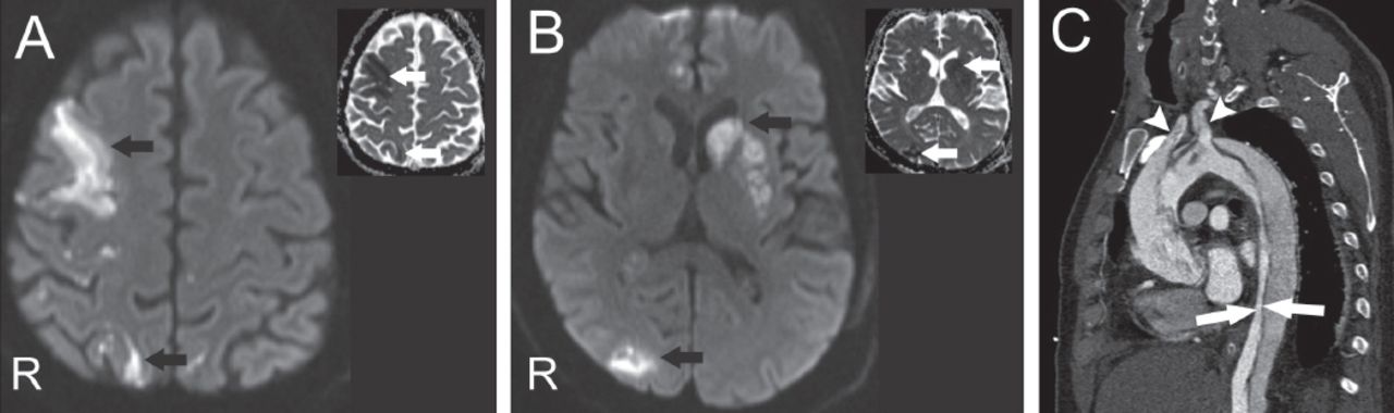
Axial diffusion weighted MRI (DWI, b=1000 sec/mm2) A & B) the brain and the corresponding apparent diffusion coefficient map (ADC map, insets in A & B) show restricted diffusion (black and white arrows), suggestive of acute infarction, in the right frontal and parietal convexities and left basal ganglia. C) Computerized tomographic angiography of the aorta showing dissection involving the whole length of aorta including the ascending segment (termed type A aortic dissection), with the arrows pointing to the true lumen in the descending aorta. Note the extension of dissection into brachiocephalic trunk (first white arrowhead) and left common carotid artery (second white arrowhead).

Footnotes

  • Notice: Authors are encouraged to submit quizzes for possible publication in the Journal. These may be in any specialty, and should approximately follow the format used here (maximum of 2 figures). Please address any submissions to: Editor, Saudi Medical Journal, Prince Sultan Military Medical City, PO Box 7897, Riyadh 11159, Kingdom of Saudi Arabia. Tel. +966 (11) 4777714 Ext. 42841. E-mail: info{at}smj.org.sa

  • Copyright: © Neurosciences

Neurosciences is an Open Access journal and articles published are distributed under the terms of the Creative Commons Attribution-NonCommercial License (CC BY-NC). Readers may copy, distribute, and display the work for non-commercial purposes with the proper citation of the original work.

References

  1. ↵
    1. Bossone E,
    2. Corteville DC,
    3. Harris KM,
    4. Suzuki T,
    5. Fattori R,
    6. Hutchison S,
    7. et al.
    (2013) Stroke and outcomes in patients with acute type A aortic dissection. Circulation 128, S175–S179.
  2. ↵
    1. Clark CE,
    2. Taylor RS,
    3. Shore AC,
    4. Ukoumunne OC,
    5. Campbell JL
    (2012) Association of a difference in systolic blood pressure between arms with vascular disease and mortality: a systematic review and meta-analysis. Lancet 379, 905–914.
  3. ↵
    1. Tsai TT,
    2. Nienaber CA,
    3. Eagle KA
    (2005) Acute aortic syndromes. Circulation 112, 3802–3813.
PreviousNext
Back to top

In this issue

Neurosciences Journal: 20 (4)
Neurosciences Journal
Vol. 20, Issue 4
1 Oct 2015
  • Table of Contents
  • Cover (PDF)
  • Index by author
Print
Download PDF
Email Article

Thank you for your interest in spreading the word on Neurosciences Journal.

NOTE: We only request your email address so that the person you are recommending the page to knows that you wanted them to see it, and that it is not junk mail. We do not capture any email address.

Enter multiple addresses on separate lines or separate them with commas.
An adult with focal neurological deficits and inter-arm blood pressure difference
(Your Name) has sent you a message from Neurosciences Journal
(Your Name) thought you would like to see the Neurosciences Journal web site.
Citation Tools
An adult with focal neurological deficits and inter-arm blood pressure difference
Ramachandiran Nandhagopal
Neurosciences Journal Oct 2015, 20 (4) 402-403; DOI: 10.17712/nsj.2015.4.20150042

Citation Manager Formats

  • BibTeX
  • Bookends
  • EasyBib
  • EndNote (tagged)
  • EndNote 8 (xml)
  • Medlars
  • Mendeley
  • Papers
  • RefWorks Tagged
  • Ref Manager
  • RIS
  • Zotero
Share
An adult with focal neurological deficits and inter-arm blood pressure difference
Ramachandiran Nandhagopal
Neurosciences Journal Oct 2015, 20 (4) 402-403; DOI: 10.17712/nsj.2015.4.20150042
Twitter logo Facebook logo Mendeley logo
  • Tweet Widget
  • Facebook Like
  • Google Plus One
Bookmark this article

Jump to section

  • Article
    • Clinical Presentation
    • Questions
    • Answers
    • Discussion
    • Footnotes
    • References
  • Figures & Data
  • eLetters
  • References
  • Info & Metrics
  • PDF

Related Articles

  • No related articles found.
  • PubMed
  • Google Scholar

Cited By...

  • No citing articles found.
  • Google Scholar

More in this TOC Section

  • Multiple cranial nerve enhancement in a case of neuronal ceroid lipofuscinosis type 8
  • Unilateral Lennox–Gastaut syndrome associated with Klippel–Trénaunay syndrome
  • A child with leukemia and behavioral changes
Show more Clinical Image

Similar Articles

Navigate

  • home

More Information

  • Help

Additional journals

  • All Topics

Other Services

  • About

© 2025 Neurosciences Journal Neurosciences is copyright under the Berne Convention and the International Copyright Convention. All rights reserved. Neurosciences is an Open Access journal and articles published are distributed under the terms of the Creative Commons Attribution-NonCommercial License (CC BY-NC). Readers may copy, distribute, and display the work for non-commercial purposes with the proper citation of the original work. Electronic ISSN 1658-3183. Print ISSN 1319-6138.

Powered by HighWire