
Main menu

  • Home
  • Content
    • Latest
    • Ahead of print
    • Archive
  • Info for
    • Authors
    • Reviewers
    • Subscribers
    • Institutions
    • Advertisers
  • About Us
    • About Us
    • Editorial Office
    • Editorial Board
  • More
    • Alerts
    • Feedback
    • Folders
    • Help
  • Other Publications
    • Saudi Medical Journal

User menu

  • My alerts
  • Log in

Search

  • Advanced search
Neurosciences Journal
  • Other Publications
    • Saudi Medical Journal
  • My alerts
  • Log in
Neurosciences Journal

Advanced Search

  • Home
  • Content
    • Latest
    • Ahead of print
    • Archive
  • Info for
    • Authors
    • Reviewers
    • Subscribers
    • Institutions
    • Advertisers
  • About Us
    • About Us
    • Editorial Office
    • Editorial Board
  • More
    • Alerts
    • Feedback
    • Folders
    • Help
  • Follow psmmc on Twitter
  • Visit psmmc on Facebook
  • RSS
OtherClinical Note
Open Access

Response to rituximab in an anti-muscle-specific receptor tyrosine kinase- and anti-acetylcholine recepto-double-positive myasthenia gravis patient concomitant with follicular dendritic cell sarcoma

Jianmeng Wang, Xiujuan Wu, Hui Deng, Ye Liu, Hongping Liu, Xueli Fan, Kangding Liu, Jiang Wu and Hong-Liang Zhang
Neurosciences Journal January 2016, 21 (1) 77-78; DOI: https://doi.org/10.17712/nsj.2016.1.20150483
Jianmeng Wang
From the Department of Neurology (Wang, Wu X, Deng, Liu H, Liu K, Wu J, Zhang H), Department of Pathology (Liu Y), the First Hospital of Jilin University, Jilin University, Changchun, China, and the Department of Neurobiology (Zhang), Care Sciences and Society, Karolinska Institute, Stockholm, Sweden
MD
  • Find this author on Google Scholar
  • Find this author on PubMed
  • Search for this author on this site
Xiujuan Wu
From the Department of Neurology (Wang, Wu X, Deng, Liu H, Liu K, Wu J, Zhang H), Department of Pathology (Liu Y), the First Hospital of Jilin University, Jilin University, Changchun, China, and the Department of Neurobiology (Zhang), Care Sciences and Society, Karolinska Institute, Stockholm, Sweden
MD
  • Find this author on Google Scholar
  • Find this author on PubMed
  • Search for this author on this site
Hui Deng
From the Department of Neurology (Wang, Wu X, Deng, Liu H, Liu K, Wu J, Zhang H), Department of Pathology (Liu Y), the First Hospital of Jilin University, Jilin University, Changchun, China, and the Department of Neurobiology (Zhang), Care Sciences and Society, Karolinska Institute, Stockholm, Sweden
MD, PhD
  • Find this author on Google Scholar
  • Find this author on PubMed
  • Search for this author on this site
Ye Liu
From the Department of Neurology (Wang, Wu X, Deng, Liu H, Liu K, Wu J, Zhang H), Department of Pathology (Liu Y), the First Hospital of Jilin University, Jilin University, Changchun, China, and the Department of Neurobiology (Zhang), Care Sciences and Society, Karolinska Institute, Stockholm, Sweden
MD, PhD
  • Find this author on Google Scholar
  • Find this author on PubMed
  • Search for this author on this site
Hongping Liu
From the Department of Neurology (Wang, Wu X, Deng, Liu H, Liu K, Wu J, Zhang H), Department of Pathology (Liu Y), the First Hospital of Jilin University, Jilin University, Changchun, China, and the Department of Neurobiology (Zhang), Care Sciences and Society, Karolinska Institute, Stockholm, Sweden
MD
  • Find this author on Google Scholar
  • Find this author on PubMed
  • Search for this author on this site
Xueli Fan
From the Department of Neurology (Wang, Wu X, Deng, Liu H, Liu K, Wu J, Zhang H), Department of Pathology (Liu Y), the First Hospital of Jilin University, Jilin University, Changchun, China, and the Department of Neurobiology (Zhang), Care Sciences and Society, Karolinska Institute, Stockholm, Sweden
MD
  • Find this author on Google Scholar
  • Find this author on PubMed
  • Search for this author on this site
Kangding Liu
From the Department of Neurology (Wang, Wu X, Deng, Liu H, Liu K, Wu J, Zhang H), Department of Pathology (Liu Y), the First Hospital of Jilin University, Jilin University, Changchun, China, and the Department of Neurobiology (Zhang), Care Sciences and Society, Karolinska Institute, Stockholm, Sweden
MD, PhD
  • Find this author on Google Scholar
  • Find this author on PubMed
  • Search for this author on this site
Jiang Wu
From the Department of Neurology (Wang, Wu X, Deng, Liu H, Liu K, Wu J, Zhang H), Department of Pathology (Liu Y), the First Hospital of Jilin University, Jilin University, Changchun, China, and the Department of Neurobiology (Zhang), Care Sciences and Society, Karolinska Institute, Stockholm, Sweden
MD, PhD
  • Find this author on Google Scholar
  • Find this author on PubMed
  • Search for this author on this site
  • For correspondence: [email protected]
Hong-Liang Zhang
From the Department of Neurology (Wang, Wu X, Deng, Liu H, Liu K, Wu J, Zhang H), Department of Pathology (Liu Y), the First Hospital of Jilin University, Jilin University, Changchun, China, and the Department of Neurobiology (Zhang), Care Sciences and Society, Karolinska Institute, Stockholm, Sweden
MD, PhD
  • Find this author on Google Scholar
  • Find this author on PubMed
  • Search for this author on this site
  • Article
  • Figures & Data
  • eLetters
  • Info & Metrics
  • References
  • PDF
Loading

Myasthenia gravis (MG) is an antibody-mediated autoimmune disorder affecting neuromuscular junctions, which is strongly associated with thymoma. Usually, the target of the autoimmune attack is the acetylcholine receptor (AChR) of the postsynaptic skeletal muscle endplate. However, non-AChR components, such as muscle-specific receptor tyrosine kinase (MuSK) and low-density lipoprotein receptor related protein 4 (LRP4), both of which are involved in the endplate maturation, may also serve as targets for autoantibodies.1 Myasthenia gravis may develop secondary to follicular dendritic cell sarcoma (FDCS), a rare malignant neoplasm of follicular dendritic cells (FDCs).2 However, therapeutic options for these MG patients are unclear. We herein illustrated the therapeutic effect of Rituximab on an anti-MuSK- and anti-AChR-positive MG patient concomitant with FDCS.

A 60-year-old woman was admitted to the in-patient department with 20 days symptoms of exertion dysphagia and dyspnea. She has a 6-month history of fatigable limb weakness, fluctuating ptosis, and diplopia, which worsened with activity and ameliorated with rest. Her fluctuating symptoms were responsive to intramuscular neostigmine. She had been diagnosed with recurrent FDCS during the past 15 years, and lymphadenectomy had been performed for 4 times. Upon physical examination, she was fully alert and oriented. Bilateral ptosis, weakness of eyelid closure without lower facial involvement, weakened lifting of bilateral soft palates, and head drop were noted while the other cranial nerves were intact. The muscle strength grade was Medical Research Council (MRC) Grade-4 in the limbs and tendon reflexes were normal. The Babinski signs were negative bilaterally. Lymph nodes were palpable in her left cervical and axillary regions. The laboratory investigations including routine blood test, liver functions, and renal, serum cancer marker (carcino-embryonic antigen, CA199, CA153, CA724, CA242, neuron-specific enolase, free beta-human chorionic gonadotrnpin, squamous cell carcinoma antigen, alpha fetoprotein) and paraneoplastic neurological syndrome (PNS) screening were unremarkable. The antibodies to AChR (optical density [OD] value=0.442, reference value: OD value <0.367) and MuSK (OD=0.846, reference value: OD <0.512) were positive. Thoracic and abdominal CT revealed no thymic abnormality, or any mediastinal or distant sites of mass suggestive of possible extranodal lesion. Instead, lymphadenectasis of her left cervical and axillary region was reported. Needle biopsy of the lymph node and pathology confirmed a diagnosis of FDCS (Figure 1). The Hematoxylin and Eosin staining revealed diffuse proliferation of spindled cells with nuclear atypia (Figure 1A) and eosinophilic cytoplasm (Figure 1B). Scattered small lymphoid cells and perivascular (arrow) clusters of small lymphocytes (Figure 1C) are also marked. Immunohistochemistry demonstrates positivity of tumor cells for CD21 (Figure 1D), CD35 (Figure 1E), vimentin (Figure 1F), S-100 (Figure 1G), CD3 (Figure 1H) and CD1a (not shown) while negativity for EMA, CK, CD20, lysozyme, MPO, CD34, CD30, and HMB45 (not shown). The Ki-67 labeling index of these cells reached approximately 20%. Based on the work-up, recurrent FDCS associated MG were diagnosed. On day 7 after admission, respiratory failure requiring intubation and ventilation indicative of myasthenic crisis led to her transfer to the Neurological Intensive Care Unit (N-ICU). Intravenous immunoglobulin (ivIg) was administered (400 mg/kg/day for 5 days) followed by tapered methylprednisolone (1000 mg, 500 mg, 250 mg, and 120 mg methylprednisolone, and each dose of steroids were used for 3 days before the one mg/kg/day oral methylprednisolone) was prescribed, which resulted in gradual recovery and extubation 30 days later. However, on day 38 of hospitalization, bilateral ptosis, weakness of eyelid closure and head drop were noted again, and the myasthenic crisis recurred subsequently without any identified causes like infections, or abrupt reduction of oral methylprednisolone. Considering the exacerbation and myasthenic crisis, together with the positive antibodies associated with MG, rituximab (375 mg/m2 iv injection per week for 4 consecutive weeks) was immediately prescribed in addition to oral methylprednisolone (one mg/kg/day). Gradually, she regained spontaneous respiration with a maximum of 10 hours after the first administration, and succeeded in extubation on day 53 of hospitalization.

Figure 1
  • Download figure
  • Open in new tab
  • Download powerpoint
Figure 1

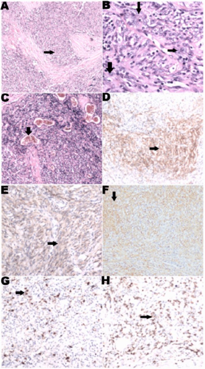
Pathological and immunohistochemical features of the follicular dendritic cell sarcoma (FDCS). The Hematoxylin and Eosin staining reveal diffuse proliferation of spindled cells with nuclear atypia (arrows, A to C, ×40). Eosinophilic cytoplasm with indistinct cytoplasmic borders and vesicular nuclei of the spindled cells are shown (B×200, arrows). Scattered small lymphoid cells and perivascular (arrow) clusters of small lymphocytes (C×40) are also marked. Immunohistochemistry demonstrates positivity of tumor cells for CD21 (D×100), CD35 (E×100), vimentin (F×100), S-100 (G×100), CD3 (H×100) and CD1a (arrows).

Our experience from this case favors use of rituximab in FDCS-associated anti-MuSK- and anti-AChR-positive MG. Interestingly, CD3 and CD1a, which are negative in most FDCS were positive in this case. This variant subtype has been scarcely reported to be related to the onset of MG.3 In the anti-AChR-positive MG, antibodies of Ig G1 and IgG3 isotype may potentially activate complement and attract lymphocytes, which results in the loss of functional AChRs.1 While in anti-MuSK-positive MG, the non-complement-binding IgG4 subclass interferes with the cellular agrin-MuSK signaling cascade and contributes to the formation of AChR-rapsyn clusters, which result in a reduction of the functional AChRs without the loss of junctional folds or AChR density.1 Moreover, patients with anti-MuSK-positive MG usually manifest with atypical severe clinical symptoms with a predilection of middle-aged woman.1 The occurrence of positive antibodies to MuSK and AChR, alleged double-positive MG, has been documented in literature.3 In this case, anti-MuSK MG is associated mostly with bulbar symptoms and limb weakness is uncommon. Also, the association between thymoma and anti-AchR antibodies is uncommon.

The pathogenesis of MG complicated with FDCS remains largely unknown. FDCS might mediate aberrant immune activation through different mechanisms, such as disturbing cytokine production and leading to the imbalance of the immunological microenvironment.

Rituximab is a monoclonal antibody targeting CD20 antigen and may deplete B cells, mainly pre-B and mature B cells, via cell-mediated or complement-dependent cytotoxicity.4 In addition to its confirmatory effects on B cell lymphoma, rituximab has been occasionally tested in various antibody-mediated autoimmune disorders, including neuromyelitis optica and MG.1 Rituximab proved to be effective in treating MG, particularly in dealing with refractory MG.3 The positive anti-MuSK- and anti-AChR autoantibodies in our patient indicated a pathogenic role of antibody-producing B cells. Given her recurrent myasthenic crisis and poor response to the methylprednisolone therapy, we prescribed rituximab which led to a marked improvement. Although the pathogenesis of MG concurrent with FDCS is unclear, rituximab appears to be a promising alternative in the treatment of these patients with refractory MG.

In summary, refractory MG might be associated with FDCS. Rituximab appears effective in dealing with patients with refractory MG.

  • Received July 26, 2015.
  • Accepted November 25, 2015.
  • Copyright: © Neurosciences

Neurosciences is an Open Access journal and articles published are distributed under the terms of the Creative Commons Attribution-NonCommercial License (CC BY-NC). Readers may copy, distribute, and display the work for non-commercial purposes with the proper citation of the original work.

References

  1. ↵
    1. Meriggioli MN,
    2. Sanders DB
    (2009) Autoimmune myasthenia gravis:emerging clinical and biological heterogeneity. Lancet Neurol 8, 475–490.
  2. ↵
    1. Kairouz S,
    2. Hashash J,
    3. Kabbara W,
    4. McHayleh W,
    5. Tabbara IA
    (2007) Dendritic cell neoplasms:an overview. Am J Hematol 82, 924–928.
  3. ↵
    1. Zouvelou V,
    2. Zisimopoulou P,
    3. Psimenou E,
    4. Matsigkou E,
    5. Stamboulis E,
    6. Tzartos SJ
    (2014) AChR-myasthenia gravis switching to double-seropositive several years after the onset. J Neuroimmunol 267, 111–112.
  4. ↵
    1. Yang CS,
    2. Yang L,
    3. Li T,
    4. Zhang DQ,
    5. Jin WN,
    6. Li MS,
    7. et al.
    (2013) Responsiveness to reduced dosage of rituximab in Chinese patients with neuromyelitis optica. Neurology 81, 710–713.
PreviousNext
Back to top

In this issue

Neurosciences Journal: 21 (1)
Neurosciences Journal
Vol. 21, Issue 1
1 Jan 2016
  • Table of Contents
  • Cover (PDF)
  • Index by author
Print
Download PDF
Email Article

Thank you for your interest in spreading the word on Neurosciences Journal.

NOTE: We only request your email address so that the person you are recommending the page to knows that you wanted them to see it, and that it is not junk mail. We do not capture any email address.

Enter multiple addresses on separate lines or separate them with commas.
Response to rituximab in an anti-muscle-specific receptor tyrosine kinase- and anti-acetylcholine recepto-double-positive myasthenia gravis patient concomitant with follicular dendritic cell sarcoma
(Your Name) has sent you a message from Neurosciences Journal
(Your Name) thought you would like to see the Neurosciences Journal web site.
Citation Tools
Response to rituximab in an anti-muscle-specific receptor tyrosine kinase- and anti-acetylcholine recepto-double-positive myasthenia gravis patient concomitant with follicular dendritic cell sarcoma
Jianmeng Wang, Xiujuan Wu, Hui Deng, Ye Liu, Hongping Liu, Xueli Fan, Kangding Liu, Jiang Wu, Hong-Liang Zhang
Neurosciences Journal Jan 2016, 21 (1) 77-78; DOI: 10.17712/nsj.2016.1.20150483

Citation Manager Formats

  • BibTeX
  • Bookends
  • EasyBib
  • EndNote (tagged)
  • EndNote 8 (xml)
  • Medlars
  • Mendeley
  • Papers
  • RefWorks Tagged
  • Ref Manager
  • RIS
  • Zotero
Share
Response to rituximab in an anti-muscle-specific receptor tyrosine kinase- and anti-acetylcholine recepto-double-positive myasthenia gravis patient concomitant with follicular dendritic cell sarcoma
Jianmeng Wang, Xiujuan Wu, Hui Deng, Ye Liu, Hongping Liu, Xueli Fan, Kangding Liu, Jiang Wu, Hong-Liang Zhang
Neurosciences Journal Jan 2016, 21 (1) 77-78; DOI: 10.17712/nsj.2016.1.20150483
Twitter logo Facebook logo Mendeley logo
  • Tweet Widget
  • Facebook Like
  • Google Plus One
Bookmark this article

Jump to section

  • Article
    • References
  • Figures & Data
  • eLetters
  • References
  • Info & Metrics
  • PDF

Related Articles

  • No related articles found.
  • PubMed
  • Google Scholar

Cited By...

  • No citing articles found.
  • Google Scholar

More in this TOC Section

  • How to make a decision on the choice of therapy for multiple sclerosis with sarcoidosis comorbidity?
  • Mycoplasma pneumonia and stroke: causation, association, or coincidence?
  • Posterior reversible encephalopathy syndrome overshadowing COVID-19: An Abstruse scenario or a conspicuous association?
Show more Clinical Note

Similar Articles

Navigate

  • home

More Information

  • Help

Additional journals

  • All Topics

Other Services

  • About

© 2025 Neurosciences Journal Neurosciences is copyright under the Berne Convention and the International Copyright Convention. All rights reserved. Neurosciences is an Open Access journal and articles published are distributed under the terms of the Creative Commons Attribution-NonCommercial License (CC BY-NC). Readers may copy, distribute, and display the work for non-commercial purposes with the proper citation of the original work. Electronic ISSN 1658-3183. Print ISSN 1319-6138.

Powered by HighWire