


Cerebral venous thrombosis (CVT) is uncommon and its clinical presentation is extremely variable and potentially life-threatening. The highly variable clinical presentation creates a diagnostic challenge for clinicians. The onset can be acute, subacute, or chronic. The symptoms and signs of CVT can be grouped into 3 major syndromes: 1) isolated intracranial hypertension syndrome (headache, intracranial hypertension features namely, papilledema, double vision and visual obscurations), 2) focal syndrome (focal neurological deficits, epileptic seizures), and 3) encephalopathy (multifocal signs, mental status changes, stupor, or coma).1,2 Many conditions have been considered as risk factors for CVT in adults. Many of these conditions are permanent, while some are transient, namely, dehydration, pregnancy, and so forth. The most frequent are prothrombotic conditions (antithrombin deficiency, protein C or S deficiency, or Factor V Leiden mutation).1 Cushing’s syndrome can be caused by oral, injected, topical, or inhaled glucocorticoids.3 Hypercortisolism produced by Cushing disease or syndrome can result in thromboembolic complications.4 Use of high dose intravenous (IV) steroids for neurological conditions such as multiple sclerosis has been associated with development of systemic as well as cerebral venous thrombosis;5 however, a similar association has not been shown with the use of topical steroids. It is not known whether topical steroids have similar prothrombotic effect as the systemic steroids. In this article, we report a case of a patient who developed CVT due to hypercoagulable state in Cushing’s syndrome, which in turn was thought to be caused by over use of topical steroids over long period. We want to draw the attention of the readers to this rare and potentially devastating condition; helping increase clinicians’ awareness of this unusual and rare cause of CVT.
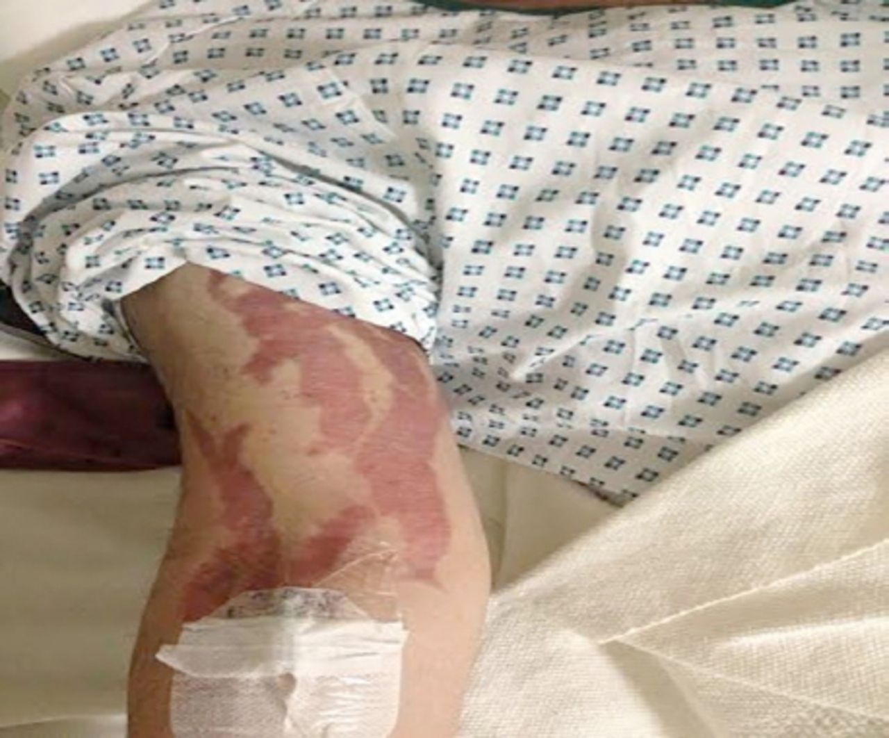
A 21-year-old gentleman presented to our emergency room complaining of headache and double vision for 3 weeks. Headache was constant and severe involving the entire head accompanied by nausea and vomiting. Eight months ago, he started to develop easy-fatigability and somnolence. He noted increase in his weight, mainly the abdomen, and upper back. This was associated with red/purple striae over his body, starting as small areas in his arm, then becoming wider; spreading over the trunk and thighs. He also complained of light bruising. He had a history of using copious amounts of topical steroids (clobetasol propionate) for more than a year; self-treating the post-shaving skin irritation. He did not have other neurological symptoms. There was no history of any oro-genital ulcers, redness of eyes, or prior systemic thrombosis. He denied any previous similar attacks. His general physical examination showed moon facies, abdominal obesity, posterior neck fat pads (buffalo hump), wide red striae over arms, abdomen, and thighs (Figure 1). Neurological examination showed he was fully conscious, alert, and oriented, with no cognitive dysfunction. Cranial nerve examination showed bilateral papilledema grade III, limited abduction of left eye, and areas of visual obscuration. Rest of cranial nerve examination was normal. The motor examination of upper limbs was normal including bulk, tone, power, and deep tendon reflexes. Lower limbs motor examination revealed mild bilateral proximal weakness grade 4/5 in the Medical Research Council Scale (MRC). There was no ataxia and no sensory deficits. Deep tendon reflexes were symmetrical, 2+ with down going plantars bilaterally.

Right arm and forearm showing broad and extensive striae.
Laboratory investigations
His routine blood investigations including complete blood count, chemistries, liver function tests, renal function tests, and coagulation profile were normal. Glycosylated hemoglobin was 6.5%, autoimmune profile including vasculitis workup, serum vitamin B12 levels, folate, and thyroid function tests were all normal. Coagulopathy workup including protein C, antithrombin III, prothrombin, and factor V Leiden mutation, factor II prothrombin analysis, C3 and C4 complement levels, homocysteine, anticardiolipin antibodies were all normal. Protein S was slightly low 41.2% (normal range 65-140) while he was taking oral anticoagulation, and was likely artifactual. Cortisol level in serum repeated many times was within normal range as well as cortisol level in 24-hour urine. Non-contrasted CT scan of brain was normal. The CT venogram showed superior sagittal, left transverse, and sigmoid venous thrombosis extending to the left jugular vein. Brain MRI revealed a normal brain parenchymal findings. He was treated with subcutaneous low molecular heparin followed by oral anticoagulation therapy with the international normalized ration target of 2-3. The initial symptoms of headache and diplopia started to decrease 7 days after admission. At 3 month follow up, his symptoms and signs had completely resolved. The diagnosis of cerebral venous thrombosis requires a high index of suspicion as the presentations are highly variable. There is no definitive way of making the diagnosis clinically only. Neuroimaging is the mainstay of confirming the clinical impression. Hyperdensity in the region of cerebral venous sinuses raises the possibility of CVT; however, the anatomic variations of cerebral venous sinuses make this finding less sensitive.1 Computerized tomography venography has evolved as a rapid and reliable method of confirming cerebral venous thrombosis. Magnetic resonance venogram (MRV) is another non-invasive method of detecting CVT. The conventional angiography with direct visualization of cerebral venous sinuses are not used as often now, due to the availability of CTV and MRV.1
Cerebral venous thrombosis is known to occur with high dose intravenous steroid use;5 however, there are no reports of cerebral venous thrombosis associated with the use of topical steroids. The extensive investigation for prothrombotic conditions was negative in our patient, we consider the use of topical steroids as the most plausible explanation of CVT in our case. Predisposing risk factors are found in 85%, these may include prothrombotic conditions, head injury, infectious diseases, dehydration and malignancy; the underlying cause in the rest 15% of cases cannot be determined.2 One of the underlying pathogenesis of cerebral venous thrombosis is occlusion of dural sinus resulting in decreased CSF absorption and elevated intracranial pressure. Our patient complained of double vision, which is not a commonly reported symptom of CVT. If the intracranial pressure is quite high, a sixth cranial nerve palsy may develop. Hypercortisolism seen with Cushing syndrome increases the risk of thromboembolic complications.4 Although the pathogenetic mechanism of this predisposition to hypercoagulation is not fully understood. Literature review shows that CVT may occur with an estimated incidence of 0.58% following steroid administration in MS patients with no other risk factors. High dose intravenous (IV) steroids are used to treat many neurological conditions; however, the reported incidence of cerebral venous thrombosis due to high dose IV steroids use is very low.5
In our case, there was no history of CVT risk factors such as smoking, diabetes mellitus, systemic venous thrombosis, or other systemic or inflammatory diseases, as well as there was absence of clinical signs of dehydration or infection. An extensive laboratory investigations for thrombophilia in our patient demonstrated no underlying risk factors associated with CVT. This excluded essentially all other etiological factors for cerebral venous thrombosis, leading us to conclude that the use of large amounts of topical steroids was the culprit for CVT in our patient. We suggest that physicians should be aware that Cushing’s syndrome can occur through topical steroids, and it is a possible cause of cerebral sinus thrombosis.
Footnotes
Disclosure
The authors declare no conflicting interests, support or funding from any drug company.
- Received August 20, 2015.
- Accepted November 25, 2015.
- Copyright: © Neurosciences
Neurosciences is an Open Access journal and articles published are distributed under the terms of the Creative Commons Attribution-NonCommercial License (CC BY-NC). Readers may copy, distribute, and display the work for non-commercial purposes with the proper citation of the original work.
In this issue


Related Articles
Cited By...
- No citing articles found.