
Main menu

  • Home
  • Content
    • Latest
    • Ahead of print
    • Archive
  • Info for
    • Authors
    • Reviewers
    • Subscribers
    • Institutions
    • Advertisers
  • About Us
    • About Us
    • Editorial Office
    • Editorial Board
  • More
    • Alerts
    • Feedback
    • Folders
    • Help
  • Other Publications
    • Saudi Medical Journal

User menu

  • My alerts
  • Log in

Search

  • Advanced search
Neurosciences Journal
  • Other Publications
    • Saudi Medical Journal
  • My alerts
  • Log in
Neurosciences Journal

Advanced Search

  • Home
  • Content
    • Latest
    • Ahead of print
    • Archive
  • Info for
    • Authors
    • Reviewers
    • Subscribers
    • Institutions
    • Advertisers
  • About Us
    • About Us
    • Editorial Office
    • Editorial Board
  • More
    • Alerts
    • Feedback
    • Folders
    • Help
  • Follow psmmc on Twitter
  • Visit psmmc on Facebook
  • RSS
OtherClinical Note
Open Access

Ependymoma in supratentorial extra-axial location

Abdulrahman Y. Alturki, Nasir R. Awan and Khaled N. Almusrea
Neurosciences Journal April 2016, 21 (2) 173-174; DOI: https://doi.org/10.17712/nsj.2016.2.20150503
Abdulrahman Y. Alturki
From the Department of Neurosurgery (Alturki, Almusrea), National Neurosciences Institute, King Fahad Medical City, Riyadh, Kingdom of Saudi Arabia, the Department of Neurosurgery (Alturki), the Montreal Neurological Institute and Hospital, Montreal, Quebec, Canada, and the Department of Neurosurgery (Awan), Sharif Medical City Hospital, Lahore, Pakistan
MSc, FRCSC
  • Find this author on Google Scholar
  • Find this author on PubMed
  • Search for this author on this site
  • For correspondence: [email protected]
Nasir R. Awan
From the Department of Neurosurgery (Alturki, Almusrea), National Neurosciences Institute, King Fahad Medical City, Riyadh, Kingdom of Saudi Arabia, the Department of Neurosurgery (Alturki), the Montreal Neurological Institute and Hospital, Montreal, Quebec, Canada, and the Department of Neurosurgery (Awan), Sharif Medical City Hospital, Lahore, Pakistan
MBBS, FCPS
  • Find this author on Google Scholar
  • Find this author on PubMed
  • Search for this author on this site
Khaled N. Almusrea
From the Department of Neurosurgery (Alturki, Almusrea), National Neurosciences Institute, King Fahad Medical City, Riyadh, Kingdom of Saudi Arabia, the Department of Neurosurgery (Alturki), the Montreal Neurological Institute and Hospital, Montreal, Quebec, Canada, and the Department of Neurosurgery (Awan), Sharif Medical City Hospital, Lahore, Pakistan
MD, FRCSC
  • Find this author on Google Scholar
  • Find this author on PubMed
  • Search for this author on this site
  • Article
  • Figures & Data
  • eLetters
  • Info & Metrics
  • References
  • PDF
Loading

Ependymoma is a common type of pediatric brain tumors (≈10% of all central nervous system tumors in the pediatric age group), it is usually located in the infratentorial compartment and a third of the ependymomas are supratentorial. Of the supratentorial ependymomas, extraventricular ones are rare and usually located in close proximity to the ventricles (some still have connection to ventricle margin). Extraventricular ependymomas with no relation to the ventricular ependyma are exceedingly rare and only few cases were reported in the literature including this case.

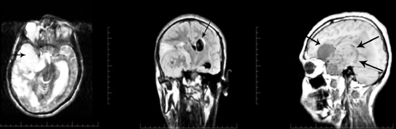
A 14-year-old boy was referred from a secondary care hospital with 4 years history of progressively worsening generalized tonic clonic seizures. There was headache, vomiting, behavioral disturbance, and deterioration in vision and level of consciousness over one month prior to admission. On examination the patient was drowsy, arousable but did not give relevant response to questions. Glasgow coma score was 12 (normal range=12-15). Pupils were equal and reacting to light and there was bilateral papilledema. There was upper motor neuron type left sided hemiparesis with increased tone and brisk reflexes. The MRI (Figure 1) showed a large mixed density extra-axial lesion in the right temporal region with a frontal cystic component and a significant midline shift.

Figure 1
  • Download figure
  • Open in new tab
  • Download powerpoint
Figure 1

Multi-sequential magnetic resonance imaging shows large mixed density extra-axial lesion in the right temporal region with a frontal cystic component and a significant midline shift.

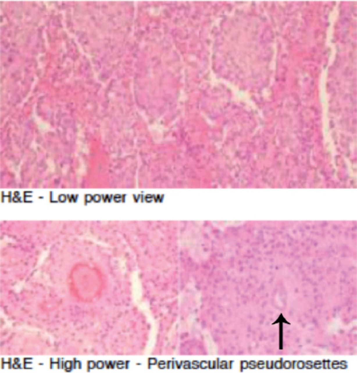
Craniotomy for excision of this highly vascular lesion was carried out, complete resection achieved. Frozen section reported to be “consistent with ependymoma” was not so consistent with our intraoperative findings of the lesion being extra-axial. Final histopathology report (including secondary opinions from other neuropathologists), was pointing towards the diagnosis of ependymoma (world health organization, grade 2), with perivascular pseudorosettes (Figure 2) as well as positive glial fibrillary acidic protein and epithelial membrane antigen staining. Clear extra-axial location of the lesion with part of the blood supply coming from dural vessels and the final histopathology report lead to appraisal of available literature. Finding very few reported cases at such location.

Figure 2
  • Download figure
  • Open in new tab
  • Download powerpoint
Figure 2

Histopathology sections (hematoxylin and eosin stain).

Ependymomas are tumors of neuroectodermal origin arising from ependymal cells within the cerebrospinal axis cavities, including the central canal of the spinal cord, the filum terminale and the ventricles.1 Ependymomas can be found in the brain parenchyma, possibly arising from ectopic fetal rests of ependymal cells although it is not clear how ependymal cell rests can get to be anywhere other than the immediate vicinity of the ventricular system.2 Cell migration away from periventricular areas during development has been proposed as a possible etiology. Extra-axial ependymomas are found in spine and occasionally in the posterior fossa extending through the basal foramina3 but supratentorial extra-axial ependymomas constitute an extremely rare entity.2 Embryonal cell migration seems an unlikely explanation for these tumors. Ependymomas are also known to spread by cerebrospinal fluid seeding,1 whether seeding can occur across the tentorium cerebelli or not is not definite.4

In conclusion, ependymomas at extra-ventricular location constitute an uncommon but known entity. Of these, supratentorial extra-axial ependymomas are extremely rare; possibly arising from ectopic fetal rests of ependymal cells.

Footnotes

  • Disclosure

    No funding for this work was received from any organization or foundation.

  • Received August 5, 2015.
  • Accepted January 20, 2016.
  • Copyright: © Neurosciences

Neurosciences is an Open Access journal and articles published are distributed under the terms of the Creative Commons Attribution-NonCommercial License (CC BY-NC). Readers may copy, distribute, and display the work for non-commercial purposes with the proper citation of the original work.

References

  1. ↵
    1. Reni M,
    2. Gatta G,
    3. Mazza E,
    4. Vecht C
    (2007) Ependymoma. Crit Rev Oncol Hematol 63, 81–89.
  2. ↵
    1. Miyazawa T,
    2. Hirose T,
    3. Nakanishi K,
    4. Uozumi Y,
    5. Tsuzuki N,
    6. Shima K
    (2007) Supratentorial ectopic cortical ependymoma occurring with intratumoral hemorrhage. Brain Tumor Pathol 24, 35–40.
  3. ↵
    1. Kasliwal MK,
    2. Chandra PS,
    3. Sharma BS
    (2007) Images in neuro oncology: primary extraaxial cerebellopontine angle ependymoma. J Neurooncol 83, 31–32.
  4. ↵
    1. Zhao C,
    2. Wang C,
    3. Zhang M,
    4. Jiang T,
    5. Liu W,
    6. Li W
    (2015) Primary cerebellopontine angle ependymoma with spinal metastasis in an adult patient: A case report. Oncol Lett 10, 1755–1758.
PreviousNext
Back to top

In this issue

Neurosciences Journal: 21 (2)
Neurosciences Journal
Vol. 21, Issue 2
1 Apr 2016
  • Table of Contents
  • Cover (PDF)
  • Index by author
Print
Download PDF
Email Article

Thank you for your interest in spreading the word on Neurosciences Journal.

NOTE: We only request your email address so that the person you are recommending the page to knows that you wanted them to see it, and that it is not junk mail. We do not capture any email address.

Enter multiple addresses on separate lines or separate them with commas.
Ependymoma in supratentorial extra-axial location
(Your Name) has sent you a message from Neurosciences Journal
(Your Name) thought you would like to see the Neurosciences Journal web site.
Citation Tools
Ependymoma in supratentorial extra-axial location
Abdulrahman Y. Alturki, Nasir R. Awan, Khaled N. Almusrea
Neurosciences Journal Apr 2016, 21 (2) 173-174; DOI: 10.17712/nsj.2016.2.20150503

Citation Manager Formats

  • BibTeX
  • Bookends
  • EasyBib
  • EndNote (tagged)
  • EndNote 8 (xml)
  • Medlars
  • Mendeley
  • Papers
  • RefWorks Tagged
  • Ref Manager
  • RIS
  • Zotero
Share
Ependymoma in supratentorial extra-axial location
Abdulrahman Y. Alturki, Nasir R. Awan, Khaled N. Almusrea
Neurosciences Journal Apr 2016, 21 (2) 173-174; DOI: 10.17712/nsj.2016.2.20150503
Twitter logo Facebook logo Mendeley logo
  • Tweet Widget
  • Facebook Like
  • Google Plus One
Bookmark this article

Jump to section

  • Article
    • Footnotes
    • References
  • Figures & Data
  • eLetters
  • References
  • Info & Metrics
  • PDF

Related Articles

  • No related articles found.
  • PubMed
  • Google Scholar

Cited By...

  • No citing articles found.
  • Google Scholar

More in this TOC Section

  • How to make a decision on the choice of therapy for multiple sclerosis with sarcoidosis comorbidity?
  • Mycoplasma pneumonia and stroke: causation, association, or coincidence?
  • Posterior reversible encephalopathy syndrome overshadowing COVID-19: An Abstruse scenario or a conspicuous association?
Show more Clinical Note

Similar Articles

Navigate

  • home

More Information

  • Help

Additional journals

  • All Topics

Other Services

  • About

© 2025 Neurosciences Journal Neurosciences is copyright under the Berne Convention and the International Copyright Convention. All rights reserved. Neurosciences is an Open Access journal and articles published are distributed under the terms of the Creative Commons Attribution-NonCommercial License (CC BY-NC). Readers may copy, distribute, and display the work for non-commercial purposes with the proper citation of the original work. Electronic ISSN 1658-3183. Print ISSN 1319-6138.

Powered by HighWire