
Main menu

  • Home
  • Content
    • Latest
    • Ahead of print
    • Archive
  • Info for
    • Authors
    • Reviewers
    • Subscribers
    • Institutions
    • Advertisers
  • About Us
    • About Us
    • Editorial Office
    • Editorial Board
  • More
    • Alerts
    • Feedback
    • Folders
    • Help
  • Other Publications
    • Saudi Medical Journal

User menu

  • My alerts
  • Log in

Search

  • Advanced search
Neurosciences Journal
  • Other Publications
    • Saudi Medical Journal
  • My alerts
  • Log in
Neurosciences Journal

Advanced Search

  • Home
  • Content
    • Latest
    • Ahead of print
    • Archive
  • Info for
    • Authors
    • Reviewers
    • Subscribers
    • Institutions
    • Advertisers
  • About Us
    • About Us
    • Editorial Office
    • Editorial Board
  • More
    • Alerts
    • Feedback
    • Folders
    • Help
  • Follow psmmc on Twitter
  • Visit psmmc on Facebook
  • RSS
Research ArticleOriginal Article
Open Access

Tumors of the lateral and third ventricle: surgical management and outcome analysis in 42 cases

Sherif M. Elwatidy, Abdulrahman A. Albakr, Abdullah A. Al Towim and Safdar H. Malik
Neurosciences Journal October 2017, 22 (4) 274-281; DOI: https://doi.org/10.17712/nsj.2017.4.20170149
Sherif M. Elwatidy
From the Division of Neurosurgery, Department of Surgery, College of Medicine, King Saud University, Riyadh, Kingdom of Saudi Arabia
FRCS (SN)
  • Find this author on Google Scholar
  • Find this author on PubMed
  • Search for this author on this site
Abdulrahman A. Albakr
From the Division of Neurosurgery, Department of Surgery, College of Medicine, King Saud University, Riyadh, Kingdom of Saudi Arabia
MD
  • Find this author on Google Scholar
  • Find this author on PubMed
  • Search for this author on this site
  • For correspondence: [email protected]
Abdullah A. Al Towim
From the Division of Neurosurgery, Department of Surgery, College of Medicine, King Saud University, Riyadh, Kingdom of Saudi Arabia
MD
  • Find this author on Google Scholar
  • Find this author on PubMed
  • Search for this author on this site
Safdar H. Malik
From the Division of Neurosurgery, Department of Surgery, College of Medicine, King Saud University, Riyadh, Kingdom of Saudi Arabia
MD, FCPS
  • Find this author on Google Scholar
  • Find this author on PubMed
  • Search for this author on this site
  • Article
  • Figures & Data
  • eLetters
  • Info & Metrics
  • References
  • PDF
Loading

Article Figures & Data

Figures

  • Tables
  • Figure 1
    • Download figure
    • Open in new tab
    • Download powerpoint
    Figure 1

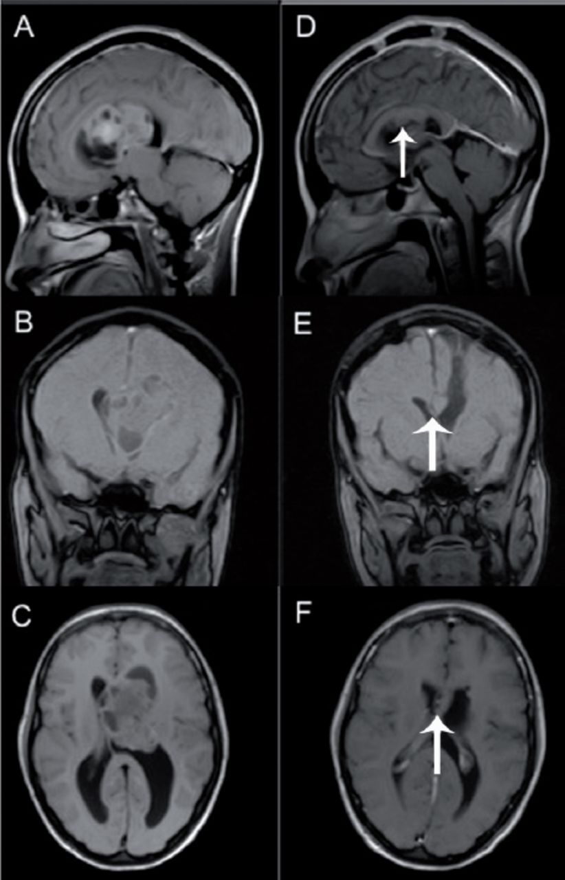
    A 30-year-old woman presenting with a headache, blurry vision, and attacks of generalized body numbness for 2 months. A-C) Sagittal, coronal, and axial T1 Magnetic Resonance Imaging (MRI) with and without contrast enhancement showing third ventricular tumor with extension to the left lateral ventricle. She underwent left frontal craniotomy, frontal transcortical approach with subtotal excision of the tumor. The pathology revealed central neurocytoma. D-F) Postoperative with and without contrast enhancement T1 sagittal, coronal, and axial scans showing small residual tumor (arrow) under the corpus callosum. She postoperatively developed right side hemiparesis. In her follow-up, 6 and 9 months following surgery, her weakness improved significantly. We think the reason for her weakness is due to the retraction on the motor control areas.

  • Figure 2
    • Download figure
    • Open in new tab
    • Download powerpoint
    Figure 2

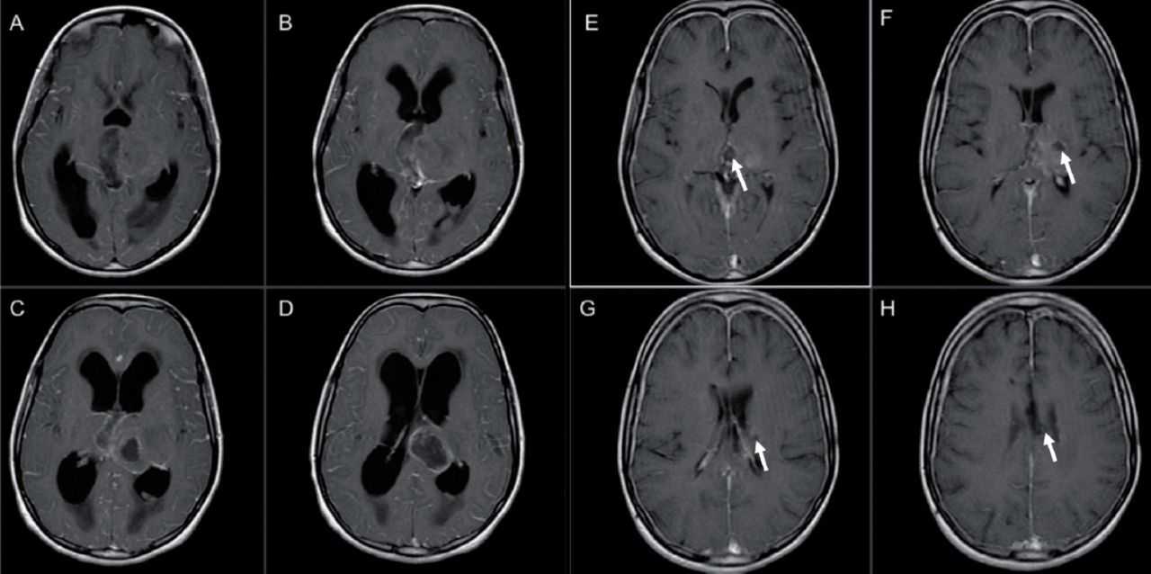
    A 10-year-old girl presenting with 2 months history of a persistent headache, vomiting, and tremor in both hands. Additionally, she had 2 episodes of seizures. A-D) Contrast-enhanced T1 axial scans showing a mass occupying the left thalamus, crossing the midline, and infiltrating the third ventricle. She underwent resection via transcallosal approach with subtotal excision of the tumor. The pathology revealed central glioblastoma multiforme. E-H) Postoperative contrast-enhanced T1 axial scans showing small residual tumor. Postoperatively, the patient recovered very well with no neurological deficits.

Tables

  • Figures
    • View popup
    Table 1

    Clinical features of patients with lateral and third ventricular tumors.

    Signs and symptomsPediatricsAdultsAll patients
    n (%)
    Headache9 (50)20 (83)29 (69)
    Nausea-vomiting8 (44)8 (33)16 (38)
    Visual deficit*2 (11)8 (33)10 (24)
    Seizures5 (28)2 (8)7 (17)
    Change in behavior2 (11)4 (17)6 (14)
    Unstable gait*3 (17)1 (4)4 (10)
    Altered mental status1 (6)3 (13)4 (10)
    Motor deficit*-3 (13)3 (7)
    Dizziness-2 (8)2 (5)
    Urine incontinence2 (11)-2 (5)
    Fever1 (6)1 (4)2 (5)
    Tremor1 (6)1 (4)2 (5)
    Neck stiffness1 (6)-1 (5)
    Tinnitus-1 (4)1 (2)
    Endocrine dysfunction-1 (4)1 (2)
    • ↵* Some patients experienced more than one symptom

    • View popup
    Table 2

    Histopathological types of lateral and third ventricular tumors.

    HistopathologyPediatricsAdultsAll patients
    n (%)
    Pineal region tumors4 (10)2 (5)6 (14)
    Colloid cyst-6 (14)6 (14)
    Pilocytic astrocytoma3 (10)2 (5)5 (12)
    Central neurocytoma1 (2)4 (10)5 (12)
    Glioblastoma1 (2)3 (7)4 (10)
    Atypical teratoid rhabdoid tumor3 (7)-3 (7)
    Ependymoma-2 (5)2 (5)
    Meningioma-2 (5)2 (5)
    Lymphoma-1 (2)1 (2)
    Choroid plexus papilloma1 (2)-1 (2)
    Anaplastic ependymoma1 (2)-1 (2)
    Anaplastic astrocytoma1 (2)-1 (2)
    Mature teratoma1 (2)-1 (2)
    Anaplastic oligodendroglioma-1 (2)1 (2)
    Craniopharyngioma-1 (2)1 (2)
    Glioneuronal tumor1 (2)-1 (2)
    Langerhans cell histiocytosis1 (2)-1 (2)
    Total182442
    • View popup
    Table 3

    Postoperative complications and mortality in patients with lateral and third ventricular tumors

    Complicationsall patients n (%)
    Seizure5 (12)
    Hydrocephalus5 (12)
    Wound site infection3 (7)
    Intraventricular hemorrhage2 (5)
    Weakness1 (2)
    Vision disturbance1 (2)
    Diabetes insipidus1 (2)
    Mortality2 (5)
    Hypothalamic syndrome1 (2)
    Brain abscess and Ventriculitis1 (2)
    • *Some patients experienced more than one complication

    • View popup
    Table 4

    Surgical approaches in patients with lateral and third ventricular tumors.

    Surgical approachesNo. of all patients
    n (%)
    Transcortical approach19 (45 %)
    Endoscopic biopsy8 (19 %)
    Transcallosal approach5 (12 %)
    Infratentorial supracerebellar approach3 (7 %)
    Pterional approach3 (7 %)
    Endoscopic resection2 (5%)
    Posterior fossa craniotomy1 (2 %)
    Subfrontal approach1 (2 %)
PreviousNext
Back to top

In this issue

Neurosciences Journal: 22 (4)
Neurosciences Journal
Vol. 22, Issue 4
1 Oct 2017
  • Table of Contents
  • Cover (PDF)
  • Index by author
Print
Download PDF
Email Article

Thank you for your interest in spreading the word on Neurosciences Journal.

NOTE: We only request your email address so that the person you are recommending the page to knows that you wanted them to see it, and that it is not junk mail. We do not capture any email address.

Enter multiple addresses on separate lines or separate them with commas.
Tumors of the lateral and third ventricle: surgical management and outcome analysis in 42 cases
(Your Name) has sent you a message from Neurosciences Journal
(Your Name) thought you would like to see the Neurosciences Journal web site.
Citation Tools
Tumors of the lateral and third ventricle: surgical management and outcome analysis in 42 cases
Sherif M. Elwatidy, Abdulrahman A. Albakr, Abdullah A. Al Towim, Safdar H. Malik
Neurosciences Journal Oct 2017, 22 (4) 274-281; DOI: 10.17712/nsj.2017.4.20170149

Citation Manager Formats

  • BibTeX
  • Bookends
  • EasyBib
  • EndNote (tagged)
  • EndNote 8 (xml)
  • Medlars
  • Mendeley
  • Papers
  • RefWorks Tagged
  • Ref Manager
  • RIS
  • Zotero
Share
Tumors of the lateral and third ventricle: surgical management and outcome analysis in 42 cases
Sherif M. Elwatidy, Abdulrahman A. Albakr, Abdullah A. Al Towim, Safdar H. Malik
Neurosciences Journal Oct 2017, 22 (4) 274-281; DOI: 10.17712/nsj.2017.4.20170149
Twitter logo Facebook logo Mendeley logo
  • Tweet Widget
  • Facebook Like
  • Google Plus One
Bookmark this article

Jump to section

  • Article
    • Abstract
    • Methods
    • Results
    • Discussion
    • Footnotes
    • References
  • Figures & Data
  • eLetters
  • References
  • Info & Metrics
  • PDF

Related Articles

  • No related articles found.
  • PubMed
  • Google Scholar

Cited By...

  • No citing articles found.
  • Google Scholar

More in this TOC Section

  • Ranking the difficulty of the cognitive tasks in Dual-Tasks during walking in healthy adults
  • Exploring physiotherapist’s ability to identify cauda equina syndrome early
  • Does the severity of facet joint osteoarthritis affect facet medial branch radiofrequency thermocoagulation results?
Show more Original Article

Similar Articles

Navigate

  • home

More Information

  • Help

Additional journals

  • All Topics

Other Services

  • About

© 2025 Neurosciences Journal Neurosciences is copyright under the Berne Convention and the International Copyright Convention. All rights reserved. Neurosciences is an Open Access journal and articles published are distributed under the terms of the Creative Commons Attribution-NonCommercial License (CC BY-NC). Readers may copy, distribute, and display the work for non-commercial purposes with the proper citation of the original work. Electronic ISSN 1658-3183. Print ISSN 1319-6138.

Powered by HighWire