
Main menu

  • Home
  • Content
    • Latest
    • Ahead of print
    • Archive
  • Info for
    • Authors
    • Reviewers
    • Subscribers
    • Institutions
    • Advertisers
  • About Us
    • About Us
    • Editorial Office
    • Editorial Board
  • More
    • Alerts
    • Feedback
    • Folders
    • Help
  • Other Publications
    • Saudi Medical Journal

User menu

  • My alerts
  • Log in

Search

  • Advanced search
Neurosciences Journal
  • Other Publications
    • Saudi Medical Journal
  • My alerts
  • Log in
Neurosciences Journal

Advanced Search

  • Home
  • Content
    • Latest
    • Ahead of print
    • Archive
  • Info for
    • Authors
    • Reviewers
    • Subscribers
    • Institutions
    • Advertisers
  • About Us
    • About Us
    • Editorial Office
    • Editorial Board
  • More
    • Alerts
    • Feedback
    • Folders
    • Help
  • Follow psmmc on Twitter
  • Visit psmmc on Facebook
  • RSS
OtherClinical Image
Open Access

Clinical Image

Asmaa Alenizi and Khalid Hundallah
Neurosciences Journal January 2019, 24 (1) 63-65;
Asmaa Alenizi
MD
  • Find this author on Google Scholar
  • Find this author on PubMed
  • Search for this author on this site
  • For correspondence: [email protected]
Khalid Hundallah
MD
  • Find this author on Google Scholar
  • Find this author on PubMed
  • Search for this author on this site
  • For correspondence: [email protected]
  • Article
  • Figures & Data
  • eLetters
  • Info & Metrics
  • References
  • PDF
Loading

Notice: Authors are encouraged to submit clinical images for possible publication in the Journal. These may be in any field of Clinical Neurosciences, and should approximately follow the format used here. Please address any submissions to the Assistant Editor, Neurosciences Journal, Prince Sultan Military Medical City, PO Box 7897, Riyadh 11159, Kingdom of Saudi Arabia. E-mail: malaskar{at}psmmc.med.sa

Joubert Syndrome

Clinical Presentation

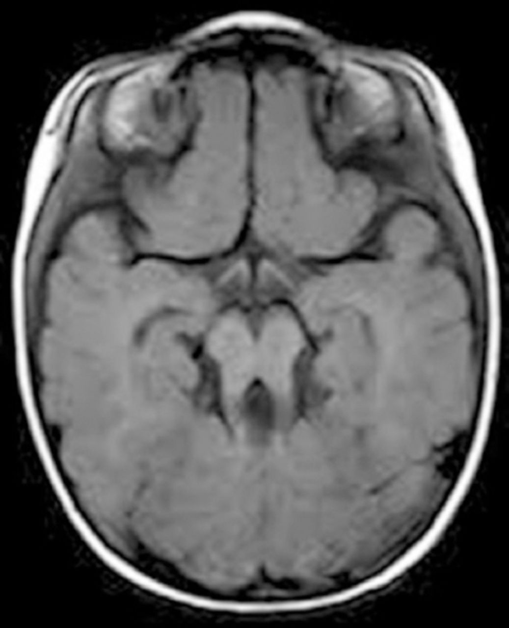
You are evaluating a 3-year-old boy with developmental delay and ataxia. On examination, he is hypotonic. He has abnormal eye movement and abnormal breathing pattern. His brain MRI is shown in Figure 1.

Figure 1
  • Download figure
  • Open in new tab
  • Download powerpoint
Figure 1

Brain MRI of a 3-year-old boy with a history of developmental delay and ataxia.

Questions:

  1. What is the most likely diagnosis:

    1. Ataxia with oculomotor apraxia type I

    2. Ataxia with oculomotor apraxia type II

    3. Joubert syndrome

    4. Leber congenital amaurosis

  2. In which condition is a molar tooth seen on brain MRI?

    1. Joubert syndrome

    2. COACH syndrome

    3. CORS syndrome

    4. All of the above

  3. Which of the following can be clinical abnormalities in Joubert Syndrome:

    1. Neonatal hypotonia

    2. Dysregulated breathing rhythms

    3. Facial dysmorphisms

    4. All of the above

  4. Which of the following genes causing Joubert syndrome is noticed to be associated with retinal blindness and renal involvement?

    1. CEP290

    2. TMEM67

    3. RPGRIP1L

    4. None of the above

  5. The overall risk of having renal disease in patients of Jeubert Syndrome:

    1. 3%

    2. 10%

    3. 15%

    4. 30%

Answers & Discussion

1. C)

Joubert syndrome is an autosomal recessive malformation of the brainstem and agenesis or hypoplasia of the cerebellar vermis. It presents with episodic hyperpnoea, abnormal eye movements, hypotonia, ataxia and mild to severe intellectual disability.4

2. (D)

Molar tooth sign (MTS) results from hypo-dysplasia of the cerebellar vermis, abnormally deep interpeduncular fossa at the level of the isthmus and upper pons, and horizontalized, thickened and elongated superior cerebellar peduncles. It was initially described in Joubert syndrome and related disorders (JSRD) but is now recognized to occur in a number of other conditions like nephronophthisis, cerebello-oculo-renal syndrome (CORS), COACH syndrome and Cogan’s syndrome.2

3. (A)

Genetic testing can predict disease severity. Mutations in certain genes causing Joubert syndrome, such as AHI1, INPP5E, CC2D2A, and ARL13B, can be detected in patients with MTS who do not have involvement of other organs, or they can be detected in patients with MTS together with retinal blindness. Mutations in TMEM216 and RPGRIP1L genes are often encountered in those patients with MTS and renal involvement. Mutations in CEP290 are encountered in half of patients with MTS together with retinal blindness and renal involvement. Mutations in TMEM67 are the most common cause of MTS with liver involvement. Thus, the specific gene mutation identified in a patient can help predict the range of organ involvement.3

4. (d)

The cardinal neurological features of Joubert Syndrome and Joubert Syndrome Related Disorders (JSRD) are hypotonia, altered respiratory pattern in the neonatal period and abnormal ocular movements. In general, the breathing abnormalities improve with age. Truncal ataxia develops over time and acquisition of gross motor milestones is delayed. Cognitive abilities are variable, ranging from severe intellectual disability to normal. Additional findings can include retinal dystrophy, renal disease, ocular colobomas, occipital encephalocele, hepatic fibrosis, polydactyly, oral hamartomas, and endocrine abnormalities. Both intra- and interfamilial variation are seen.1

5. (D)

Renal disease is reported in 25-30% of patients with JS varying from chronic tubulointerstitial nephropathy in the form of juvenile nephronophthisis to nephrocystic disease which is clinically and morphologically similar to autosomal recessive polycystic kidney disease. Acute or chronic renal insufficiency manifests either late in the first decade of life or early in the second. End stage renal failure is usually reached by the end of the second decade, requiring dialysis or kidney transplantation.1

  • Copyright: © Neurosciences

Neurosciences is an Open Access journal and articles published are distributed under the terms of the Creative Commons Attribution-NonCommercial License (CC BY-NC). Readers may copy, distribute, and display the work for non-commercial purposes with the proper citation of the original work.

References

  1. ↵
    1. Ben-Salem S,
    2. Al-Shamsi AM,
    3. Gleeson JG,
    4. Ali BR,
    5. Al-Gazali L
    (2014) Mutation spectrum of Joubert syndrome and related disorders among Arabs. Hum Genome Var 1, 14020.
  2. ↵
    1. Brancati F,
    2. Dallapiccola B,
    3. Valente EM
    (2010) Joubert Syndrome and related disorders. Orphanet Journal of Rare Diseases 5, 20.
  3. ↵
    1. Swaiman KF,
    2. Ashwal S,
    3. Ferriero DM
    1. Millen KJ,
    2. Gleeson JG
    (2016) Disorders of Cerebellar and Brainstem Development. in Swaiman's Pediatric Neurology: Principles and Practice, eds Swaiman KF, Ashwal S, Ferriero DM (Elsevier, Edinburgh), 6th edition.
  4. ↵
    1. Elhassanien AF,
    2. Alghaiaty HA
    (2013) Joubert syndrome:Clinical and radiological characteristics of nine patients. Ann Indian Acad Neurol 16, 239–244.
PreviousNext
Back to top

In this issue

Neurosciences Journal: 24 (1)
Neurosciences Journal
Vol. 24, Issue 1
1 Jan 2019
  • Table of Contents
  • Cover (PDF)
  • Index by author
Print
Download PDF
Email Article

Thank you for your interest in spreading the word on Neurosciences Journal.

NOTE: We only request your email address so that the person you are recommending the page to knows that you wanted them to see it, and that it is not junk mail. We do not capture any email address.

Enter multiple addresses on separate lines or separate them with commas.
Clinical Image
(Your Name) has sent you a message from Neurosciences Journal
(Your Name) thought you would like to see the Neurosciences Journal web site.
Citation Tools
Clinical Image
Asmaa Alenizi, Khalid Hundallah
Neurosciences Journal Jan 2019, 24 (1) 63-65;

Citation Manager Formats

  • BibTeX
  • Bookends
  • EasyBib
  • EndNote (tagged)
  • EndNote 8 (xml)
  • Medlars
  • Mendeley
  • Papers
  • RefWorks Tagged
  • Ref Manager
  • RIS
  • Zotero
Share
Clinical Image
Asmaa Alenizi, Khalid Hundallah
Neurosciences Journal Jan 2019, 24 (1) 63-65;
Twitter logo Facebook logo Mendeley logo
  • Tweet Widget
  • Facebook Like
  • Google Plus One
Bookmark this article

Jump to section

  • Article
    • References
  • Figures & Data
  • eLetters
  • References
  • Info & Metrics
  • PDF

Related Articles

  • No related articles found.
  • Google Scholar

Cited By...

  • No citing articles found.
  • Google Scholar

More in this TOC Section

  • Multiple cranial nerve enhancement in a case of neuronal ceroid lipofuscinosis type 8
  • Unilateral Lennox–Gastaut syndrome associated with Klippel–Trénaunay syndrome
  • A child with leukemia and behavioral changes
Show more Clinical Image

Similar Articles

Navigate

  • home

More Information

  • Help

Additional journals

  • All Topics

Other Services

  • About

© 2025 Neurosciences Journal Neurosciences is copyright under the Berne Convention and the International Copyright Convention. All rights reserved. Neurosciences is an Open Access journal and articles published are distributed under the terms of the Creative Commons Attribution-NonCommercial License (CC BY-NC). Readers may copy, distribute, and display the work for non-commercial purposes with the proper citation of the original work. Electronic ISSN 1658-3183. Print ISSN 1319-6138.

Powered by HighWire