
Main menu

  • Home
  • Content
    • Latest
    • Ahead of print
    • Archive
  • Info for
    • Authors
    • Reviewers
    • Subscribers
    • Institutions
    • Advertisers
  • About Us
    • About Us
    • Editorial Office
    • Editorial Board
  • More
    • Alerts
    • Feedback
    • Folders
    • Help
  • Other Publications
    • Saudi Medical Journal

User menu

  • My alerts
  • Log in

Search

  • Advanced search
Neurosciences Journal
  • Other Publications
    • Saudi Medical Journal
  • My alerts
  • Log in
Neurosciences Journal

Advanced Search

  • Home
  • Content
    • Latest
    • Ahead of print
    • Archive
  • Info for
    • Authors
    • Reviewers
    • Subscribers
    • Institutions
    • Advertisers
  • About Us
    • About Us
    • Editorial Office
    • Editorial Board
  • More
    • Alerts
    • Feedback
    • Folders
    • Help
  • Follow psmmc on Twitter
  • Visit psmmc on Facebook
  • RSS
Review ArticleReview Article
Open Access

Metabolic and genetic disorders mimicking cerebral palsy

Wejdan S. Hakami, Khaled J. Hundallah and Brahim M. Tabarki
Neurosciences Journal July 2019, 24 (3) 155-163; DOI: https://doi.org/10.17712/nsj.2019.3.20190045
Wejdan S. Hakami
Division of Pediatric Neurology, Department of Pediatrics, Prince Sultan Military Medical City, Riyadh, Kingdom of Saudi Arabia
MD
  • Find this author on Google Scholar
  • Find this author on PubMed
  • Search for this author on this site
  • For correspondence: [email protected]
Khaled J. Hundallah
Division of Pediatric Neurology, Department of Pediatrics, Prince Sultan Military Medical City, Riyadh, Kingdom of Saudi Arabia
MD
  • Find this author on Google Scholar
  • Find this author on PubMed
  • Search for this author on this site
Brahim M. Tabarki
Division of Pediatric Neurology, Department of Pediatrics, Prince Sultan Military Medical City, Riyadh, Kingdom of Saudi Arabia
MD
  • Find this author on Google Scholar
  • Find this author on PubMed
  • Search for this author on this site
  • Article
  • Figures & Data
  • eLetters
  • Info & Metrics
  • References
  • PDF
Loading

Article Figures & Data

Figures

  • Tables
  • Figure 1
    • Download figure
    • Open in new tab
    • Download powerpoint
    Figure 1

    Axial T2-weighted brain magnetic resonance image of a 5-year-old child with Pelizaeus-Merzbacher disease shows bilateral symmetrical hyperintensity of the deep, periventricular and subcortical white matter including the posterior limbs of the internal capsules.
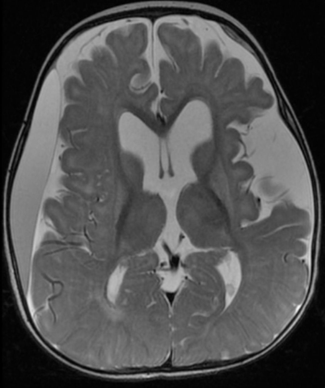
  • Figure 2
    • Download figure
    • Open in new tab
    • Download powerpoint
    Figure 2

    Axial T2-weighted brain magnetic resonance image of a 9-months-old child with glutaric aciduria type1 shows subacute right frontoparietotemporal subdural hematoma with mass effect, diffuse brain atrophy with widening of sylvian fissures bilaterally and secondary ventriculomegaly and increased signal intensity of basal ganglia bilaterally.
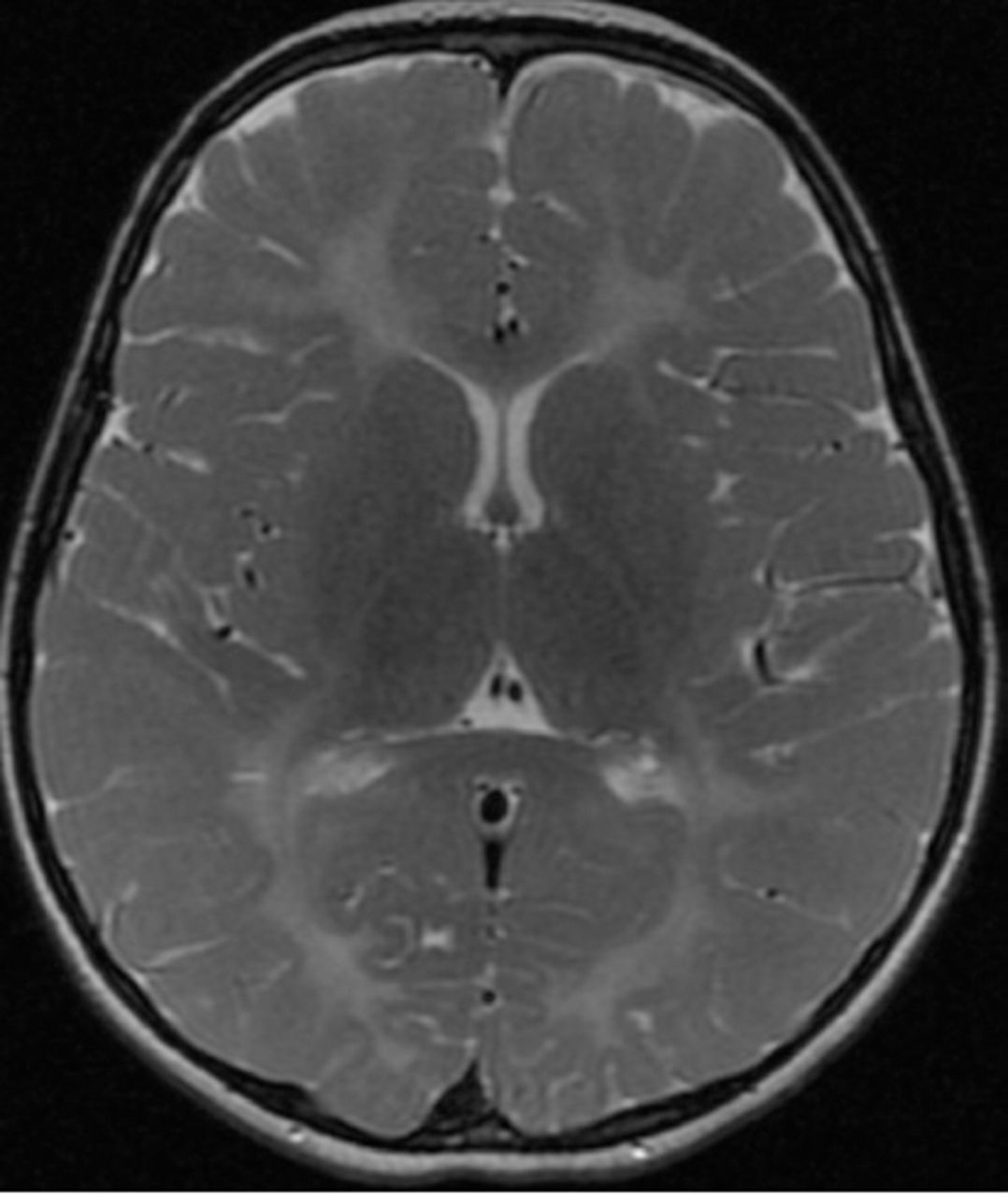
  • Figure 3
    • Download figure
    • Open in new tab
    • Download powerpoint
    Figure 3

    Axial T2-weighted brain magnetic resonance image of a 2-year-old child with molybdenum cofactor deficiency shows extensive brain damage, signifiant loss of white matter with cystic changes, ex vacuo dilatation of ventricles and mushrom-shaped gyri.
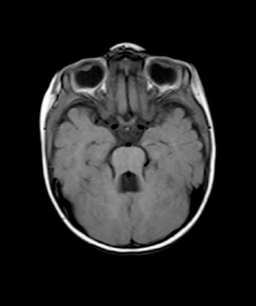
  • Figure 4
    • Download figure
    • Open in new tab
    • Download powerpoint
    Figure 4

    Axial T1-weighted brain magnetic resonance image of a 1-year-old child with Joubert syndrome shows the classical molar tooth sign.
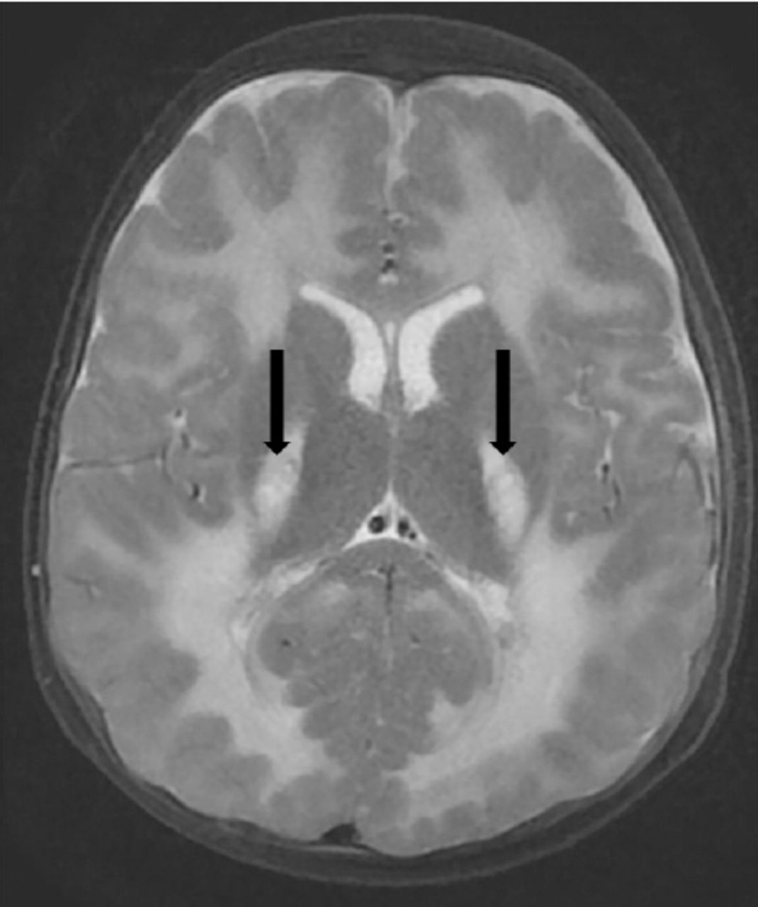
  • Figure 5
    • Download figure
    • Open in new tab
    • Download powerpoint
    Figure 5

    Axial T2-weighted brain magnetic resonance images of a 2-year-old child with mitochondrial leukoencephalopathy, caused by ISCA2 gene mutation shows leukodystrophy with supra and infra tentorial predominantly deep white matter involvement and central areas of cavitation (arrows).

Tables

  • Figures
    • View popup
    Table 1

    Red flags in the history and examination that should prompt the consideration of another diagnosis

    History
     No risk factors for CP such as: prematurity, low birthweight, multiple births, hypoglycemia, jaundice and kernicterus, intrapartum asphyxia, intracranial hemorrhage, infection, stroke, or head injuries
     Positive family history of CP
     Fluctuation in motor symptoms
     Paroxysmal symptoms in relation to time of day, diet/fasting, or activity
     Progressive neurological symptoms
     Regression of milestones
    Examination
     Dysmorphic features
     Isolated motor dysfunction such as isolated ataxia or isolated hypotonia without dystonia or spasticity
     Peripheral nervous system abnormalities: absent reflexes, sensory signs
     Eye movement abnormalities (e.g., oculogyria, oculomotor apraxia, or paroxysmal saccadic eye-head movements)
     Optic atrophy/retinopathy
    • CP- cerebral palsy

    • View popup
    Table 2

    Red flags in the MRI brain findings that should prompt consideration of another diagnosis.

    Normal neuroimaging
    Nonspecific abnormalities, such as isolated globus pallidus involvement, which can suggest methylmalonic aciduria
    Imaging may demonstrate specific lesions that are inconsistent with perinatal brain injury, but characteristic of a particular genetic disorder, such as leukodystrophies, features of glutaric aciduria type 1, or features of Joubert syndrome
    • View popup
    Table 3

    Common metabolic and genetic disorders that mimic CP according to the prominent motor dysfunction.

    Disorders with prominent spasticityDisorders with prominent dyskinesiaDisorders with prominent ataxia
    -  Hereditary spastic paraglegias
    -  Arginase deficiency
    -  COL4A1-Related spastic CP
    -  Biotinidase deficiency
    -  Aicardi-Goutières syndrome
    -  Sulfite oxidase deficiency/Molybdenum cofactor deficiency22
    -  Leukodystrophies, such as metachromatic leukodystrophy,23 adrenoleukodystrophy,24 Sjorgen Larsson syndrome25
    -  Dopa-responsive dystonia
    -  Sepiapterin reductase deficiency
    -  Glutaric aciduria type 1
    -  Glucose transporter deficiency type 1
    -  Neurodegeneration with brain iron accumulation
    -  Cerebral creatine deficiency syndrome
    -  Lesch Nyhan syndrome
    -  Cerebral folate deficiency
    -  ADCY5-related dyskinesia
    -  PCDH12-related dyskinesia34
    -  NKX2-1 related ataxic dyskinetic CP35
    -  TSEN54 Gene-related pontocerebellar hypoplasia type 236
    -  Glucose transporter deficiency type 1
    -  Ataxia telangiectasia
    -  Pelizaeus-Merzbacher disease
    -  Hereditary ataxias
    -  Joubert syndrome
    -  Mitochondrial cytopathies (mainly 8993 mutation)42
    -  Pontocerebellar hypoplasia36
    -  Cockayne syndrome43
    -  Niemann-Pick disease type C44
    -  Angelman syndrome12
    -  Gangliosidosis type 1, juvenile and adult forms45
    -  Non-ketotic hyperglycinemia3
    -  Maple syrup urine disease3
    -  NKX2-1 related ataxic dyskinetic CP35
    • CP- cerebral palsy

PreviousNext
Back to top

In this issue

Neurosciences Journal: 24 (3)
Neurosciences Journal
Vol. 24, Issue 3
1 Jul 2019
  • Table of Contents
  • Cover (PDF)
  • Index by author
Print
Download PDF
Email Article

Thank you for your interest in spreading the word on Neurosciences Journal.

NOTE: We only request your email address so that the person you are recommending the page to knows that you wanted them to see it, and that it is not junk mail. We do not capture any email address.

Enter multiple addresses on separate lines or separate them with commas.
Metabolic and genetic disorders mimicking cerebral palsy
(Your Name) has sent you a message from Neurosciences Journal
(Your Name) thought you would like to see the Neurosciences Journal web site.
Citation Tools
Metabolic and genetic disorders mimicking cerebral palsy
Wejdan S. Hakami, Khaled J. Hundallah, Brahim M. Tabarki
Neurosciences Journal Jul 2019, 24 (3) 155-163; DOI: 10.17712/nsj.2019.3.20190045

Citation Manager Formats

  • BibTeX
  • Bookends
  • EasyBib
  • EndNote (tagged)
  • EndNote 8 (xml)
  • Medlars
  • Mendeley
  • Papers
  • RefWorks Tagged
  • Ref Manager
  • RIS
  • Zotero
Share
Metabolic and genetic disorders mimicking cerebral palsy
Wejdan S. Hakami, Khaled J. Hundallah, Brahim M. Tabarki
Neurosciences Journal Jul 2019, 24 (3) 155-163; DOI: 10.17712/nsj.2019.3.20190045
Twitter logo Facebook logo Mendeley logo
  • Tweet Widget
  • Facebook Like
  • Google Plus One
Bookmark this article

Jump to section

  • Article
    • Abstract
    • Acknowledgements
    • Footnotes
    • References
  • Figures & Data
  • eLetters
  • References
  • Info & Metrics
  • PDF

Related Articles

  • No related articles found.
  • PubMed
  • Google Scholar

Cited By...

  • No citing articles found.
  • Google Scholar

More in this TOC Section

  • Structural and functional changes in the hippocampus induced by environmental exposures
  • Tumefactive demyelinating lesions: A literature review of recent findings
  • Epilepsia partialis continua: A review
Show more Review Article

Similar Articles

Navigate

  • home

More Information

  • Help

Additional journals

  • All Topics

Other Services

  • About

© 2025 Neurosciences Journal Neurosciences is copyright under the Berne Convention and the International Copyright Convention. All rights reserved. Neurosciences is an Open Access journal and articles published are distributed under the terms of the Creative Commons Attribution-NonCommercial License (CC BY-NC). Readers may copy, distribute, and display the work for non-commercial purposes with the proper citation of the original work. Electronic ISSN 1658-3183. Print ISSN 1319-6138.

Powered by HighWire