
Main menu

  • Home
  • Content
    • Latest
    • Ahead of print
    • Archive
  • Info for
    • Authors
    • Reviewers
    • Subscribers
    • Institutions
    • Advertisers
  • About Us
    • About Us
    • Editorial Office
    • Editorial Board
  • More
    • Alerts
    • Feedback
    • Folders
    • Help
  • Other Publications
    • Saudi Medical Journal

User menu

  • My alerts
  • Log in

Search

  • Advanced search
Neurosciences Journal
  • Other Publications
    • Saudi Medical Journal
  • My alerts
  • Log in
Neurosciences Journal

Advanced Search

  • Home
  • Content
    • Latest
    • Ahead of print
    • Archive
  • Info for
    • Authors
    • Reviewers
    • Subscribers
    • Institutions
    • Advertisers
  • About Us
    • About Us
    • Editorial Office
    • Editorial Board
  • More
    • Alerts
    • Feedback
    • Folders
    • Help
  • Follow psmmc on Twitter
  • Visit psmmc on Facebook
  • RSS
OtherClinical Image
Open Access

Clinical Image

Ismail I. Ismail, Jasem Y. Al-Hashel, John K. John and Mohamed Ibrahim
Neurosciences Journal July 2019, 24 (3) 245-246;
Ismail I. Ismail
MSc
  • Find this author on Google Scholar
  • Find this author on PubMed
  • Search for this author on this site
Jasem Y. Al-Hashel
MD
  • Find this author on Google Scholar
  • Find this author on PubMed
  • Search for this author on this site
John K. John
MD
  • Find this author on Google Scholar
  • Find this author on PubMed
  • Search for this author on this site
Mohamed Ibrahim
MD
  • Find this author on Google Scholar
  • Find this author on PubMed
  • Search for this author on this site
  • Article
  • Figures & Data
  • eLetters
  • Info & Metrics
  • References
  • PDF
Loading

Notice: Authors are encouraged to submit clinical images for possible publication in the Journal. These may be in any field of Clinical Neurosciences, and should approximately follow the format used here. Please address any submissions to the Assistant Editor, Neurosciences Journal, Prince Sultan Military Medical City, PO Box 7897, Riyadh 11159, Kingdom of Saudi Arabia. E-mail: malaskar{at}psmmc.med.sa

Isolated oculomotor nerve palsy secondary to fascicular midbrain infarction

Clinical Presentation

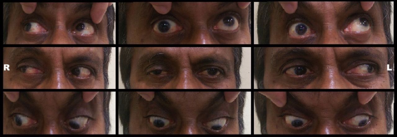
A 52-year-old man with a past medical history of uncontrolled hypertension and diabetes mellitus presented with acute onset of drooping of the right upper eyelid and double vision on looking to the left side. He had no other associated ocular or neurological manifestations. Neurological examination showed a fully conscious and oriented gentleman with normal speech and higher mental functions. Cranial nerves assessment showed ptosis of the right upper eyelid with downward and outward deviation in primary gaze. Extraocular movements assessment is shown in Figure 1. Right pupil was dilated with very sluggish reaction to direct and consensual light; 7.5 mm (Dark), 7 mm (Light). He did not exhibit Bell’s phenomenon. Otherwise, his motor, sensory, cerebellar and gait assessment were normal. Magnetic resonance imaging (MRI) of the brain is shown in Figure 2.

Figure 1
  • Download figure
  • Open in new tab
  • Download powerpoint
Figure 1

Photographs of 9 cardinal positions of gaze showing right upper eyelid partial ptosis and exotropia in primary gaze, limited adduction and impaired vertical voluntary gaze (upward gaze more affected than downward gaze) on the right side. Dilated right pupil cannot be seen.

Figure 2
  • Download figure
  • Open in new tab
  • Download powerpoint
Figure 2

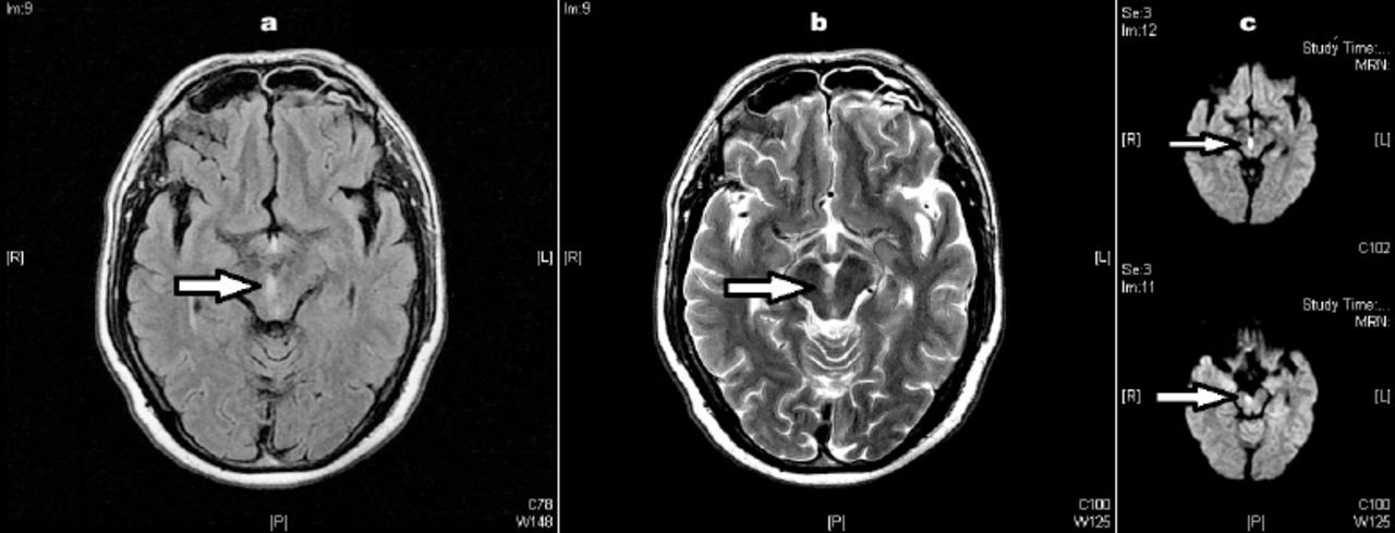
MRI of the brain at the level of the midbrain; a) axial FLAIR, b) axial T2-weighted images and c) diffusion-weighted images (DWI) showing an ischemic lesion (arrow) in the paramedian upper midbrain tegmentum involving the oculomotor fascicles on right side.

Questions:

  1. What are the clinical features of oculomotor nerve palsy?

  2. What are the differences between nuclear and fascicular midbrain lesions?

  3. What are the common syndromes usually associated with fascicular midbrain lesions?

Answers & Discussion

  1. Oculomotor nerve palsy is an important and common clinical finding. Its main features are diplopia and ptosis secondary to affection of the levator palpebrae superioris, superior rectus, medial rectus, inferior rectus, and inferior oblique muscles. It also innervates the iris sphincter and the annular portion of the ciliary muscle through its parasympathetic fibers resulting in fixed dilated pupil if affected.1 The nuclei of the third cranial nerve originate at the level of the superior colliculus. The oculomotor fascicles run ventrally and laterally from the oculomotor complex, pass through and medial to the red nucleus and exit the brainstem medial to the cerebral peduncles.2

  2. A third nerve palsy due to midbrain infarction results from either nuclear or fascicular lesion. Such lesions can usually be differentiated clinically. The subnucleus of the levator palpebrae superioris, the central caudate nucleus, is single and midline, and the fibers to the superior rectus muscles originate in paramedian nuclei and provide crossed innervation. Lesions of the oculomotor nucleus would result in bilateral ptosis and contralateral upward gaze palsy. However, lesion affecting fascicular fibers, as in our patient, will result in unilateral palsy mimicking a peripheral third nerve lesion. Pupillary affection in such cases is variable, but its involvement is more common.3

  3. Several syndromes have been recognized with fascicular lesions including: Weber syndrome, Benedikt syndrome, Nothnagel syndrome and Claude syndrome. Rarely, fascicular lesion of the third cranial nerve develop in isolation without involvement of adjacent midbrain structures as in our patient.4

Acknowledgment

The authors would like to thank Dr Ehab Abdelnabi, Neuroradiologist, Ibn Sina Hospital, for clarity and correctness.

  • Copyright: © Neurosciences

Neurosciences is an Open Access journal and articles published are distributed under the terms of the Creative Commons Attribution-NonCommercial License (CC BY-NC). Readers may copy, distribute, and display the work for non-commercial purposes with the proper citation of the original work.

References

  1. ↵
    1. Schwartz TH,
    2. Lycette CA,
    3. Yoon SS,
    4. Kargman DE
    (1995) Clinicoradiographic evidence for oculomotor fascicular anatomy. J Neurol Neurosurg Psychiatry 59, 338.
  2. ↵
    1. Ogawa K,
    2. Suzuki Y,
    3. Oishi M,
    4. Kamei S
    (2012) Clinical study of twenty-one patients with pure midbrain infarction. Eur Neurol 67, 81–89.
  3. ↵
    1. Fang C,
    2. Leavitt JA,
    3. Hodge DO,
    4. Holmes JM,
    5. Mohney BG,
    6. Chen JJ
    (2017) Incidence and Etiologies of Acquired Third Nerve Palsy Using a Population-Based Method. JAMA Ophthalmol 135, 23–28.
  4. ↵
    1. Ruchalski K,
    2. Hathout GM
    (2012) A medley of midbrain maladies:a brief review of midbrain anatomy and syndromology for radiologists. Radiol Res Pract 2012, 258524.
PreviousNext
Back to top

In this issue

Neurosciences Journal: 24 (3)
Neurosciences Journal
Vol. 24, Issue 3
1 Jul 2019
  • Table of Contents
  • Cover (PDF)
  • Index by author
Print
Download PDF
Email Article

Thank you for your interest in spreading the word on Neurosciences Journal.

NOTE: We only request your email address so that the person you are recommending the page to knows that you wanted them to see it, and that it is not junk mail. We do not capture any email address.

Enter multiple addresses on separate lines or separate them with commas.
Clinical Image
(Your Name) has sent you a message from Neurosciences Journal
(Your Name) thought you would like to see the Neurosciences Journal web site.
Citation Tools
Clinical Image
Ismail I. Ismail, Jasem Y. Al-Hashel, John K. John, Mohamed Ibrahim
Neurosciences Journal Jul 2019, 24 (3) 245-246;

Citation Manager Formats

  • BibTeX
  • Bookends
  • EasyBib
  • EndNote (tagged)
  • EndNote 8 (xml)
  • Medlars
  • Mendeley
  • Papers
  • RefWorks Tagged
  • Ref Manager
  • RIS
  • Zotero
Share
Clinical Image
Ismail I. Ismail, Jasem Y. Al-Hashel, John K. John, Mohamed Ibrahim
Neurosciences Journal Jul 2019, 24 (3) 245-246;
Twitter logo Facebook logo Mendeley logo
  • Tweet Widget
  • Facebook Like
  • Google Plus One
Bookmark this article

Jump to section

  • Article
    • Acknowledgment
    • References
  • Figures & Data
  • eLetters
  • References
  • Info & Metrics
  • PDF

Related Articles

  • No related articles found.
  • Google Scholar

Cited By...

  • No citing articles found.
  • Google Scholar

More in this TOC Section

  • Multiple cranial nerve enhancement in a case of neuronal ceroid lipofuscinosis type 8
  • Unilateral Lennox–Gastaut syndrome associated with Klippel–Trénaunay syndrome
  • A child with leukemia and behavioral changes
Show more Clinical Image

Similar Articles

Navigate

  • home

More Information

  • Help

Additional journals

  • All Topics

Other Services

  • About

© 2025 Neurosciences Journal Neurosciences is copyright under the Berne Convention and the International Copyright Convention. All rights reserved. Neurosciences is an Open Access journal and articles published are distributed under the terms of the Creative Commons Attribution-NonCommercial License (CC BY-NC). Readers may copy, distribute, and display the work for non-commercial purposes with the proper citation of the original work. Electronic ISSN 1658-3183. Print ISSN 1319-6138.

Powered by HighWire