


Article Figures & Data
Figures
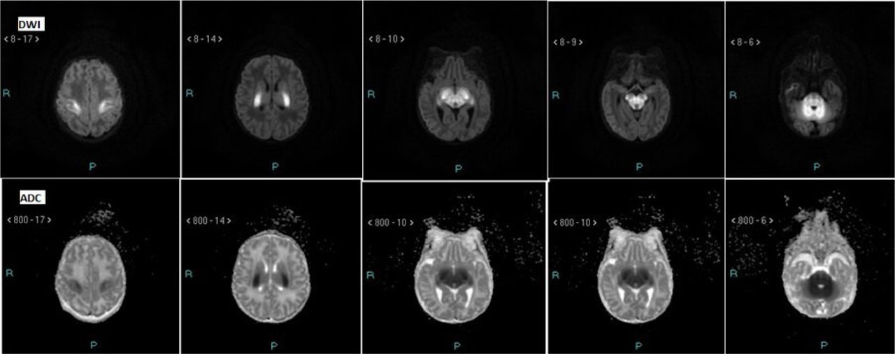
- Figure 1
Selected brain MRI images of Maple Syrup Urine Disease (MSUD) demonstrating diffusion restriction along bilateral perirolandic white matter, posterior limb internal capsule, thalami, brainstem, and cerebellum as evident by abnormal high (bright or white) signal on DWI (upper row) and low (dark or black) signal on ADC (bottom row).
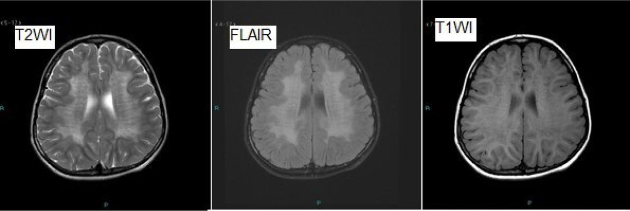
- Figure 2
A case of Metachromatic Leukodystrophy (MLD) with MR images showing low T1, high T2 and FLAIR periventricular white matter signal abnormality (tigroid appearance), attributed to both widened perivascular spaces and abnormal white matter.
- Figure 3
A case of Neuronal Ceroid Lipofuscinosis (NCL), with upper row images A (axial T2WI), B (Axial FLAIR) and C (Sagittal T1WI) and similar sequence images in down row 1.5 years apart, showing interval progressive loss of brain tissue and prominent cerebellar atrophy, besides abnormal high signal of periventricular white matter.
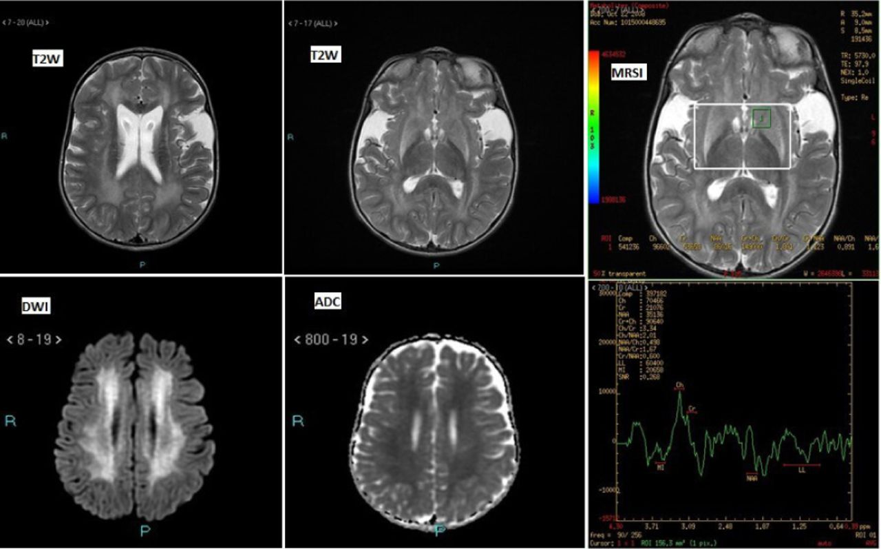
- Figure 4
Selected MRI images in child with Glutaric aciduria showing abnormal T2W periventricular high signal (upper row left corner image) with relatively smaller basal ganglia and widened sylvian fissures and anterior temporal pole extra-axial CSF spaces (upper row middle image), diffusion restriction along periventricular white matter (bottom row left corner and middle images). MR spectroscopic imaging (MRSI) with region of interest (at left basal ganglia as shown in upper row right corner image) and peaks of metabolites (decreased NAA/Cr, increased lactate) in spectroscopic image (bottom row right corner image).
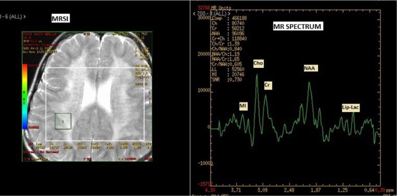
- Figure 5
MR spectroscopic imaging (left image) in a case of Metachromatic Leukodystrophy (MLD) showing reduced NAA, increased Cho/Cr ratio and lip-lactate peaks in MR spectroscopic image (right image).


Tables
MRI findings Neurometabolic Disorder (Metabolic & Genetic testing) Total Positive Negative Positive 59 (74.7) 20 (25.3) 79 (100) Negative 13 (24.1) 41 (75.9) 54 (100) Total 72 (54.1) 61 (45.9) 133 (100) Chi square = 33.09
p-value = 0.0001
- Table 2
The MRI pattern and specific imaging findings in neurometabolic diseases and other inherited metabolic disorders with profound CNS manifestations.
Neurometabolic Disorders & other Inherited Metabolic disorders (with profound CNS manifestations) Inheritance Brain Magnetic Resonance Imaging Findings Magnetic Resonance Spectroscopy Disorders of Protein metabolism Organic aciduria (glutaric aciduria) AR Bilateral basal ganglia lesions with restricted diffusion during acute encephalopathy, cerebral dysgenesis (enlarged Sylvian fissures, wide CSF spaces anterior to the temporal poles), macrocephaly Decreased N-acetyl-aspartate (NAA)/ creatine (Cr) ratio, lactate peak within basal ganglia acutely Urea cycle defect(Citrullinemia) AR White matter edema as result of hyperammonemia, basal ganglia involvement Prominent increase of glutamine/ glutamate and lipid/lactate complexes, decrease of NAA Aminoacidopathy (MSUD) AR Vacuolating myelinopathy (intramyelinic edema), edematous lesions with restricted diffusion in the peri-rolandic white matter, posterior limb of the internal capsules, cerebral peduncles, brainstem, deep cerebellar white matter, and globi pallidi Decreased N-acetylaspartate, methyl resonances of branched amino acids at 0.9–1.0 ppm, and lactate in acute metabolic decompensation Carbohydrate metabolism Galactosemia AR Diffuse edema, diffusion restriction Galactitol (Gal-ol) doublet peak at 3.7 parts per million G6PD deficiency X-linked Symmetrical lesions in bilateral globus pallidus, hyperintense on T2/FLAIR Kernicterus; increased levels of glutamine and glutamate along with decreased levels of choline and N-acetyl-aspartate Fatty Acid Oxidation Disorder Carnitine deficiency, Acyl-Coenzyme Dehydrogenase Deficiency, glutaric aciduria type 2 AR Underdeveloped frontal and temporal lobes with enlarged sylvian fissures, delayed myelination,Multifocal parenchymal and intraventricular hemorrhages as well as white matter signal intensity changes Normal N-acetylaspartate and an increased choline-creatine ratio Peroxisomal disorders Adrenoleukodystrophy (ALD) X-linked Deep white matter in the parieto-occipital lobes and splenium of the corpus callosum, cortical and subcortical U-fiber sparing, enhancement in 50% Neuronal loss manifested by a decrease in the NAA peak and an elevation in the lactate peak Zellweger Syndrome AR Ventricular enlargement, abnormal gyration patterns (pachygyria; especially medial gyri around peri-rolandic regions, polymicrogyria; laterally), cerebral periventricular pseudocysts Nonspecific reduction of N-acetylaspartate with lipid accumulation Lysosomal disorders Mucopolysaccharidosis AR (except Hunter, MP-II; X-linked) Enlarged perivascular spaces (“cribriform” or “spindle-like”), white matter lesions, hydrocephalus, cortical atrophy, ‘honeycomb-like’ appearance in the basal ganglia and thalami Decreased N-acetylaspartate, total choline and glutamate in the white matter, and an elevation of myo-inositol, glucose-aminoglycans (GAG) peak at 3.7 ppm (higher than myoinositol) Metachromatic Leucodystrophy (MLD) AR Bilateral symmetrical abnormal high SI on T2 and FLAIR images in the deep periventricular white matter, with sparing of the subcortical U fibers and peripheral “tigroid” or “leopard pattern” of dysmyelination Reduced N-acetyl aspartate (NAA), increased myoinositol, increased lactate Krabbe disease(Globoid cell Leucodystrophy) AR High signal involving periventricular white matter, centrum semiovale and deep grey matter, sparing of subcortical U-fibres Markedly reduced NAA, Abnormally high Cho/Cr ratio Neuronal Ceroid Lipofuscinosis (NCL) AR Generalized brain atrophy and hyperintense white matter changes Reduced NAA, elevated lipids Mitochondrial Disorders Leigh Syndrome AR, X-linked Symmetrical hyperintense lesions in the basal ganglia and/or brain stem on T2-weighted MR images Elevated choline, occasionally elevated lactate, reduced NAA Disorders of purine and pyrimidine metabolism Lesch-Nyhan disease X-linked Decreased cerebral volume with a predilection for white matter, cortical and/or brainstem atrophy Decreased metabolites, especially N-acetylaspartate and glutamate/glutamine, only in the prefrontal cortex


Jump to section
Related Articles
Cited By...
- No citing articles found.