
Main menu

  • Home
  • Content
    • Latest
    • Ahead of print
    • Archive
  • Info for
    • Authors
    • Reviewers
    • Subscribers
    • Institutions
    • Advertisers
  • About Us
    • About Us
    • Editorial Office
    • Editorial Board
  • More
    • Alerts
    • Feedback
    • Folders
    • Help
  • Other Publications
    • Saudi Medical Journal

User menu

  • My alerts
  • Log in

Search

  • Advanced search
Neurosciences Journal
  • Other Publications
    • Saudi Medical Journal
  • My alerts
  • Log in
Neurosciences Journal

Advanced Search

  • Home
  • Content
    • Latest
    • Ahead of print
    • Archive
  • Info for
    • Authors
    • Reviewers
    • Subscribers
    • Institutions
    • Advertisers
  • About Us
    • About Us
    • Editorial Office
    • Editorial Board
  • More
    • Alerts
    • Feedback
    • Folders
    • Help
  • Follow psmmc on Twitter
  • Visit psmmc on Facebook
  • RSS
Case ReportCase Reports
Open Access

Extrahepatic pseudocyst in an adult

Yi-An Chen, Cheng-Ta Hsieh and Jui-Ming Sun
Neurosciences Journal January 2021, 26 (1) 97-102; DOI: https://doi.org/10.17712/nsj.2021.1.20200117
Yi-An Chen
From the Department of Neurological Surgery (Chen), Tri-Service General Hospital, National Defense Medical Center, from the Department of Medicine (Hsieh), School of Medicine, Fu Jen Catholic University, Taipei, from the Division of Neurosurgery (Hsieh), Department of Surgery, Sijhih Cathay General Hospital, from the Department of Medicine (Hsieh), School of Medicine, Fu Jen Catholic University, New Taipei, from the Section of Neurosurgery (Sun), Department of Surgery, Ditmanson Medical Foundation, Chia-Yi Christian Hospital, and the Department of Biotechnology (Sun), Asia University, Taichung, Taiwan
MD
  • Find this author on Google Scholar
  • Find this author on PubMed
  • Search for this author on this site
Cheng-Ta Hsieh
From the Department of Neurological Surgery (Chen), Tri-Service General Hospital, National Defense Medical Center, from the Department of Medicine (Hsieh), School of Medicine, Fu Jen Catholic University, Taipei, from the Division of Neurosurgery (Hsieh), Department of Surgery, Sijhih Cathay General Hospital, from the Department of Medicine (Hsieh), School of Medicine, Fu Jen Catholic University, New Taipei, from the Section of Neurosurgery (Sun), Department of Surgery, Ditmanson Medical Foundation, Chia-Yi Christian Hospital, and the Department of Biotechnology (Sun), Asia University, Taichung, Taiwan
MD
  • Find this author on Google Scholar
  • Find this author on PubMed
  • Search for this author on this site
Jui-Ming Sun
From the Department of Neurological Surgery (Chen), Tri-Service General Hospital, National Defense Medical Center, from the Department of Medicine (Hsieh), School of Medicine, Fu Jen Catholic University, Taipei, from the Division of Neurosurgery (Hsieh), Department of Surgery, Sijhih Cathay General Hospital, from the Department of Medicine (Hsieh), School of Medicine, Fu Jen Catholic University, New Taipei, from the Section of Neurosurgery (Sun), Department of Surgery, Ditmanson Medical Foundation, Chia-Yi Christian Hospital, and the Department of Biotechnology (Sun), Asia University, Taichung, Taiwan
MD
  • Find this author on Google Scholar
  • Find this author on PubMed
  • Search for this author on this site
  • For correspondence: [email protected]
  • Article
  • Figures & Data
  • eLetters
  • Info & Metrics
  • References
  • PDF
Loading

Article Figures & Data

Figures

  • Tables
  • Figure 1
    • Download figure
    • Open in new tab
    • Download powerpoint
    Figure 1

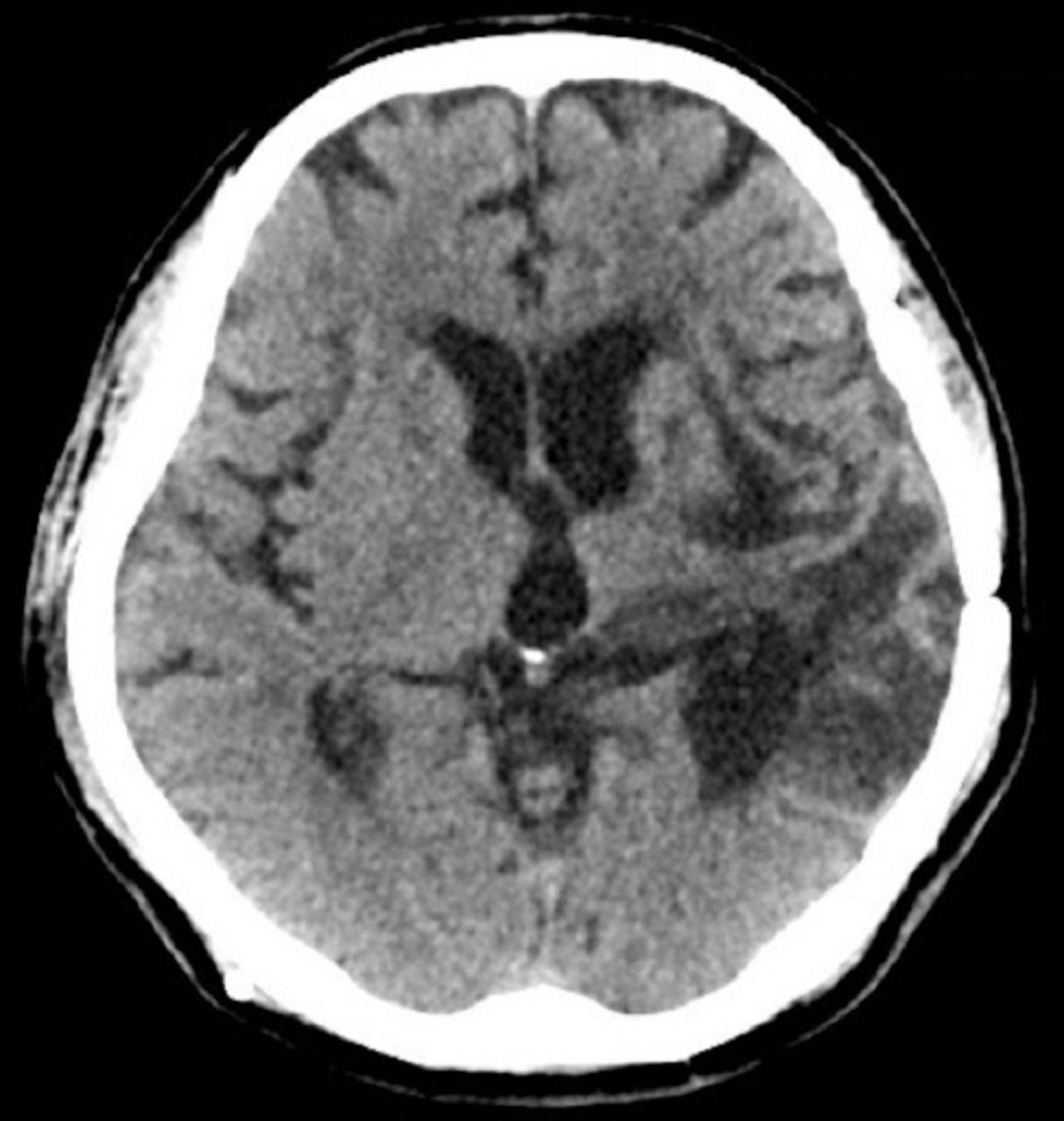
    Axial view of computed tomographic scan of brain revealed brain atrophy, large encephalomalacia at left basal ganglion, and mild hydrocephalus.

  • Figure 2
    • Download figure
    • Open in new tab
    • Download powerpoint
    Figure 2

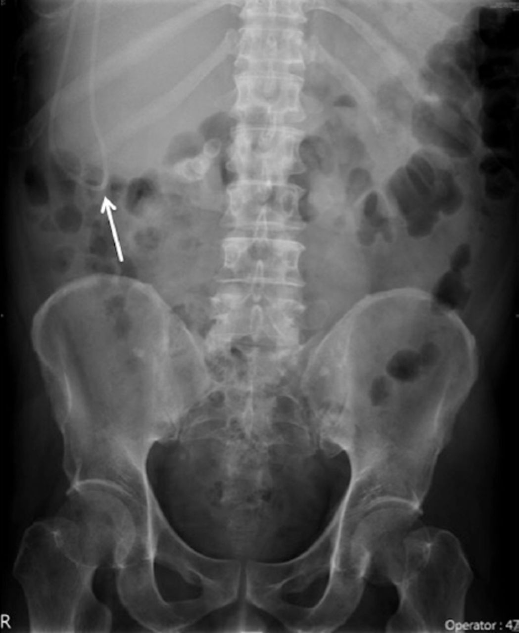
    Abdominal X-ray shows the peritoneal tip of the shunt in the right upper quadrant of the abdomen with a tube loop (white arrow).
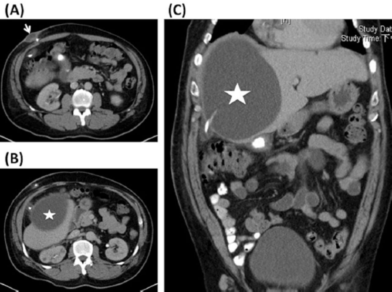
  • Figure 3
    • Download figure
    • Open in new tab
    • Download powerpoint
    Figure 3

    Enhanced computed tomographic scan of the abdomen. (A) Axial view shows the subcutaneous cyst (white arrow) at the abdominal entry site of the shunt. (B) Axial view shows the peritoneal tube penetrating the extrahepatic pseudocyst (white asterisk). (C) Reconstructed coronal view shows the shunt tip (white arrow) penetrating the extrahepatic pseudocyst (white asterisk).
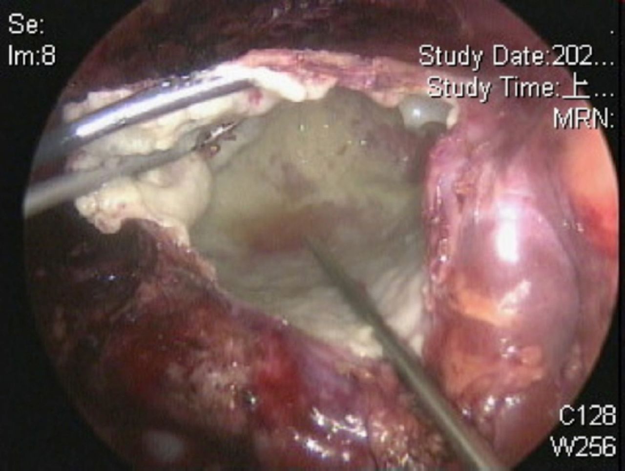
  • Figure 4
    • Download figure
    • Open in new tab
    • Download powerpoint
    Figure 4

    Intraoperative laparoscopic view reveals the unroofing of the pseudocyst.

  • Figure 5
    • Download figure
    • Open in new tab
    • Download powerpoint
    Figure 5

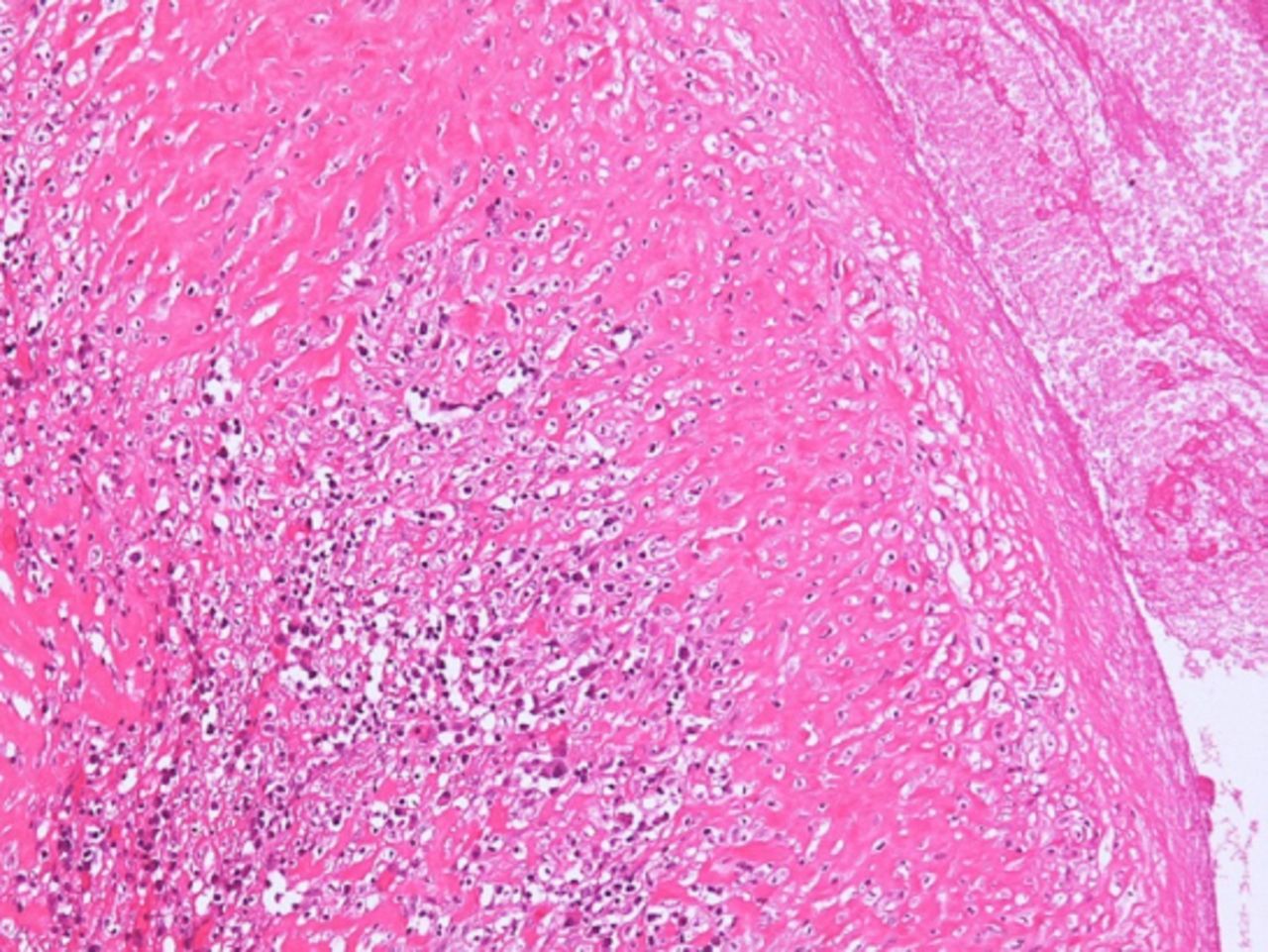
    The microscopic view of this cystic lesion demonstrates a dense thick fibrous wall and chronic granulomatous inflammation

  • Figure 6
    • Download figure
    • Open in new tab
    • Download powerpoint
    Figure 6

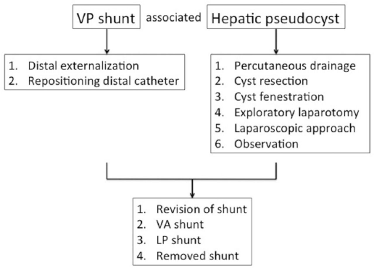
    The treatment algorithm for ventriculoperitoneal (VP) shunt-associated hepatic pseudocyst. For shunt, the distal externalization or repositioning distal catheter is initially suggested. For pseudocyst, the treatments include (1) percutaneous drainage, (2) cyst resection, (3) cyst fenestration, (4) exploratory laparotomy, (5) laparoscopic approach, or (6) observation. When the cerebral spinal fluid is sterile and hydrocephalus still exists, the revision of shunt, VA (ventriculoatrial) shunt, or lumboperitoneal (LP) shunt can be performed. Shunt may be removed when no hydrocephalus develops.

Tables

  • Figures
    • View popup
    Table 1

    The timeline for the presented case.

    DateRelevant Past Medical History and Interventions
    He had a 4-year history of spontaneous intracerebral hemorrhage at the left basal ganglion with ventricular extension and underwent VP shunt surgery with medium pressure reservoir implantation (Medtronic, USA) because of posthemorrhagic hydrocephalus
    DateSummaries from Initial and Follow-up VisitsDiagnostic Testing (including dates)Interventions
    2020/5/11• A progressive bulging mass around the previous surgical site at the right upper quadrant of the abdomen 2 weeks before admission• Laboratory examination showed white blood cell count of 5570/µL and a C-reactive protein level of 0.34 mg/µL. (2020/5/11)• Distal externalization of the VP shunt (2020/5/12)
    • Laparoscopic unroofing of the cystic lesion (2020/5/14)
    • Abdominal radiography showed a loop of the peritoneal tube around the right upper quadrant of the abdomen. (2020/5/11)• The distal shunt was ligated at the right anterior chest region (2020/5/22)
    • Computed tomography (CT) scan of the abdomen showed a large cystic lesion at the right extrahepatic region was also discovered, containing the peritoneal tube of the VP shunt. (2020/5/12)• Flomoxef 1gm i.v. q8h (2020/5/12 to 2020/5/22)
    • CSF culture: no bacterial growth (2020/5/12 to 2020/5/14)
    2020/5/23Discharge without symptoms of hydrocephalus or fever
    2020/7/28Alert and orientated. No recurrent symptoms.
PreviousNext
Back to top

In this issue

Neurosciences Journal: 26 (1)
Neurosciences Journal
Vol. 26, Issue 1
1 Jan 2021
  • Table of Contents
  • Cover (PDF)
  • Index by author
Print
Download PDF
Email Article

Thank you for your interest in spreading the word on Neurosciences Journal.

NOTE: We only request your email address so that the person you are recommending the page to knows that you wanted them to see it, and that it is not junk mail. We do not capture any email address.

Enter multiple addresses on separate lines or separate them with commas.
Extrahepatic pseudocyst in an adult
(Your Name) has sent you a message from Neurosciences Journal
(Your Name) thought you would like to see the Neurosciences Journal web site.
Citation Tools
Extrahepatic pseudocyst in an adult
Yi-An Chen, Cheng-Ta Hsieh, Jui-Ming Sun
Neurosciences Journal Jan 2021, 26 (1) 97-102; DOI: 10.17712/nsj.2021.1.20200117

Citation Manager Formats

  • BibTeX
  • Bookends
  • EasyBib
  • EndNote (tagged)
  • EndNote 8 (xml)
  • Medlars
  • Mendeley
  • Papers
  • RefWorks Tagged
  • Ref Manager
  • RIS
  • Zotero
Share
Extrahepatic pseudocyst in an adult
Yi-An Chen, Cheng-Ta Hsieh, Jui-Ming Sun
Neurosciences Journal Jan 2021, 26 (1) 97-102; DOI: 10.17712/nsj.2021.1.20200117
Twitter logo Facebook logo Mendeley logo
  • Tweet Widget
  • Facebook Like
  • Google Plus One
Bookmark this article

Jump to section

  • Article
    • Abstract
    • Case Report
    • Discussion
    • Footnotes
    • References
  • Figures & Data
  • eLetters
  • References
  • Info & Metrics
  • PDF

Related Articles

  • No related articles found.
  • PubMed
  • Google Scholar

Cited By...

  • No citing articles found.
  • Google Scholar

More in this TOC Section

  • A novel variant of RBCK1 gene causes mild polyglucosan myopathy
  • Unusual presentation of neurobrucellosis in Jeddah, Kingdom of Saudi Arabia
  • Sjogren’s syndrome associated with bilateral peripheral facial paralysis
Show more CASE REPORTS

Similar Articles

Navigate

  • home

More Information

  • Help

Additional journals

  • All Topics

Other Services

  • About

© 2025 Neurosciences Journal Neurosciences is copyright under the Berne Convention and the International Copyright Convention. All rights reserved. Neurosciences is an Open Access journal and articles published are distributed under the terms of the Creative Commons Attribution-NonCommercial License (CC BY-NC). Readers may copy, distribute, and display the work for non-commercial purposes with the proper citation of the original work. Electronic ISSN 1658-3183. Print ISSN 1319-6138.

Powered by HighWire