
Main menu

  • Home
  • Content
    • Latest
    • Ahead of print
    • Archive
  • Info for
    • Authors
    • Reviewers
    • Subscribers
    • Institutions
    • Advertisers
  • About Us
    • About Us
    • Editorial Office
    • Editorial Board
  • More
    • Alerts
    • Feedback
    • Folders
    • Help
  • Other Publications
    • Saudi Medical Journal

User menu

  • My alerts
  • Log in

Search

  • Advanced search
Neurosciences Journal
  • Other Publications
    • Saudi Medical Journal
  • My alerts
  • Log in
Neurosciences Journal

Advanced Search

  • Home
  • Content
    • Latest
    • Ahead of print
    • Archive
  • Info for
    • Authors
    • Reviewers
    • Subscribers
    • Institutions
    • Advertisers
  • About Us
    • About Us
    • Editorial Office
    • Editorial Board
  • More
    • Alerts
    • Feedback
    • Folders
    • Help
  • Follow psmmc on Twitter
  • Visit psmmc on Facebook
  • RSS
Case ReportCase Reports
Open Access

Unique challenges in the management of a Giant retroperitoneal Schwannoma associated with vertebral body destruction and spinal canal invasion

Mubarak A. Algahtany, Ahmad M. Almalki, Ismaeel D. Albeshre and Rabab N. Badri
Neurosciences Journal July 2021, 26 (3) 289-294; DOI: https://doi.org/10.17712/nsj.2021.3.20200184
Mubarak A. Algahtany
From the Division of Neurosurgery (Algahtany), Department of Surgery, College of Medicine, King Khalid University, from the Department of Surgery (Almalki), Department of Neurosurgery (Albshre), Department of Histopathology (Badri), Asir Central Hospital, Abha, Kingdom of Saudi Arabia
MD, FRCSC
  • Find this author on Google Scholar
  • Find this author on PubMed
  • Search for this author on this site
  • For correspondence: [email protected]
Ahmad M. Almalki
From the Division of Neurosurgery (Algahtany), Department of Surgery, College of Medicine, King Khalid University, from the Department of Surgery (Almalki), Department of Neurosurgery (Albshre), Department of Histopathology (Badri), Asir Central Hospital, Abha, Kingdom of Saudi Arabia
MD, SB-Surg
  • Find this author on Google Scholar
  • Find this author on PubMed
  • Search for this author on this site
Ismaeel D. Albeshre
From the Division of Neurosurgery (Algahtany), Department of Surgery, College of Medicine, King Khalid University, from the Department of Surgery (Almalki), Department of Neurosurgery (Albshre), Department of Histopathology (Badri), Asir Central Hospital, Abha, Kingdom of Saudi Arabia
MD
  • Find this author on Google Scholar
  • Find this author on PubMed
  • Search for this author on this site
Rabab N. Badri
From the Division of Neurosurgery (Algahtany), Department of Surgery, College of Medicine, King Khalid University, from the Department of Surgery (Almalki), Department of Neurosurgery (Albshre), Department of Histopathology (Badri), Asir Central Hospital, Abha, Kingdom of Saudi Arabia
MD
  • Find this author on Google Scholar
  • Find this author on PubMed
  • Search for this author on this site
  • Article
  • Figures & Data
  • eLetters
  • Info & Metrics
  • References
  • PDF
Loading

Article Figures & Data

Figures

  • Figure 1
    • Download figure
    • Open in new tab
    • Download powerpoint
    Figure 1

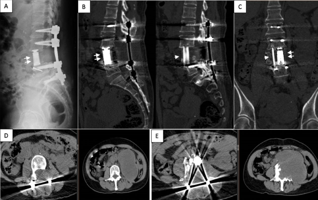
    - Preoperative CT and MRI scan images demonstrate the extent of the retroperitoneal tumor (single white arrows), L4 destruction (double white arrows), and spinal canal invasion (double black arrows). A-C) Plain CT scan; A) Soft tissue and B) bone window axial images of the abdomen at the L4 pedicle level, C) bone window sagittal image of the lumbar spine. D-F) Contrast-enhanced T1-weighted MRI scan images of the lumbar spine; D) Axial with fat saturation, E) sagittal, and F) coronal.

  • Figure 2
    • Download figure
    • Open in new tab
    • Download powerpoint
    Figure 2

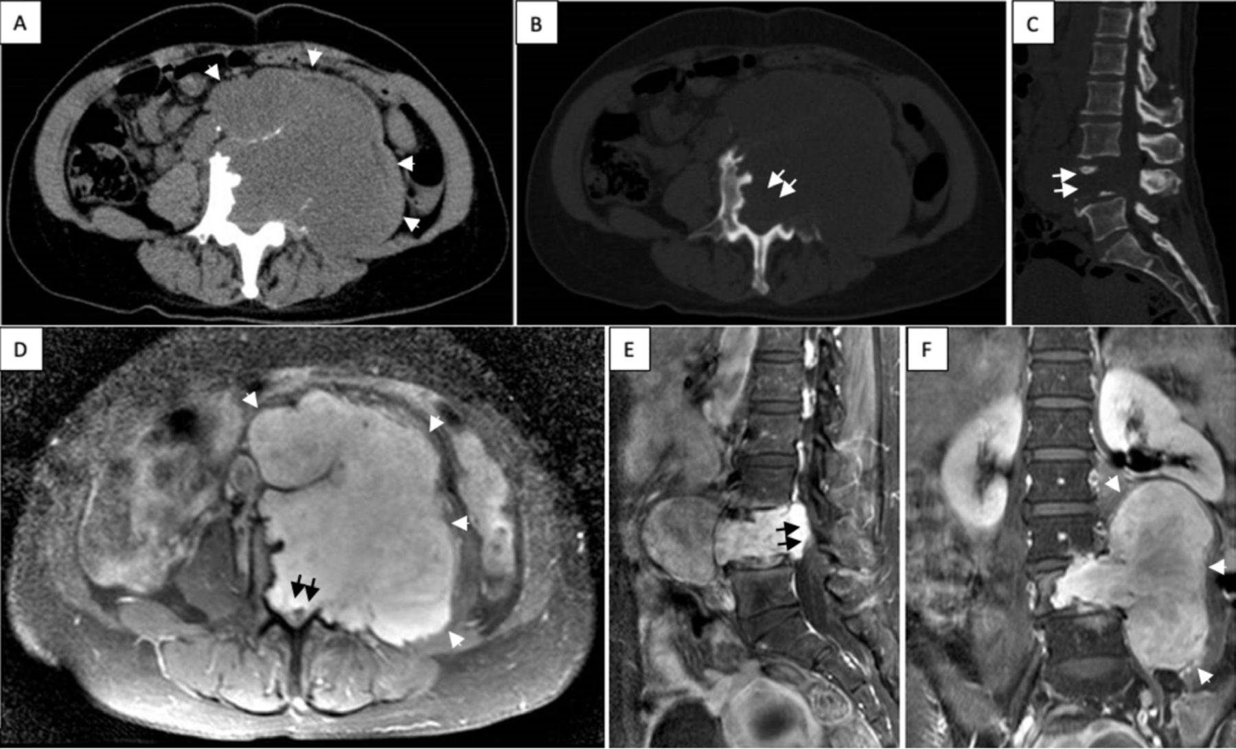
    - Postoperative 6 months follow-up, plain x-ray A) and CT scan images B-E) demonstrate a good fusion of the cage (double white arrows) and the fibular graft (single white arrows) A-C), as well as total tumor resection and lack of recurrence (D and E). A) standing lateral radiograph of the lumbar spine, B) sagittal, C) coronal bone window images. D) Axial soft tissue window image at the L3 pedicle level, E) at the L4 pedicle level with preoperative control images on the right side.

  • Figure 3
    • Download figure
    • Open in new tab
    • Download powerpoint
    Figure 3

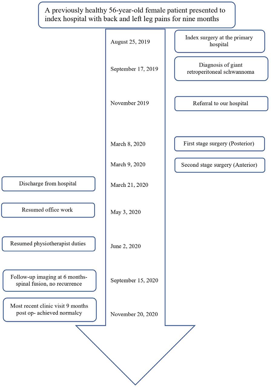
    - Timeline of the presented case.

  • Figure 4
    • Download figure
    • Open in new tab
    • Download powerpoint
    Figure 4

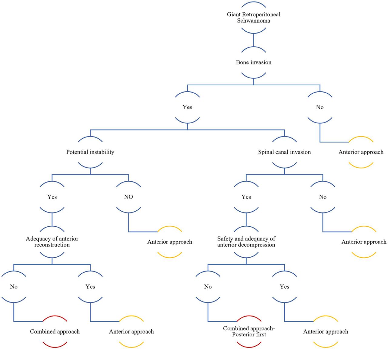
    - A flow chart for choosing the most appropriate surgical approach.

PreviousNext
Back to top

In this issue

Neurosciences Journal: 26 (3)
Neurosciences Journal
Vol. 26, Issue 3
1 Jul 2021
  • Table of Contents
  • Cover (PDF)
  • Index by author
Print
Download PDF
Email Article

Thank you for your interest in spreading the word on Neurosciences Journal.

NOTE: We only request your email address so that the person you are recommending the page to knows that you wanted them to see it, and that it is not junk mail. We do not capture any email address.

Enter multiple addresses on separate lines or separate them with commas.
Unique challenges in the management of a Giant retroperitoneal Schwannoma associated with vertebral body destruction and spinal canal invasion
(Your Name) has sent you a message from Neurosciences Journal
(Your Name) thought you would like to see the Neurosciences Journal web site.
Citation Tools
Unique challenges in the management of a Giant retroperitoneal Schwannoma associated with vertebral body destruction and spinal canal invasion
Mubarak A. Algahtany, Ahmad M. Almalki, Ismaeel D. Albeshre, Rabab N. Badri
Neurosciences Journal Jul 2021, 26 (3) 289-294; DOI: 10.17712/nsj.2021.3.20200184

Citation Manager Formats

  • BibTeX
  • Bookends
  • EasyBib
  • EndNote (tagged)
  • EndNote 8 (xml)
  • Medlars
  • Mendeley
  • Papers
  • RefWorks Tagged
  • Ref Manager
  • RIS
  • Zotero
Share
Unique challenges in the management of a Giant retroperitoneal Schwannoma associated with vertebral body destruction and spinal canal invasion
Mubarak A. Algahtany, Ahmad M. Almalki, Ismaeel D. Albeshre, Rabab N. Badri
Neurosciences Journal Jul 2021, 26 (3) 289-294; DOI: 10.17712/nsj.2021.3.20200184
Twitter logo Facebook logo Mendeley logo
  • Tweet Widget
  • Facebook Like
  • Google Plus One
Bookmark this article

Jump to section

  • Article
    • Abstract
    • Case Report
    • Acknowledgements
    • Footnotes
    • References
  • Figures & Data
  • eLetters
  • References
  • Info & Metrics
  • PDF

Related Articles

  • No related articles found.
  • PubMed
  • Google Scholar

Cited By...

  • No citing articles found.
  • Google Scholar

More in this TOC Section

  • A novel variant of RBCK1 gene causes mild polyglucosan myopathy
  • Unusual presentation of neurobrucellosis in Jeddah, Kingdom of Saudi Arabia
  • Sjogren’s syndrome associated with bilateral peripheral facial paralysis
Show more CASE REPORTS

Similar Articles

Navigate

  • home

More Information

  • Help

Additional journals

  • All Topics

Other Services

  • About

© 2025 Neurosciences Journal Neurosciences is copyright under the Berne Convention and the International Copyright Convention. All rights reserved. Neurosciences is an Open Access journal and articles published are distributed under the terms of the Creative Commons Attribution-NonCommercial License (CC BY-NC). Readers may copy, distribute, and display the work for non-commercial purposes with the proper citation of the original work. Electronic ISSN 1658-3183. Print ISSN 1319-6138.

Powered by HighWire