
Main menu

  • Home
  • Content
    • Latest
    • Ahead of print
    • Archive
  • Info for
    • Authors
    • Reviewers
    • Subscribers
    • Institutions
    • Advertisers
  • About Us
    • About Us
    • Editorial Office
    • Editorial Board
  • More
    • Alerts
    • Feedback
    • Folders
    • Help
  • Other Publications
    • Saudi Medical Journal

User menu

  • My alerts
  • Log in

Search

  • Advanced search
Neurosciences Journal
  • Other Publications
    • Saudi Medical Journal
  • My alerts
  • Log in
Neurosciences Journal

Advanced Search

  • Home
  • Content
    • Latest
    • Ahead of print
    • Archive
  • Info for
    • Authors
    • Reviewers
    • Subscribers
    • Institutions
    • Advertisers
  • About Us
    • About Us
    • Editorial Office
    • Editorial Board
  • More
    • Alerts
    • Feedback
    • Folders
    • Help
  • Follow psmmc on Twitter
  • Visit psmmc on Facebook
  • RSS
Case SeriesCase Series
Open Access

Cerebral phaeohyphomycosis at a tertiary healthcare center in Saudi Arabia

Mayyadah H. Alabdely, Abdullah S. Alolayan, Reem S. Almaghrabi and Hail M. Al-Abdely
Neurosciences Journal April 2023, 28 (2) 136-142; DOI: https://doi.org/10.17712/nsj.2023.2.20220118
Mayyadah H. Alabdely
From the Department of Critical Care (Alolayan), Department of Medicine (Almaghrabi, Al-Abdely), King Faisal Specialist Hospital and Research Center, Riyadh, Kingdom of Saudi Arabia
MD
  • Find this author on Google Scholar
  • Find this author on PubMed
  • Search for this author on this site
  • For correspondence: [email protected]
Abdullah S. Alolayan
From the Department of Critical Care (Alolayan), Department of Medicine (Almaghrabi, Al-Abdely), King Faisal Specialist Hospital and Research Center, Riyadh, Kingdom of Saudi Arabia
MD
  • Find this author on Google Scholar
  • Find this author on PubMed
  • Search for this author on this site
Reem S. Almaghrabi
From the Department of Critical Care (Alolayan), Department of Medicine (Almaghrabi, Al-Abdely), King Faisal Specialist Hospital and Research Center, Riyadh, Kingdom of Saudi Arabia
MD
  • Find this author on Google Scholar
  • Find this author on PubMed
  • Search for this author on this site
Hail M. Al-Abdely
From the Department of Critical Care (Alolayan), Department of Medicine (Almaghrabi, Al-Abdely), King Faisal Specialist Hospital and Research Center, Riyadh, Kingdom of Saudi Arabia
MD
  • Find this author on Google Scholar
  • Find this author on PubMed
  • Search for this author on this site
  • Article
  • Figures & Data
  • eLetters
  • Info & Metrics
  • References
  • PDF
Loading

Abstract

Objectives: To report cases of cerebral phaeohyphomycosis at a tertiary hospital in Riyadh, Saudi Arabia. Phaeohyphomycetes are a widely distributed group of fungi whose cell walls contain 1,8 dihydroxynaphthalene-melanin. Cerebral infections caused by these fungi are uncommon and primarily associated with neurotrophic phaeohyphomycetes.

Methods: In January of 2020 we looked back to identify cases of culture-positive cerebral phaeohyphomycosis from our medical records at King Faisal Specialist Hospital and Research Center in Riyadh, Saudi Arabia. Data on demographics, potential risk factors, clinical presentation, treatment, and outcomes were analyzed.

Results: Twelve cases of cerebral phaeohyphomycosis were identified, of which 4 were caused by Rhinocladiella mackenziei and the other 8 were caused by various phaeohyphomycetes. There were 2 cases caused by Neoscytalidium dimidiatum, and one case each caused by the following: Acrophialophora fusispora, Chaetomium atrobrunneum, Exophiala dermatitidis, Exerohilum rostratum, Fonsecaea pedrosoi, and Cladophialophora bantiana. Most patients (10 of 12) had underlying immunosuppression. R. mackenziei caused a brain-only infection manifesting as abscess formation. Four patients survived for more than a year after therapy. Surgical evacuation and triazole therapy with posaconazole or itraconazole, alone or in combination with other antifungal agents, were associated with success.

Conclusion: Cerebral phaeohyphomycosis is an uncommon fungal infection that primarily affects immunocompromised patients and is associated with poor prognosis. R. mackenziei is the most prevalent fungus in our facility and has been linked to a universal mortality.

Fungi are widespread, are present in the soil, and decompose organic matter. The presence of 1,8 dihydroxynaphthalene-melanin in the cell walls of phaeohyphomycetes is characteristic of many different genera of fungi. Phaeohyphomycosis refers to various diseases, such as cutaneous, subcutaneous, cerebral, and disseminated diseases.1

In 1974, the term phaeohyphomycosis was coined by Ajello et al2 to describe subcutaneous and systemic infections caused by fungi that produce dark-brown septate hyphae in lesions.2 Melanin has been proposed to act as a virulence factor in these species through various mechanisms. By neutralizing free radicals and hypochlorite, it protects fungi from phagocytic cells and exerts oxidative burst effects. Furthermore, melanin can bind to hydrolytic enzymes, preventing their action on the fungal plasma membrane.1 However, factors other than melanin levels may be at play. Chitin has also been proposed as a possible virulence factor for cell-wall enrichment.1

Certain species of these fungi have been reported to cause brain abscesses with rare involvement of other sites and are considered neurotrophic.1 Cladophialophora bantiana is the most common dematiaceous fungus reported to cause brain infection.2 Rhinocladiella mackenziei has been detected in Middle Eastern residents, particularly those in the Gulf region.1 In addition to a few imported cases in the United Kingdom and France, Pakistan and Iran have reported more recent cases.3-6 In the United States, an outbreak of meningeal phaeohyphomycosis caused by Exerohilum rostratum was linked to contaminated methylprednisolone solution injected primarily into the back.7 However, reporting infections caused by these fungi is not required; thus, there is no accurate estimate of their incidence and prevalence.8 Here, we report our experience with several cases of cerebral phaeohyphomycosis from a single center.

Methods

A retrospective, single-center cohort study was conducted at King Faisal Specialist Hospital and Research Center in Riyadh, Saudi Arabia from January 1999 to December 2000 based on a review of electronic medical records and medical charts. Cases with a positive culture of phaeohyphomycetes recovered from brain biopsies or cerebrospinal fluid (CSF) were identified using a microbiology database. The diagnosis was based on fungal microbiological recovery. Morphological identification of the growth characteristics and slide culture served as the primary diagnostic test at the species level. Two additional cases were confirmed via polymerase chain reaction (PCR) amplification and ribosomal RNA sequencing. A review of the data included the following: demographics; potential risk factors including underlying condition, surgeries, steroid therapy, and other immunosuppressive therapies; and clinical presentation, treatment, and outcome. For the descriptive analysis, Microsoft Excel and IBM Statistical Package for Social Sciences (SPSS) Statistics version 22 (IBMCorp, Armonk, NY, USA) for Mac were used. This study was approved by the review board of our institution. The authors confirm that the ethical policies of the journal, as outlined on the author guidelines page, have been adhered to. A literature review of similar cases was also conducted.

Results

We identified 12 cases of cerebral phaeohyphomycosis through brain tissue cultures and one patient with persistently positive CSF cultures. The histopathology of all patients, except one, was positive for fungal hyphae. Two cases were confirmed via PCR amplification and RNA sequencing (Table 1). The patients were from various regions of Saudi Arabia, with the exception of 3 patients from the same region.

View this table:
  • View inline
  • View popup
Table 1

- Characteristics of phaeohyphomycosis cases at our tertiary hospital in Riyadh, Saudi Arabia.26,31,32*

View this table:
  • View inline
  • View popup
Table 1 continued

- Characteristics of phaeohyphomycosis cases at our tertiary hospital in Riyadh, Saudi Arabia.26,31,32*

Of the 12 patients, 8 were men and 4 were women. In terms of the causative agent, four cases were caused by Rhinocladiella mackenziei, and 2 by Neoscytalidium dimidiatum. Meanwhile, the remaining six cases were caused by various phaeohyphomycetes: Acrophialophora fusispora, Chaetomium atrobrunneum, Exophiala dermatitidis, Exerohilum rostratum, Fonsecaea pedrosoi, and Cladophialophora bantiana (Table 1).

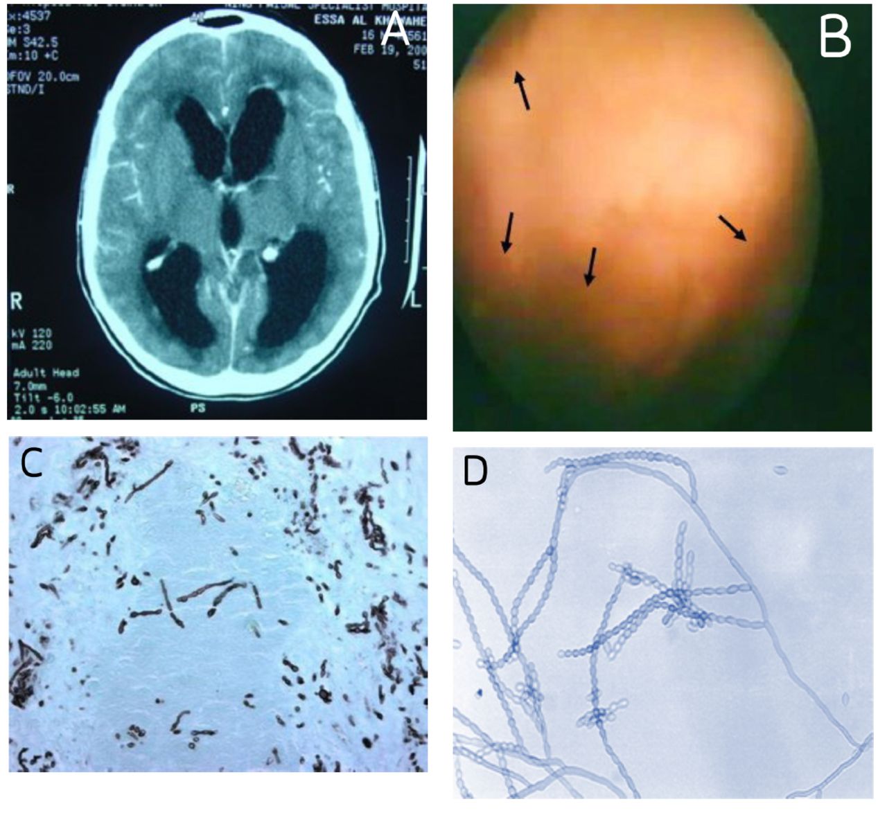
Ten patients were immunocompromised because of organ transplantation or cancer, and one was diagnosed with chronic granulomatous disease (CGD). A young patient with C. bantiana infection had no discernible immune deficiency despite a comprehensive immunological evaluation (Figure 1).

Figure 1
  • Download figure
  • Open in new tab
  • Download powerpoint
Figure 1

- Radiographic, ventriculoscopic, and histopathological morphologies of Cladophialophora bantiana A) A patient with hydrocephalus due to a brain infection caused by Cladophialophora bantiana. B) Ventriculoscopy revealed extensive ventriculitis, with fungus incorporating the lateral ventricles (arrows). C) Gomori methenamine silver histopathology stain of the brain revealed septate hyphae, and D) fungal culture revealed the characteristic morphology of C. bantiana.

Of the 4 patients infected with R. mackenziei, 2 received kidney transplants, one received a liver transplant, and one was immunocompetent. R. mackenziei caused an infection that occurred solely in the brain, resulting in abscess formation. Other phaeohyphomycetes involved different organs, such as sinuses, or disseminated multiorgan involvement.

After diagnosis, 4 patients survived for more than a year. One patient with CGD survived for 10 years after receiving suppressive antifungal therapy. One of the 4 patients with R. mackenziei infection survived for over 3 years on antifungal therapy. Nine of the 12 patients were treated with medical therapy and surgical resection. Most patients underwent surgery and received antifungal combination therapy (Figure 2). Surgery was combined with the triazole antifungal agents posaconazole or itraconazole to treat survivors.

Discussion

Phaeohyphomycosis causes a wide range of diseases, including those that are cutaneous, subcutaneous, cerebral, and disseminated,8 with a mortality rate of 70% in cerebral and disseminated diseases.9 C. bantiana is the most common phaeohyphomycete that causes cerebral infections. More than 80 cases of cerebral phaeohyphomycosis caused by C. bantiana infection have been reported worldwide, and nearly half of these cases involve immunocompetent individuals.8 E. dermatitidis is the second most common phaeohyphomycete that causes cerebral infections.9 Only one case of each was identified in our study. One case was caused by E. rostratum, a common saprobic fungus on plant debris that rarely causes clinically significant infections, except in immunocompromised patients, although localized infections have been reported.10

In 2012, the Centers for Disease Control and Prevention (CDC) in the United States reported an outbreak of fungal meningitis caused by direct inoculation of methylprednisolone contaminated with E. rostratum into human tissue. The contaminated injection was identified and removed from clinical use, ending the outbreak.7 Our E. rostratum-infected patient was pregnant and had no history of injections or immunocompromised status. We also encountered a case caused by F. pedrosoi, a well-known cause of chromoblastomycosis in tropical climate zones, particularly in South America.11 N. dimidiatum (syn. Scytalidium dimidiatum) is a fungus that causes onychomycosis and tinea pedis but rarely causes cerebral infection.12 It caused a fatal infection in 2 of our patients who had recently received kidney transplants. C. atrobrunneum has been reported to cause cerebral infection in patients with hematological malignancies, as observed in our patient with acute myeloid leukemia. In addition, the fungus was repeatedly cultured in the CSF.13 Meanwhile, A. fusispora is a rare cause of phaeohyphomycosis, with only 5 cases and 2 cases of brain abscesses reported in medical literature, including our case.14 Cases of cerebral phaeohyphomycosis are typically sporadic and unrestricted by geography, except for those caused by R. mackenziei, which are typically restricted to the Middle East. Recently, Iran, Pakistan, the United Kingdom, and France have reported cases.3-6 To the best of our knowledge, as of July 10, 2022, 37 cases of R. mackenziei have been reported,15,16 12 of which originated in Saudi Arabia (Table 2).17-26

View this table:
  • View inline
  • View popup
Table 2

- Cases of R. mackenziei reported in Saudi Arabia.

R. mackenziei affects immunocompromised and immunocompetent individuals. Nine of the 12 reported cases in Saudi Arabia were immunocompetent. In our cases, 3 patients were immunocompromised and one was immunocompetent. Recent reports have primarily focused on immunocompromised individuals. Only 2 cases of prolonged survival with R. mackenziei have been reported in Saudi Arabia and the United Kingdom.5,26 The patients in both cases underwent surgical excision and posaconazole antifungal treatment.

The geographical restriction of R. mackenziei remains unclear. The fungus has not been isolated from nature, and its Middle Eastern distribution suggests a preference for the saprophytic phase in arid climates.21 In addition, diagnosis of phaeohyphomycosis is challenging because it depends on the histopathology and culture. Phaeohyphomycosis is characterized by darkly pigmented hyphae, which can be observed more clearly with Masson-Fontana melanin staining. However, this stain can also be positive for hyaline fungi, indicating a low specificity. PCR amplification and molecular sequencing have assisted in the classification and identification of fungi including black molds. In contrast, application of serology in the diagnosis of phaeohyphomycosis has not been reported. Cases with favorable outcomes suggest that medical and surgical treatment are the treatment of choice for cerebral phaeohyphomycosis.1

Typically, amphotericin B and the triazoles itraconazole and posaconazole are used for treatment. Microdilution methods for in vitro antifungal susceptibility testing have not been standardized, and it is difficult to determine the best therapy without validated minimal inhibitory concentration breakpoints. The comparison of various antifungals has only been performed in animal models. In a study conducted on murine models, posaconazole was superior to amphotericin B and itraconazole.28 Amphotericin B treatment is generally ineffective. When surgery is impossible, combination therapy with extended-spectrum triazole and flucytosine, which has in vitro activity against many black molds, has been suggested.14 There is limited data on echinocandins as antiphaeohyphomycete agents.

Few animal studies of combination antifungal therapies have demonstrated positive outcomes.29,30 In most of our patients, combination therapy was used; however, surgical resection was necessary for success.

Despite the findings, our study has some limitations. First, it is based on a case series; therefore, its findings cannot be generalized. Additionally, no comparison group was included in the description of the findings. However, we believe that our case series adds to the limited knowledge on intractable serious infections, significantly contributing to medical literature.

Conclusion

Rare and fatal cerebral phaeohyphomycosis is a fungal infection with a poor prognosis. R. mackenziei is the predominant fungus at our facility and is associated with a universal mortality, except in some cases treated with a combination of surgical and antifungal therapies.

  • Received October 23, 2022.
  • Accepted March 15, 2023.
  • Copyright: © Neurosciences

Neurosciences is an Open Access journal and articles published are distributed under the terms of the Creative Commons Attribution-NonCommercial License (CC BY-NC). Readers may copy, distribute, and display the work for non-commercial purposes with the proper citation of the original work.

References

  1. 1.↵
    1. Al-Abdely, HM.
    Phaeohyphomycosis: A Dark Question Mark in Clinical Disease. J Invasive Fungal Infect 2009; 3: 82–88.
    OpenUrl
  2. 2.↵
    1. Ajello L,
    2. Georg LK,
    3. Steigbigel RT,
    4. Wang CJ.
    A case of phaeohyphomycosis caused by a new species of Phialophora. Mycologia 1974; 66: 490–498.
    OpenUrlCrossRefPubMed
  3. 3.↵
    1. Jabeen K,
    2. Farooqi J,
    3. Zafar A,
    4. Jamil B,
    5. Mahmood SF,
    6. Ali F, et al.
    Rhinocladiella mackenziei as an emerging cause of cerebral Phaeohyphomycosis in Pakistan: a case series. Clin Infect Dis 2011; 52: 213–217.
    OpenUrlCrossRefPubMed
  4. 4.
    1. Mohammadi R,
    2. Mohammadi A,
    3. Ashtari F,
    4. Khorvash F,
    5. Hakamifard A,
    6. Vaezi A, et al.
    Cerebral Phaeohyphomycosis due to Rhinocladiella mackenziei in Persian Gulf region: A case and review. Mycoses 2018; 61: 261–265.
    OpenUrl
  5. 5.↵
    1. Hardman N,
    2. Young N,
    3. Hobson R,
    4. Sandoe J,
    5. Wellbery-Smith M,
    6. Thomson S, et al.
    Prolonged survival after disseminated Rhinocladiella infection treated with surgical excision and posaconazole. Transpl Infect Dis 2020; 22: e13264.
    OpenUrl
  6. 6.↵
    1. Cristini A,
    2. Garcia-Hermoso D,
    3. Celard M,
    4. Albrand G,
    5. Lortholary O.
    Cerebral Phaeohyphomycosis caused by Rhinocladiella mackenziei in a woman native to Afghanistan. J Clin Microbiol 2010; 48: 3451–3454.
    OpenUrlAbstract/FREE Full Text
  7. 7.↵
    1. Kontoyiannis DP,
    2. Perlin DS,
    3. Roilides E,
    4. Walsh TJ.
    What can we learn and what do we need to know amidst the iatrogenic outbreak of Exserohilum rostratum meningitis? Clin Infect Dis 2013; 57: 853–859.
    OpenUrlCrossRefPubMed
  8. 8.↵
    1. Brandt ME,
    2. Warnock DW. Epidemiology
    , clinical manifestations, and therapy of infections caused by dematiaceous fungi. J Chemother 2003; 15: 36–47.
    OpenUrlCrossRefPubMed
  9. 9.↵
    1. Horré R,
    2. de Hoog GS.
    Primary cerebral infections by melanized fungi: a review. Stud Mycol 1999; 43: 176–193.
    OpenUrl
  10. 10.↵
    1. Adler A,
    2. Yaniv I,
    3. Samra Z,
    4. Yacobovich J,
    5. Fisher S,
    6. Avrahami G, et al.
    Exserohilum: an emerging human pathogen. Eur J Clin Microbiol Infect Dis 2006; 25: 247–253.
    OpenUrlCrossRefPubMed
  11. 11.↵
    1. de Hoog GS,
    2. Attili-Angelis D,
    3. Vicente VA,
    4. Van Den Ende AH,
    5. Queiroz-Telles F.
    Molecular ecology and pathogenic potential of Fonsecaea species. Med Mycol 2004; 42: 405–416.
    OpenUrlCrossRefPubMed
  12. 12.↵
    1. Madrid H,
    2. Ruíz-Cendoya M,
    3. Cano J,
    4. Stchigel A,
    5. Orofino R,
    6. Guarro J.
    Genotyping and in vitro antifungal susceptibility of Neoscytalidium dimidiatum isolates from different origins. Int J Antimicrob Agents 2009; 34: 351–354.
    OpenUrlCrossRefPubMed
  13. 13.↵
    1. Yeghen T,
    2. Fenelon L,
    3. Campbell CK,
    4. Warnock DW,
    5. Hoffbrand AV,
    6. Prentice HG, et al.
    Chaetomium pneumonia in patient with acute myeloid leukaemia. J Clin Pathol 1996; 49: 184–186.
    OpenUrlAbstract/FREE Full Text
  14. 14.↵
    1. Chowdhary A,
    2. Meis JF,
    3. Guarro J,
    4. de Hoog GS,
    5. Kathuria S,
    6. Arendrup MC, et al.
    ESCMID and ECMM joint clinical guidelines for the diagnosis and management of systemic Phaeohyphomycosis: diseases caused by black fungi. Clin Microbiol Infect 2014; 20 Suppl 3: 47–75.
    OpenUrl
  15. 15.↵
    1. Al Otaibi TM,
    2. Gheith OA,
    3. Alobaid K,
    4. Nair P,
    5. Eldein SMZ,
    6. Mahmoud TS, et al.
    Disseminated Rhinocladiella mackenziei infection in a kidney transplant recipient: A case report and literature review. J Mycol Med 2021; 31: 101196.
    OpenUrl
  16. 16.↵
    1. Lafont Rapnouil B,
    2. Cohen JF,
    3. Bailly E,
    4. Bernard L,
    5. Garcia-Hermoso D,
    6. Lanternier F, et al.
    Morocco as a possible source for acquisition of Rhinocladiella mackenziei. PLoS Negl Trop Dis 2021; 15: e0009563.
    OpenUrl
  17. 17.↵
    1. Naim-ur-Rahman, Mahgoub ES,
    2. Chagla AH.
    Fatal brain abscesses caused by Ramichloridium obovoideum: report of three cases. Acta Neurochir (Wien) 1988; 93: 92–95.
    OpenUrlCrossRefPubMed
  18. 18.
    1. Campbell CK,
    2. Al-Hedaithy SSA.
    Phaeohyphomycosis of the brain caused by Ramichloridium mackenziei sp. nov. in middle eastern countries. J Med Vet Mycol 1993; 31: 325–332.
    OpenUrlCrossRef
  19. 19.
    1. Sutton DA,
    2. Slifkin M,
    3. Yakulis R,
    4. Rinaldi MG.
    U.S. case report of cerebral Phaeohyphomycosis caused by Ramichloridium obovoideum (R. mackenziei): criteria for identification, therapy, and review of other known dematiaceous neurotropic taxa. J Clin Microbiol 1998; 36: 708–715.
    OpenUrlAbstract/FREE Full Text
  20. 20.
    1. Podnos YD,
    2. Anastasio P,
    3. De La Maza L,
    4. Kim RB.
    Cerebral Phaeohyphomycosis caused by Ramichloridium obovoideum (Ramichloridium mackenziei): case report. Neurosurgery 1999; 45: 372–375.
    OpenUrlCrossRefPubMed
  21. 21.↵
    1. Kanj SS,
    2. Amr SS,
    3. Roberts GD.
    Ramichloridium mackenziei brain abscess: report of two cases and review of the literature. Med Mycol 2001; 39: 97–102.
    OpenUrlCrossRefPubMed
  22. 22.
    1. Kashgari TQ,
    2. Al-Miniawi H,
    3. Moawad Hanna MK.
    Cerebral Phaeohyphomycosis caused by Ramichloridium mackenziei in the Eastern Province of Saudi Arabia. Ann Saudi Med 2000; 20: 457–460.
    OpenUrlPubMed
  23. 23.
    1. Taj-Aldeen SJ,
    2. Almaslamani M,
    3. Alkhalf A,
    4. Al Bozom I,
    5. Romanelli AM,
    6. Wickes BL, et al.
    Cerebral Phaeohyphomycosis due to Rhinocladiella mackenziei (formerly Ramichloridium mackenziei): a taxonomic update and review of the literature. Med Mycol 2010; 48: 546–556.
    OpenUrlCrossRefPubMed
  24. 24.
    1. Amr SS,
    2. Al-Tawfiq JA.
    Aspiration cytology of brain abscess from a fatal case of cerebral Phaeohyphomycosis due to Ramichloridium mackenziei. Diagn Cytopathol 2007; 35: 695–699.
    OpenUrlCrossRefPubMed
  25. 25.
    1. Al-Tawfiq JA,
    2. Boukhamseen A.
    Cerebral phaeohyphomycosis due to Rhinocladiella mackenziei (formerly Ramichloridium mackenziei): case presentation and literature review. J Infect Public Health 2011; 4: 96–102.
    OpenUrlCrossRefPubMed
  26. 26.↵
    1. Al-Abdely HM,
    2. Alkhunaizi AM,
    3. Al-Tawfiq JA,
    4. Hassounah M,
    5. Rinaldi MG,
    6. Sutton DA.
    Successful therapy of cerebral Phaeohyphomycosis due to Ramichloridium mackenziei with the new triazole posaconazole. Med Mycol 2005; 43: 91–95.
    OpenUrlCrossRefPubMed
  27. 27.
    1. Hardman N,
    2. Young N,
    3. Hobson R,
    4. Sandoe J,
    5. Wellberry-Smith M,
    6. Thomson S, et al.
    Prolonged survival after disseminated Rhinocladiella infection treated with surgical excision and posaconazole. Transpl Infect Dis 2020; 22: e13264.
    OpenUrl
  28. 28.↵
    1. Al-Abdely HM,
    2. Najvar L,
    3. Bocanegra R,
    4. Fothergill A,
    5. Loebenberg D,
    6. Rinaldi MG, et al.
    SCH 56592, amphotericin B, or itraconazole therapy of experimental murine cerebral Phaeohyphomycosis due to Ramichloridium obovoideum (“Ramichloridium mackenziei”). Antimicrob Agents Chemother 2000; 44: 1159–1162.
    OpenUrlAbstract/FREE Full Text
  29. 29.↵
    1. Sun Y,
    2. Liu W,
    3. Wan Z,
    4. Wang X,
    5. Li R.
    Antifungal activity of antifungal drugs, as well as drug combinations against Exophiala dermatitidis. Mycopathologia 2011; 171: 111–117.
    OpenUrlCrossRefPubMed
  30. 30.↵
    1. Mariné M,
    2. Pastor FJ,
    3. Guarro J.
    Combined antifungal therapy in a murine model of disseminated infection by Cladophialophora bantiana. Med Mycol 2009; 47: 45–49.
    OpenUrlCrossRefPubMed
  31. 31.↵
    1. Al-Mohsen IZ,
    2. Sutton DA,
    3. Sigler L,
    4. Almodovar E,
    5. Mahgoub N,
    6. Frayha H, et al.
    Acrophialophora fusispora brain abscess in a child with acute lymphoblastic leukemia: review of cases and taxonomy. J Clin Microbiol 2000; 38: 4569–4576.
    OpenUrlAbstract/FREE Full Text
  32. 32.↵
    1. Alamri M,
    2. Alghamdi H,
    3. Althawadi S,
    4. Mutabaggani M,
    5. Dababo MA,
    6. Alajlan F, et al.
    Invasive fungal infection of the brain caused by Neoscytalidium dimidiatum in a post-renal transplant patient: A case report. Med Mycol Case Rep 2021; 34: 27–31.
    OpenUrl
PreviousNext
Back to top

In this issue

Neurosciences Journal: 28 (2)
Neurosciences Journal
Vol. 28, Issue 2
1 Apr 2023
  • Table of Contents
  • Cover (PDF)
  • Index by author
Print
Download PDF
Email Article

Thank you for your interest in spreading the word on Neurosciences Journal.

NOTE: We only request your email address so that the person you are recommending the page to knows that you wanted them to see it, and that it is not junk mail. We do not capture any email address.

Enter multiple addresses on separate lines or separate them with commas.
Cerebral phaeohyphomycosis at a tertiary healthcare center in Saudi Arabia
(Your Name) has sent you a message from Neurosciences Journal
(Your Name) thought you would like to see the Neurosciences Journal web site.
Citation Tools
Cerebral phaeohyphomycosis at a tertiary healthcare center in Saudi Arabia
Mayyadah H. Alabdely, Abdullah S. Alolayan, Reem S. Almaghrabi, Hail M. Al-Abdely
Neurosciences Journal Apr 2023, 28 (2) 136-142; DOI: 10.17712/nsj.2023.2.20220118

Citation Manager Formats

  • BibTeX
  • Bookends
  • EasyBib
  • EndNote (tagged)
  • EndNote 8 (xml)
  • Medlars
  • Mendeley
  • Papers
  • RefWorks Tagged
  • Ref Manager
  • RIS
  • Zotero
Share
Cerebral phaeohyphomycosis at a tertiary healthcare center in Saudi Arabia
Mayyadah H. Alabdely, Abdullah S. Alolayan, Reem S. Almaghrabi, Hail M. Al-Abdely
Neurosciences Journal Apr 2023, 28 (2) 136-142; DOI: 10.17712/nsj.2023.2.20220118
Twitter logo Facebook logo Mendeley logo
  • Tweet Widget
  • Facebook Like
  • Google Plus One
Bookmark this article

Jump to section

  • Article
    • Abstract
    • Methods
    • Results
    • Discussion
    • Conclusion
    • References
  • Figures & Data
  • eLetters
  • References
  • Info & Metrics
  • PDF

Related Articles

  • No related articles found.
  • PubMed
  • Google Scholar

Cited By...

  • No citing articles found.
  • Google Scholar

More in this TOC Section

  • Autoimmune epilepsy, retrospective case series of clinical features, management, and outcomes
  • Expanding the Allelic spectrum in ATP1A3-related disorders with 3 novel mutations and clinic features
  • Inherited CD59 deficiency, where neurology and genetics intertwine
Show more Case Series

Similar Articles

Navigate

  • home

More Information

  • Help

Additional journals

  • All Topics

Other Services

  • About

© 2025 Neurosciences Journal Neurosciences is copyright under the Berne Convention and the International Copyright Convention. All rights reserved. Neurosciences is an Open Access journal and articles published are distributed under the terms of the Creative Commons Attribution-NonCommercial License (CC BY-NC). Readers may copy, distribute, and display the work for non-commercial purposes with the proper citation of the original work. Electronic ISSN 1658-3183. Print ISSN 1319-6138.

Powered by HighWire