


Article Figures & Data
Figures
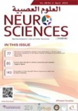
- Figure 1
- Comparison of the connections between the mPFC and amygdala, as well as the amygdala and the locus coeruleus, during normal sleep vs SD. SD - Sleep deprivation, mPFC - medial prefrontal cortex
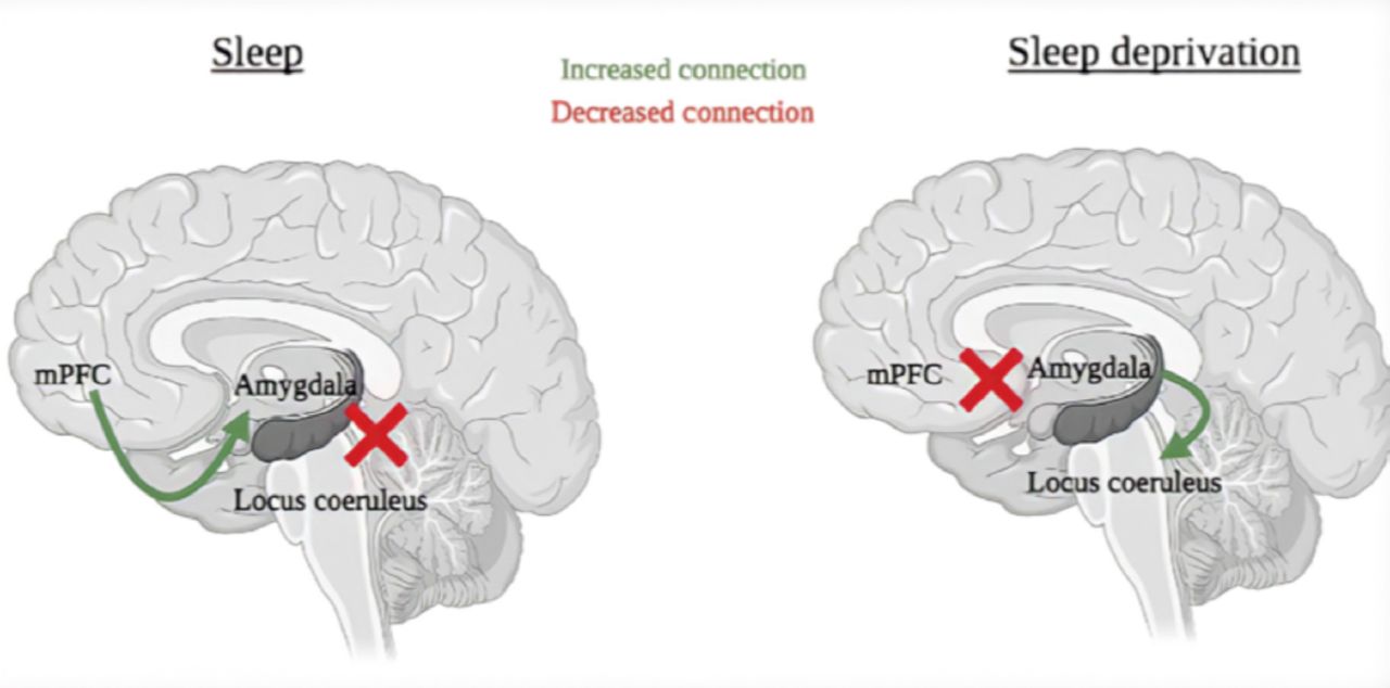
- Figure 2
- An overview of signaling pathways in the hippocampus following sleep deprivation. The dashed arrows demonstrate a reduction of a signaling pathway. (A) Altered glutamatergic signaling. disrupted cAMP signaling. (C) Down regulated mTOR signaling. Figure adapted from Prince and Abel14
- Figure 3
- Areas affected by SD (A) Areas of the brain affected by sleep deprivation, affecting alertness, memory and attentional performance. (B) In a SD state, there is unstable inhibition concerning task-related DMN and FPN activity, as well as inconsistent increasing arousal influencing activity in the thalamus. This leads to irregular signals of DMN activity and reduced FPN activity during tasks. This can lead to weakened attentiveness and working-memory functioning, becoming better with greater thalamic activity and poorer with reduced thalamic activity. Figure adapted from Krause et al54
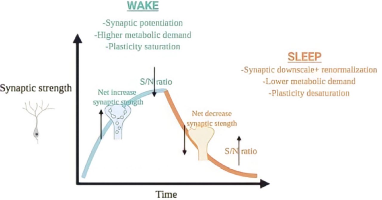
- Figure 4
- The synaptic homeostasis hypothesis. Throughout wakefulness, cortical synapses potentiate relative to activity, giving a net rise in synaptic power and a reduction is signal/noise ratio. This synaptic potentiation is linked with an escalation in slow-wave activity (SWA) in NREM sleep, where synaptic renormalization and downscaling occurs, with an increase in signal/noise ratio. This mechanism can permit additional synaptic plasticity to happen the resulting day alongside avoiding the metabolic downsides linked with excitability and extreme potentiation through wakefulness. Figure adapted from Rantamäki and Kohtala.37
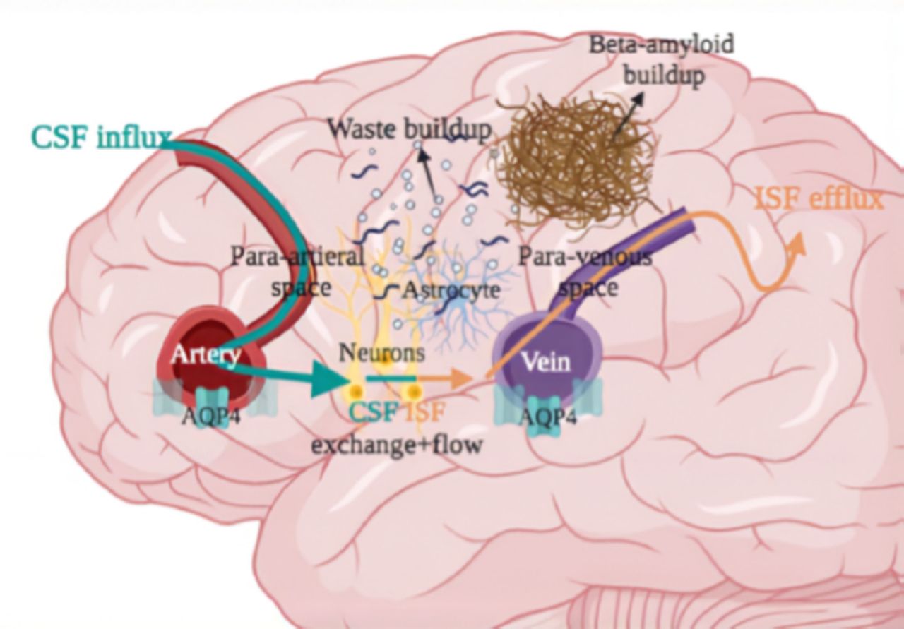
- Figure 5
- Impaired glymphatic system in the brain after sleep deprivation. Lower AQP4 expression, altered glymphatic clearance, toxic waste buildup, and higher beta-amyloid levels occur, leading to a dysfunction in cognitive performance.


In this issue


Jump to section
Related Articles
Cited By...
- No citing articles found.