


Article Figures & Data
Figures
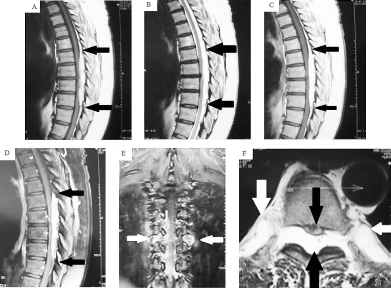
- Figure 1
Pre and post MRIs of the spine showing: A) Non-enhanced T1-weighted sagittal MRI of the mid-thoracic spine demonstrating an inhomogeneous mass, extending from T6-T9. The tumor shows predominant hyperintense fat component with small hypo-intense foci at the periphery. B) T2-weighted sagittal MRI again demonstrates predominant fat component of the lesion (between the black arrows). C) and D) Post-contrast T1-weighted sagittal MRI without and with fat-saturation showed avid inhomogeneous enhancement of the lesion. E) T2-weighted axial illustrates the mass along the posterior epidural spinal canal of the thoracic spine (lower black arrow), compressing and displacing the spinal cord anteriorly (upper black arrow), with bilateral extra-spinal extensions of the tumor (white arrows). F) Post-contrast coronal T1-weighted MRI with fat-suppression shows the bilateral extra-spinal extensions of the tumor (white arrows).
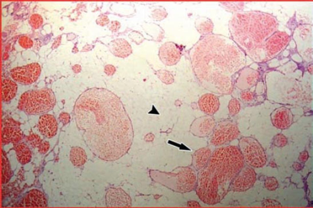
- Figure 2
Surgical specimen. Hematoxylin and eosin stain; magnification × 40; microscopically, the tumor showed mature adipose tissue (arrowhead) interlaced with numerous blood vessels ranging from capillary to cavernous in size, and a few are irregular in shape (arrow).


Tables
In this issue


Jump to section
Related Articles
Cited By...
- No citing articles found.