
Main menu

  • Home
  • Content
    • Latest
    • Ahead of print
    • Archive
  • Info for
    • Authors
    • Reviewers
    • Subscribers
    • Institutions
    • Advertisers
  • About Us
    • About Us
    • Editorial Office
    • Editorial Board
  • More
    • Alerts
    • Feedback
    • Folders
    • Help
  • Other Publications
    • Saudi Medical Journal

User menu

  • My alerts
  • Log in

Search

  • Advanced search
Neurosciences Journal
  • Other Publications
    • Saudi Medical Journal
  • My alerts
  • Log in
Neurosciences Journal

Advanced Search

  • Home
  • Content
    • Latest
    • Ahead of print
    • Archive
  • Info for
    • Authors
    • Reviewers
    • Subscribers
    • Institutions
    • Advertisers
  • About Us
    • About Us
    • Editorial Office
    • Editorial Board
  • More
    • Alerts
    • Feedback
    • Folders
    • Help
  • Follow psmmc on Twitter
  • Visit psmmc on Facebook
  • RSS
Case ReportCase Report
Open Access

Subdural hematoma caused by rupture of a posterior cerebral artery aneurysm

Zhou Feng, Qiang Tan, Lin Li and Zhi Chen
Neurosciences Journal April 2016, 21 (2) 161-163; DOI: https://doi.org/10.17712/nsj.2016.2.20150332
Zhou Feng
From the Department of Neurosurgery, Southwest Hospital, Third Military Medical University, Chongqing, China
MD
  • Find this author on Google Scholar
  • Find this author on PubMed
  • Search for this author on this site
Qiang Tan
From the Department of Neurosurgery, Southwest Hospital, Third Military Medical University, Chongqing, China
MD
  • Find this author on Google Scholar
  • Find this author on PubMed
  • Search for this author on this site
Lin Li
From the Department of Neurosurgery, Southwest Hospital, Third Military Medical University, Chongqing, China
MD
  • Find this author on Google Scholar
  • Find this author on PubMed
  • Search for this author on this site
Zhi Chen
From the Department of Neurosurgery, Southwest Hospital, Third Military Medical University, Chongqing, China
MD, PhD
  • Find this author on Google Scholar
  • Find this author on PubMed
  • Search for this author on this site
  • For correspondence: [email protected]
  • Article
  • Figures & Data
  • eLetters
  • Info & Metrics
  • References
  • PDF
Loading

Abstract

Subdural hematoma (SDH) caused by rupture of a cerebral aneurysm is rare and is usually associated with delayed diagnosis and treatment. We present a patient of a posterior cerebral artery aneurysm presenting as subacute SDH. The incidence, mechanisms and treatment of this condition are discussed.

In most cases, subdural hematoma (SDHs) are caused by head trauma. Subdural hematoma secondary to intracranial aneurysm rupture is uncommon, which account for 0.5-7.9% of all intracranial aneurysm rupture.1,2 Aneurysms of the posterior cerebral artery (PCA) comprise approximately 0.7-2.9% of all intracranial aneurysms.3,4 We report a case of subacute subdural hematoma secondary to PCA aneurysm rupture. Stent-assisted coil embolization is thought to be a safe and effective treatment for this situation. Also, the reason for presenting this case is to remind us of this condition, avoiding diagnostic and therapeutic pitfalls.

Case Report

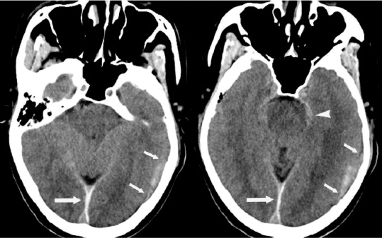
A 60-year-old hypertensive man with no history of head trauma presented with sudden headache and vomiting. four days later his symptoms suddenly worsened. He was admitted to our Department 5 days after the on-set of the symptoms. His past neurological history was unrevealing and there was no history of head trauma, blood clot disturbances, or anticoagulant therapy. His blood pressure was 172/100 mm Hg. Cranial computed tomography revealed subacute subdural hematoma in the left hemisphere, as well as interhemispheric subdural hemorrhage and minimal subarachnoid hemorrhage in the left perimesencephalic space (Figure 1). Computed tomography angiography (CTA) showed a fusiform aneurysm at the P2 segment of the left posterior cerebral artery (Figure 2A). He underwent stent-assisted coil embolization and post-embolization angiogram of the left vertebral artery showed the aneurysm was successfully occluded (Figures 2B&2C). But 3 days after the embolization of the aneurysm, he developed a change in mental status accompanied by slurred speech and narcolepsy. Emergency CT scan revealed enlargement of the left-sided subacute subdural hematoma with a new slight midline shift. Emergency drainage of the subdural hematoma SDH with a burr hole was performed. The patient’s neurological and functional status gradually improved and was transferred to local hospital for further rehabilitation 2 weeks after admission. Computed tomography scans 1 month after admission showing no abnormalities (Figure 3). And he was in further follow-up.

Figure 1
  • Download figure
  • Open in new tab
  • Download powerpoint
Figure 1

Computed tomography scans showing subacute subdural hematoma in the left hemisphere (short arrow), as well as interhemispheric subdural hemorrhage (long arrow) and minimal subarachnoid hemorrhage in the left perimesencephalic space (arrowhead).

Figure 2
  • Download figure
  • Open in new tab
  • Download powerpoint
Figure 2

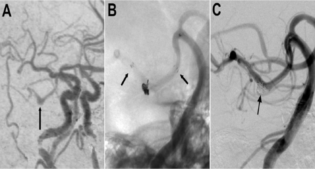
Computed tomography angiogram showing A) A fusiform aneurysm of the left posterior cerebral artery (arrow). B) Unsubtracted image before embolization showing the aneurysm treated by stent assisted coiling (arrows indicating the proximal and distal ends of the stent). C) Post-embolization angiogram of the left vertebral artery showing the occlusion of the aneurysm.

Figure 3
  • Download figure
  • Open in new tab
  • Download powerpoint
Figure 3

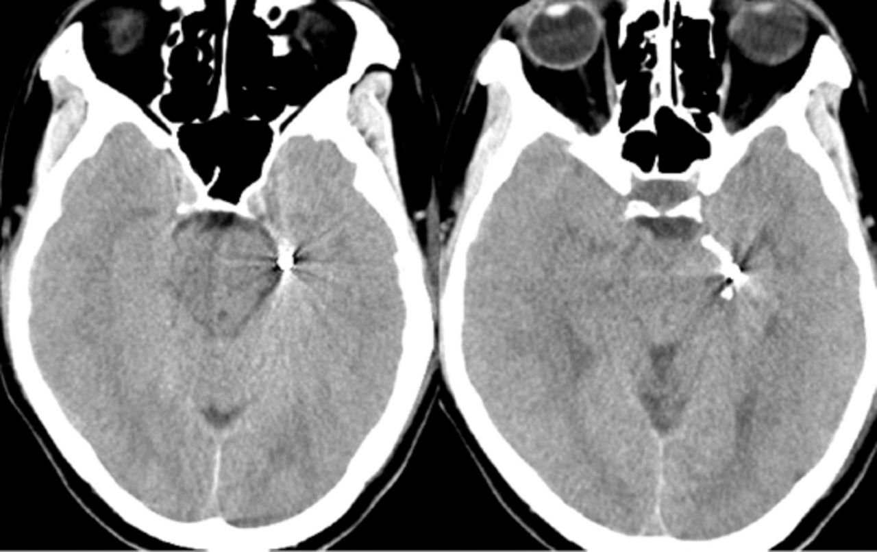
Computed tomography scans 1 month after admission showing no abnormalities.

Discussion

Intracranial aneurysm rupture is classically characterized by subarachnoid hemorrhage (SAH), and it is often complicated by intracerebral hematoma (ICH) and intraventricular hematoma (IVH). But the clinical incidence of SDH complication is very low, which was reported to be 0.5-7.9%.1,2 And the most frequently reported site of aneurysm location is the internal carotid artery (ICA)-posterior communicating artery, which is followed by aneurysms of middle cerebral artery and distal anterior cerebral artery.2,5 We described a case of ruptured PCA aneurysm presenting as subdural hematoma, which is relatively rare.

Several mechanisms have been proposed to explain the occurrence of SDH after aneurysm rupture:1,2,5,6 1)Successive minor bleedings case the developing of adhesions, resulting in final rupture into the subdural space; 2) high-pressure hemorrhage may lead to rapid accumulation of blood from the leaking aneurysm, rupturing the arachnoid membrane; 3) intracerebral hemorrhage may rupture through the cortex and cause a tear of the arachnoid membrane; 4) a carotid artery aneurysm arising from the part of carotid artery within the subdural space may rupture and cause subdural hematoma directly; and 5) the acute enlargement of the intracavernous aneurysm may erode the cavernous sinus wall. As in this case, the patient had suffered headache since 4 days before he was admitted to hospital, this warning sign may reflect the presence of successive small bleedings.

Whatever the cause, most cases of aneurysmal SDH show SAH on the CT scans, and this will address the diagnosis upon a vascular cause of hemorrhage. In a small percentage of cases, however, aneurysmal rupture can cause a subdural blood collection without SAH, a rare condition called pure SDH, which may lead to inappropriate diagnosis and management.1,7 Since cranial CT cannot reveal the cause of these pathologies, it is potential for such patients to undergo CTA or digital subtraction angiography as soon as possible to reveal the source of bleeding. Then appropriate therapy should be conducted depending on the location of the aneurysm and the clinical situation promptly.

Aneurysms of the PCA are rare. Because of the presence of complex perforating branches and adjacent to the cranial nerves, the surgical approach and exposure of the PCA is technically challenging.8 So endovascular treatment is relatively a safe and effective approach with low mortality and morbidity for these aneurysms.8,9 In this case we performed stent-assisted coil embolization to treat the aneurysm in the P2 segment of the PCA. Compared with the parent artery occlusion, the advantage of stent-assisted techniques is not only that the tight aneurysm packing with coils is attainable with less risk of coil migration or coil bulging into the parent artery, but that can preserve the parent artery, avoiding ischemic neurological events.10

Even though the patient did not undergo direct surgery, the subarachnoid hemorrhage in the left perimesencephalic space and the extravasation of the blood along the interhemispheric subdural space all suggest a causal association between the subdural bleeding and the aneurysm.

In conclusion, spontaneous arterial SDH due to a P2 segment of PCA has been rare reported earlier. We should be reminded that cerebral aneurysm rupture should be considered in spontaneous subdural hematoma, especially those with no evidence of subarachnoid hemorrhage, and the appropriate treatment should be planned with expediency

Footnotes

  • Disclosure

    Authors have no conflict of interests, and the work was not supported or funded by any drug company.

  • Received May 12, 2015.
  • Accepted July 8, 2015.
  • Copyright: © Neurosciences

Neurosciences is an Open Access journal and articles published are distributed under the terms of the Creative Commons Attribution-NonCommercial License (CC BY-NC). Readers may copy, distribute, and display the work for non-commercial purposes with the proper citation of the original work.

References

  1. ↵
    1. Inamasu J,
    2. Saito R,
    3. Nakamura Y,
    4. Ichikizaki K,
    5. Suga S,
    6. Kawase T,
    7. et al.
    (2002) Acute subdural hematoma caused by ruptured cerebral aneurysms: diagnostic and therapeutic pitfalls. Resuscitation 52, 71–76.
  2. ↵
    1. Gong J,
    2. Sun H,
    3. Shi XY,
    4. Liu WX,
    5. Shen Z
    (2014) Pure subdural haematoma caused by rupture of middle cerebral artery aneurysm: Case report and literature review. J Int Med Res 42, 870–878.
  3. ↵
    1. Honda M,
    2. Tsutsumi K,
    3. Yokoyama H,
    4. Yonekura M,
    5. Nagata I
    (2004) Aneurysms of the Posterior Cerebral Artery: Retrospective Review of Surgical Treatment. Neurol Med Chir (Tokyo) 44, 164–168.
  4. ↵
    1. Zhao ZW,
    2. Deng JP,
    3. Gao L,
    4. Gao GD
    (2008) Endovascular Management of Posterior Cerebral Artery Aneurysms. Interv Neuroradiol 14, 253–258.
  5. ↵
    1. Takada T,
    2. Yamamoto T,
    3. Ishikawa E,
    4. Zaboronok A,
    5. Kujiraoka Y,
    6. Akutsu H,
    7. et al.
    (2012) Acute Subdural Hematoma without Subarachnoid Hemorrhage Caused by Ruptured A1-A2. Junction Aneurysm. Neurol Med Chir (Tokyo) 52, 430–434.
  6. ↵
    1. Marinelli L,
    2. Parodi RC,
    3. Renzetti P,
    4. Bandini F
    (2005) Interhemispheric subdural haematoma from ruptured aneurysm: a case report. J Neurol 252, 364–366.
  7. ↵
    1. Blasi RD,
    2. Salvati A,
    3. Renna M,
    4. Chiumarulo L
    (2010) Pure subdural hematoma due to cerebral aneurysmal rupture: an often delayed diagnosis. Cardiovasc Intervent Radiol 33, 870–873.
  8. ↵
    1. Ciceri EF,
    2. Klucznik RP,
    3. Grossman RG,
    4. Rose JE,
    5. Mawad ME
    (2001) Aneurysms of the Posterior Cerebral Artery: Classification and Endovascular Treatment. AJNR Am J Neuroradiol 22, 27–34.
  9. ↵
    1. Connolly ES,
    2. Rabinstein AA,
    3. Carhuapoma JR,
    4. Derdeyn CP,
    5. Dion J,
    6. Higashida RT,
    7. et al.
    (2012) Guidelines for the Management of Aneurysmal Subarachnoid Hemorrhage. Stroke 43, 1711–1737.
  10. ↵
    1. Irie K,
    2. Negoro M,
    3. Hayakawa M,
    4. Hayashi J,
    5. Kanno T
    (2003) Treatment of a spontaneous intracranial dissecting aneurysm with stent-assisted coil embolization. Neuroradiology 45, 825–829.
PreviousNext
Back to top

In this issue

Neurosciences Journal: 21 (2)
Neurosciences Journal
Vol. 21, Issue 2
1 Apr 2016
  • Table of Contents
  • Cover (PDF)
  • Index by author
Print
Download PDF
Email Article

Thank you for your interest in spreading the word on Neurosciences Journal.

NOTE: We only request your email address so that the person you are recommending the page to knows that you wanted them to see it, and that it is not junk mail. We do not capture any email address.

Enter multiple addresses on separate lines or separate them with commas.
Subdural hematoma caused by rupture of a posterior cerebral artery aneurysm
(Your Name) has sent you a message from Neurosciences Journal
(Your Name) thought you would like to see the Neurosciences Journal web site.
Citation Tools
Subdural hematoma caused by rupture of a posterior cerebral artery aneurysm
Zhou Feng, Qiang Tan, Lin Li, Zhi Chen
Neurosciences Journal Apr 2016, 21 (2) 161-163; DOI: 10.17712/nsj.2016.2.20150332

Citation Manager Formats

  • BibTeX
  • Bookends
  • EasyBib
  • EndNote (tagged)
  • EndNote 8 (xml)
  • Medlars
  • Mendeley
  • Papers
  • RefWorks Tagged
  • Ref Manager
  • RIS
  • Zotero
Share
Subdural hematoma caused by rupture of a posterior cerebral artery aneurysm
Zhou Feng, Qiang Tan, Lin Li, Zhi Chen
Neurosciences Journal Apr 2016, 21 (2) 161-163; DOI: 10.17712/nsj.2016.2.20150332
Twitter logo Facebook logo Mendeley logo
  • Tweet Widget
  • Facebook Like
  • Google Plus One
Bookmark this article

Jump to section

  • Article
    • Abstract
    • Case Report
    • Discussion
    • Footnotes
    • References
  • Figures & Data
  • eLetters
  • References
  • Info & Metrics
  • PDF

Related Articles

  • No related articles found.
  • PubMed
  • Google Scholar

Cited By...

  • No citing articles found.
  • Google Scholar

More in this TOC Section

  • Unmasking the mimic: Leprosy neuropathy misdiagnosed as chronic inflammatory demyelinating polyneuropathy: A case report from Saudi Arabia
  • Seminal vesicle schwannoma with chronic hemorrhage
  • Marchiafava-Bignami disease post-bariatric surgery: A case report and review of similar cases
Show more Case Report

Similar Articles

Navigate

  • home

More Information

  • Help

Additional journals

  • All Topics

Other Services

  • About

© 2025 Neurosciences Journal Neurosciences is copyright under the Berne Convention and the International Copyright Convention. All rights reserved. Neurosciences is an Open Access journal and articles published are distributed under the terms of the Creative Commons Attribution-NonCommercial License (CC BY-NC). Readers may copy, distribute, and display the work for non-commercial purposes with the proper citation of the original work. Electronic ISSN 1658-3183. Print ISSN 1319-6138.

Powered by HighWire