


Article Figures & Data
Figures
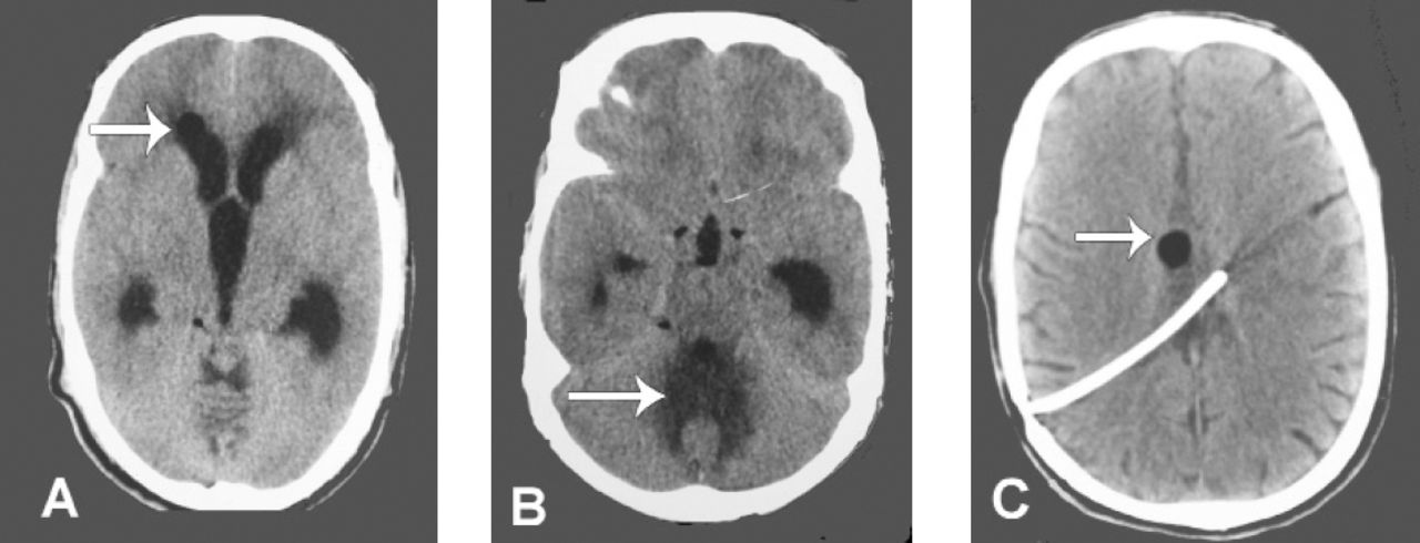
- Figure 1
Pre-op non-contrast CT brain a) Features of hydrocephalus with marker showing fat in lateral ventricle, b) Changes in the intensity in vermis, possible site of dermoid cyst after rupture and C) Post-op CT scan decompressed ventricle post–operatively with shunt in situ and marker showing fat.
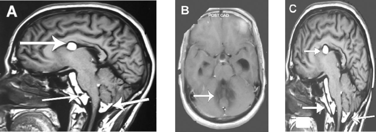
- Figure 2
Pre-op MRI with contrast a) Pre–op (T1 MRI)- Fat in the prepontine, cerebellomedullary cistern and fourth ventricle with hydrocephalus, b) Marked area shows intensity changes in vermis (non enhancement on contrast) possible site of dermoid cyst rupture c) post-op MRI (T1) fat in the same regions with ventricle decompression. Also fat visualised in the lateral ventricle.

In this issue


Jump to section
Related Articles
Cited By...
- No citing articles found.