


Article Figures & Data
Figures
- Figure 1
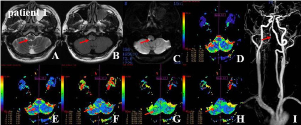
Magnetic resonance imaging-T2 weighted imaging, fluid attenuated inversion recovery (Flair) and diffusion weighted imaging showed a-c) high signal intensities in the right lateral medulla, d-h) Dynamic susceptibility contrast-enhanced-perfusion weighted imaging demonstrated a lesion ROI high cerebral blood flow, high cerebral blood flow, delayed mean transit time, decreased time to peak, and decreased time of maximum concentration on the VAH side. The representative rigid quantitative thresholds in the ROIs of 7 and 8 are shown in Table 2. i) constrast-enhanced megnetic resonance artery indicated right VAH and moderate stricture and rosary in the V4 segment of the right vertebral artery and minor kinking of the left vertebral artery. VAH - vertebral artery hypoplasia, ROI - region of interest
- Figure 2
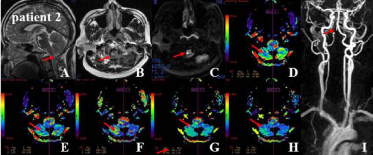
MRI-T2WI, Flair and DWI demonstrated a-c) high signal intensities in the right lateral medulla. d-h) Dynamic susceptibility contrast-enhanced-perfusion weighted imaging demonstrated a right ROI low CBF, low CBV, delayed TTP, delayed MTT, and delayed time maximum on the VAH side in Case 2. The representative rigid quantitative thresholds in the ROIs of 5 and 6 are shown in Table 2. i) constrast-enhanced megnetic resonance artery indicated right VAH and severe stricture and dilated region in the V4 segment of the right vertebral artery and moderate kinking of the left vertebral artery. VAH - vertebral artery hypoplasia, CBF - cerebral blood flow, CBV - cerebral blood volume, TTP - time to peak, MTT - mean transit time
- Figure 3
MRI-T2WI, Flair and DWI demonstrated a-c) high signal intensities in the left lateral medulla. d-h) Dynamic susceptibility contrast-enhanced-perfusion weighted imaging demonstrated left lesion ROI low CBF, low CBV, delayed MTT, and deceased Tmax on the VAH side. The representative rigid quantitative thresholds in the ROIs of 11 and 12 are shown in Table 2. i) Constrast-enhanced magnetic resonance artery indicated left VAH and minor stricture in the V4 segment of the left vertebral artery. VAH - vertebral artery hypoplasia, CBF - cerebral blood flow, CBV - cerebral blood volume, TTP - time to peak, MTT - mean transit time, ROI - region of interest
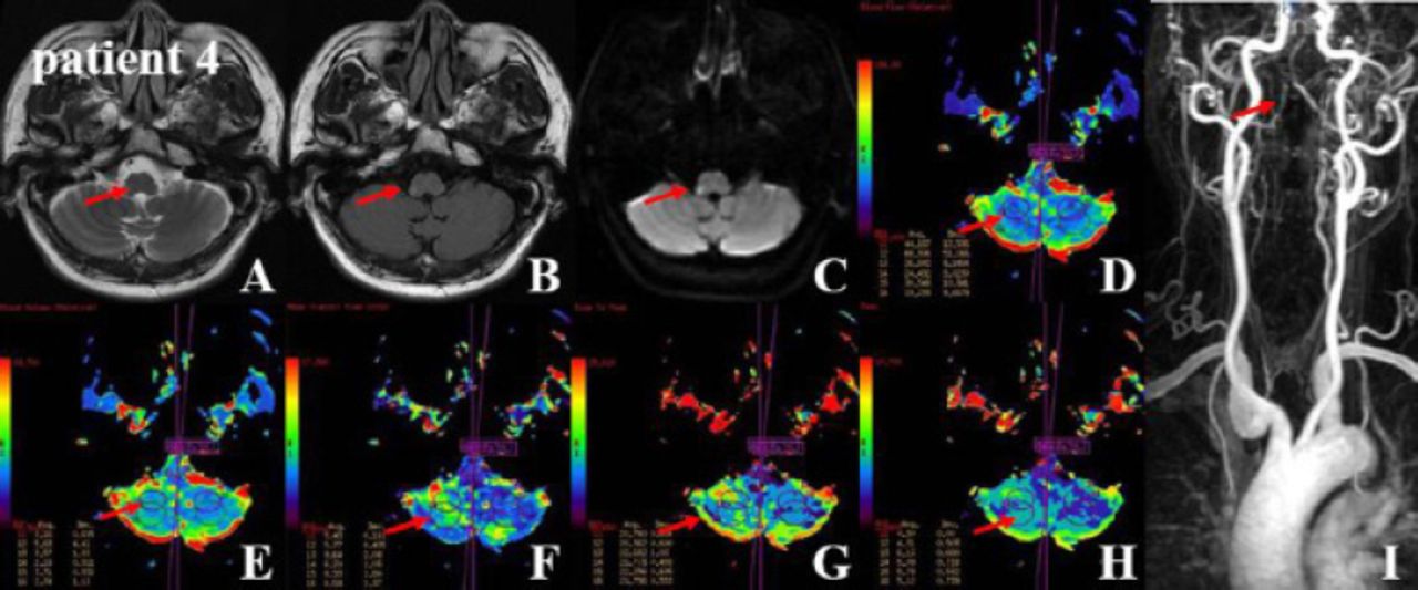
- Figure 4
MRI-T2WI, Flair and DWI demonstrated a-c) high signal intensities in the right lateral medulla. d-h) DSC-PWI demonstrated the lesion ROI low CBF, low CBV, decreased TTP, delayed MTT, and delayed Tmax on the VAH side. The representative rigid quantitative thresholds in the ROIs of 11 and 12 are shown in Table 2. i) CEMRA indicated basilar artery hypoplasia, right vertebral artery hypoplasia, and severe kinking of the left vertebral V1 segment. VAH - vertebral artery hypoplasia, CBF - cerebral blood flow, CBV - cerebral blood volume, TTP - time to peak, MTT - mean transit time


Tables
Variables Case 1 Case 2 Case 3 Case 4 Age (years) 36 45 36 54 Gender male male male male Hypertension history - - - + Diabetes history - - - - Coronary heart disease history - - - - Smoking + + + + Drinking + + + + Homocysteine (umol/L) 50.88 15.57 39.75 16.14 Triglycerides (mmol/L) 1.37 1.81 3.17 3.64 Total cholesterol (mmol/L) 3.68 4.77 4.83 6.31 Low density lipoprotein (mmol/L) 1.94 3.55 3.46 4.01 Fasting blood glucose (mmol/L) 4.90 4.60 6.06 5.24 Glycosylated hemoglobin (%) 4.84 5.26 6.05 5.10 VAH diameter (mm) 0.9 1.7 1.9 0.4 LMI side right right left right Horner’s sign + - + - Dysarthria + + + - Attenuated sensation + + + + Limb paralysis - + - + Ataxia + + + + NIHSS day 1 4 8 4 6 NIHSS day 7 1 6 1 4 NIHSS day 30 1 4 1 4 Barthel index day 7 100 70 100 80 Barthel index day 30 100 80 100 90 CBF ratio 1.28 0.49 0.89 0.65 CBV ratio 1.29 0.69 0.89 0.56 VAH - vertebral artery hypoplasia; LMI - lateral medullary infarction, Horner’s sign - ptosis, miosis, ipsilateral anhidrosis, and apparent enophthalmos, NIHSS - National Institutes of Health Stroke Scale, CBF (cerebral blood flow)ratio - between the ischemic lesion and the contralateral mirror regions of interest, CBV (cerebral blood volum)ratio - between the ischemic lesion and the contralateral mirror regions of interest
Parameters of PWI Case 1 Case 2 Case 3 Case 4 Left Right Left Right Left Right Left Right CBF 41.63±7.59 53.48±21.26 57.04±41.46 28.04±12.96 46.46±9.58 52.27±11.39 68.31±51.01 44.19±12.60 CBV 2.63±0.45 3.40±1.17 3.71±2.38 2.57±1.67 4.15±1.44 4.68±2.21 5.63±4.41 3.16±0.82 TTP 18.16±0.75 17.57±0.49 15.72±0.73 16.71±1.07 21.02±0.47 20.21±0.92 20.36±0.60 20.76±0.63 MTT 3.79±0.25 3.87±0.26 4.08±0.57 5.66±2.49 5.28±0.85 5.23±1.20 5.77±0.44 5.43±0.16 Tmax 4.50±0.00 4.32±0.49 3.30±0.71 4.30±1.08 4.67±0.47 4.73±0.54 4.31±0.50 4.50±0.00 CBF - cerebral blood flow, CBV - cerebral blood volume, TTP - time to peak, MTT - mean transit time, Tmax - time maximum, PWI - perfusion weighted imaging.
In this issue


Jump to section
Related Articles
Cited By...
- No citing articles found.