


Article Figures & Data
Figures
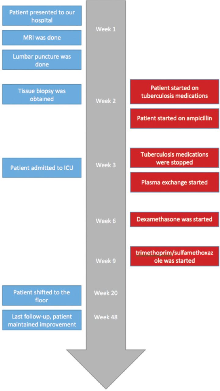
- Figure 1
Timeline figure of the clinical event.
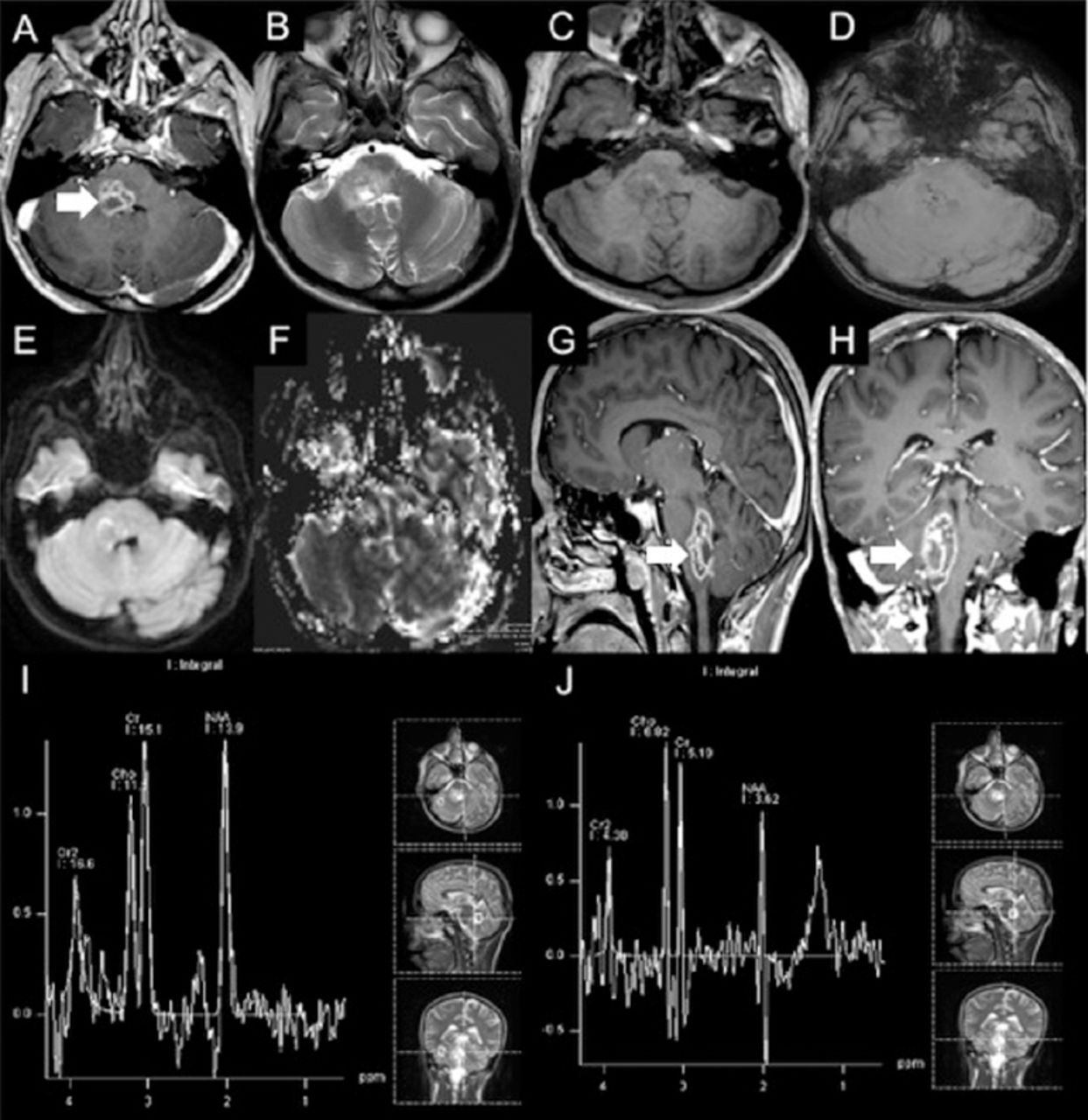
- Figure 2
Initial brain MRI showing A, G & H) focal lesion in the right side of medulla oblongata posteriorly with extension into middle cerebellar peduncle and posterior pons. Post contrast images show multiple ring-like enhancing areas are around the central hemorrhagic focus. D) Susceptibility weighted image (SWI) shows multiple foci of dark signal indicating hemorrhagic foci. E) Diffusion weighted image (DWI) shows no restricted diffusion in central non enhancing area to suggest any purulent material. F) Central hemmohagic areas give relatively increased rCBV values. The surrounding FLAIR hyperintensity show slightly lower rCBV values compared to normal appearing cerebral white matter. I) Single voxel MR spectroscopy at TE of 135 with region of interest in normal appearing tissue around the lesion shows nearly normal NAA, choline and J) creatine peaks and at TE of 135 with region of interest in the lesion shows decreased NAA and increased choline peaks. There is also increased lipid-lactate peak around 0.9 to 1.5 ppm.
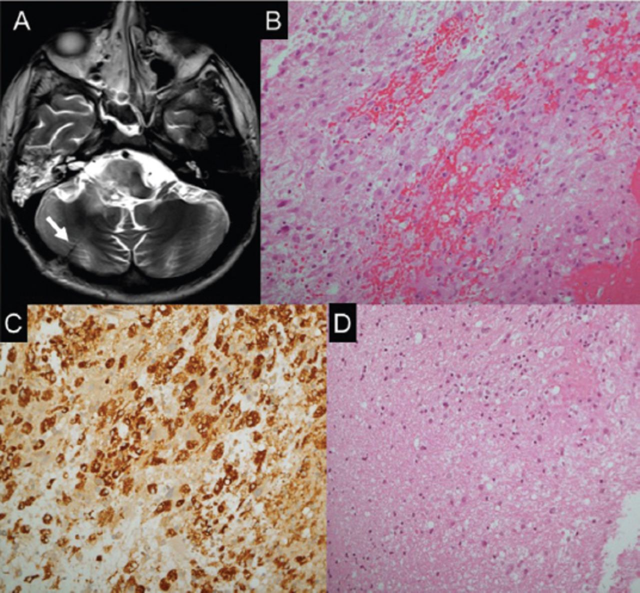
- Figure 3
Histopathological findings. A) Axial T2 weighted brain MRI showing post biopsy changes at the right cerebellar hemisphere. B) The biopsies exhibit fragments with cellular areas, formed by mixture of chronic inflammatory cells, with dominance of histiocytes, in addition to lymphocytes, plasma cells and glial cells. C) A major component of the cellular infiltrate reacts to CD 68, confirming histiocytic nature. D) In areas, the CNS tissue sampled exhibit gliosis and foci of chronic inflammation, in keeping with encephalitis.
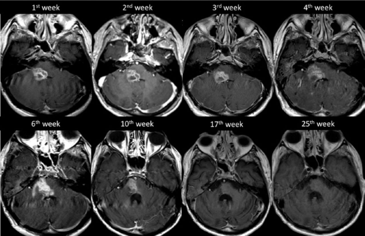
- Figure 4
The progression of the disease demonstrated on axial T1 weighted post contrast imaging.
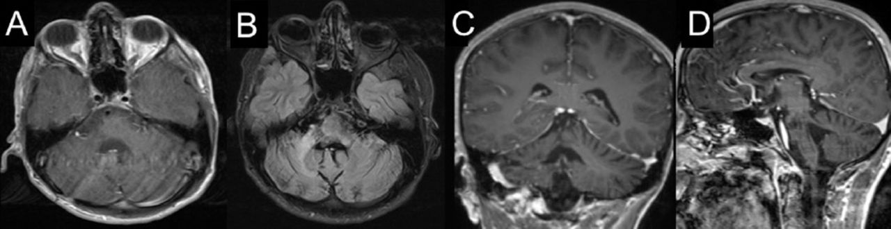
- Figure 5
Follow-up brain MRI at 57th week of presentation. (A) axial, (C) coronal, (D) sagittal T1 weighted post contrast imaging and (B) axial flair imaging show regression in the size of the lesion at the right aspect of the pons and right middle cerebellar peduncle with a mild volume loss of the right side of pons.


Tables
Variables Results CSF chemistry Glucose 4.4 Protein 0.5 g/L WBC 0 cells RBC 30 Blood cultures Negative Cryptococcus antigen Negative Asperigillous galactomannan antigen Negative Mycoplasm IgM Negative Herpes simplex virus PCR Negative HIV Negative Autoimmune markers ESR 3 Complement 3 level 137 ANA Negative Anti ds DNA Negative Jo 1 antibody Negative Tuberculosis investigations Acid fast bacilli culture Negative Acid fast bacilli PCR Negative Quantiferon Negative CSF cultures & gram stain Negative Pneumocystitis pneumonia Negative Toxoplasma IgG Negative Brucella serology Negative Hepatitis B & C Non reactive CRP 2.6 Complement 4 level 45 ANCA Negative SS-A and SS-B antibodies Negative Cryoglobulin Negative CSF - cerebrospinal fluid, WBC - white blood cells, RBC - red blood cells, PCR - polymerase chain reaction, ESR - erythrocyte sedimentation rate, ANA - antinuclear antibody, CRP - C reactive protein, ANCA - antineutrophil cytoplasmic antibodies
In this issue


Jump to section
Related Articles
Cited By...
- No citing articles found.