
Main menu

  • Home
  • Content
    • Latest
    • Ahead of print
    • Archive
  • Info for
    • Authors
    • Reviewers
    • Subscribers
    • Institutions
    • Advertisers
  • About Us
    • About Us
    • Editorial Office
    • Editorial Board
  • More
    • Alerts
    • Feedback
    • Folders
    • Help
  • Other Publications
    • Saudi Medical Journal

User menu

  • My alerts
  • Log in

Search

  • Advanced search
Neurosciences Journal
  • Other Publications
    • Saudi Medical Journal
  • My alerts
  • Log in
Neurosciences Journal

Advanced Search

  • Home
  • Content
    • Latest
    • Ahead of print
    • Archive
  • Info for
    • Authors
    • Reviewers
    • Subscribers
    • Institutions
    • Advertisers
  • About Us
    • About Us
    • Editorial Office
    • Editorial Board
  • More
    • Alerts
    • Feedback
    • Folders
    • Help
  • Follow psmmc on Twitter
  • Visit psmmc on Facebook
  • RSS
Case ReportCase Report
Open Access

Ventriculoperitoneal shunt-associated abdominal cerebrospinal fluid pseudocysts and the role of laparoscopy and a proposed management algorithm in its treatment

A report of 2 cases

Ghaida M. Fatani, Nasir M. Bustangi, Jamal S. Kamal and Abdulrahman J. Sabbagh
Neurosciences Journal August 2020, 25 (4) 320-326; DOI: https://doi.org/10.17712/nsj.2020.4.20200053
Ghaida M. Fatani
From the Faculty of Medicine (Fatani); from the Division of Pediatric Surgery (Bustangi, Kamal), and from the Division of Neurosurgery (Sabbagh), King Abdulaziz University, Jeddah, Kingdom of Saudi Arabia.
MBBS
  • Find this author on Google Scholar
  • Find this author on PubMed
  • Search for this author on this site
Nasir M. Bustangi
From the Faculty of Medicine (Fatani); from the Division of Pediatric Surgery (Bustangi, Kamal), and from the Division of Neurosurgery (Sabbagh), King Abdulaziz University, Jeddah, Kingdom of Saudi Arabia.
DES, DESC
  • Find this author on Google Scholar
  • Find this author on PubMed
  • Search for this author on this site
Jamal S. Kamal
From the Faculty of Medicine (Fatani); from the Division of Pediatric Surgery (Bustangi, Kamal), and from the Division of Neurosurgery (Sabbagh), King Abdulaziz University, Jeddah, Kingdom of Saudi Arabia.
FRCSI, FACS
  • Find this author on Google Scholar
  • Find this author on PubMed
  • Search for this author on this site
Abdulrahman J. Sabbagh
From the Faculty of Medicine (Fatani); from the Division of Pediatric Surgery (Bustangi, Kamal), and from the Division of Neurosurgery (Sabbagh), King Abdulaziz University, Jeddah, Kingdom of Saudi Arabia.
MBChB, FRCSC
  • Find this author on Google Scholar
  • Find this author on PubMed
  • Search for this author on this site
  • Article
  • Figures & Data
  • eLetters
  • Info & Metrics
  • References
  • PDF
Loading

Article Figures & Data

Figures

  • Tables
  • Figure 1
    • Download figure
    • Open in new tab
    • Download powerpoint
    Figure 1

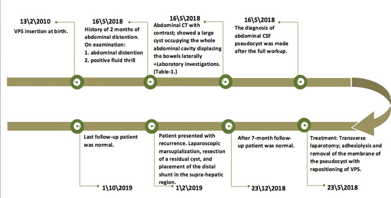
    Timeline of the first case. The patient shunted since infancy, presented later with abdominal distension, headache and decrease oral intake.Repositioning of ventriculoperitoneal shunt (VPS) to virgin area in the abdomen. CSF: cerebrospinal fluid

  • Figure 2
    • Download figure
    • Open in new tab
    • Download powerpoint
    Figure 2

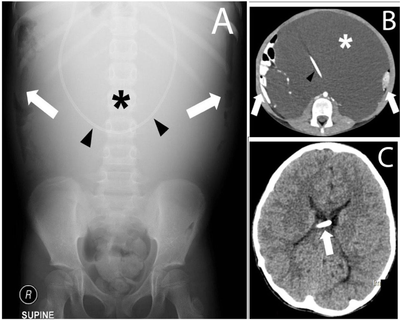
    Seven-year-old female with progressive abdominal distension. A) Frontal radiograph showing large mid-abdominal opacity (asterisk) displacing the air-filled bowel laterally (arrows). Part of the shunt is in the mid abdomen (arrowhead). B) Axial enhanced computed tomography showing a large loculated fluid (asterisk) displacing the bowels laterally (arrows). The shunt is seen within the fluid (arrowhead). C) Axial unenhanced computed tomography showing intraventricular location of the shunt (arrow) with no hydrocephalus.

  • Figure 3
    • Download figure
    • Open in new tab
    • Download powerpoint
    Figure 3

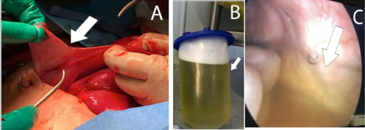
    Intraoperative images for first case. A) walls of the pseudocyst (arrowhead). B) The collected fluid evacuated from the pseudocyst (indicated by the arrow) C) Laparoscopic view from inside the cyst with remaining fluid after aspiration (arrowhead).

  • Figure 4
    • Download figure
    • Open in new tab
    • Download powerpoint
    Figure 4

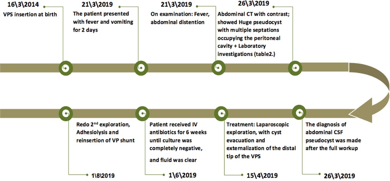
    Timeline of the second case. Five years old child shunted since birth, presented with fever and vomiting for 2 days. Laparoscopic externalization of the distal tip of ventriculoperitoneal shunt (VPS).

  • Figure 5
    • Download figure
    • Open in new tab
    • Download powerpoint
    Figure 5

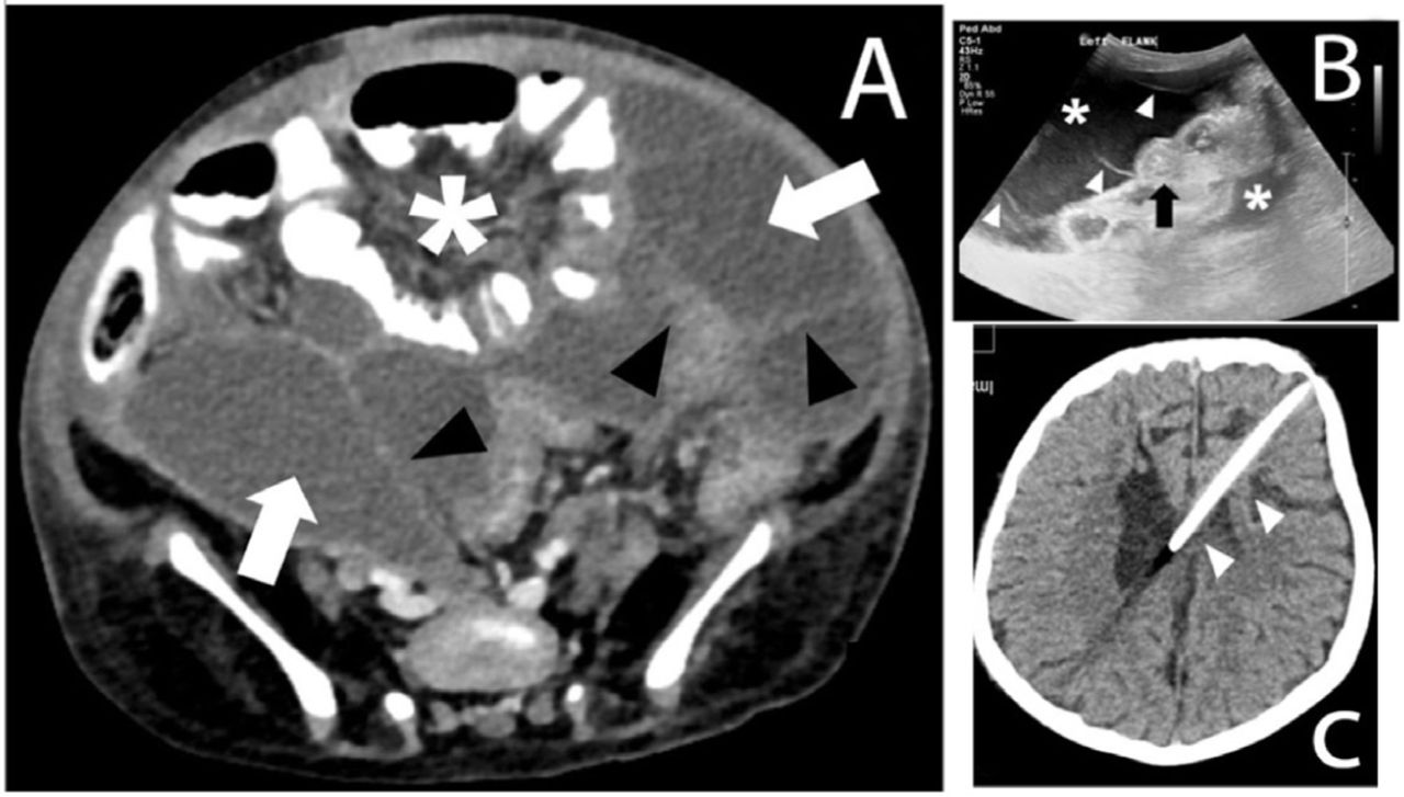
    Five-year-old female with fever and vomiting. A) Axial enhanced computed tomography showing complex fluid collections (arrows) with multiple septa (arrowheads) displacing the bowel loops (asterisk). B) Ultrasound image of the left flank showing complex fluid (asterisks) with multiple septa (arrowheads) surrounding the collapsed bowel loops (arrow). C) Axial unenhanced computed tomography showing intraventricular location of the shunt (arrow) with no hydrocephalus.

  • Figure 6
    • Download figure
    • Open in new tab
    • Download powerpoint
    Figure 6

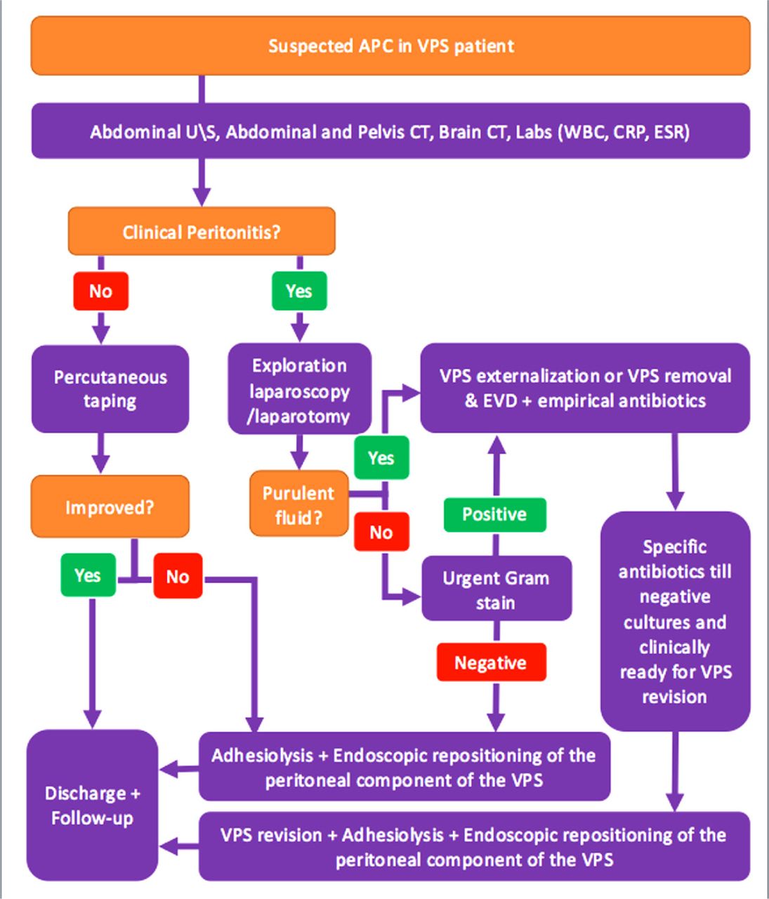
    An algorithm that summarizes the approach for patient with abdominal CSF pseudocyst.

Tables

  • Figures
    • View popup
    TABLE 1

    Laboratory results for the first case.

    LaboratoryResultsNormal range
    White blood counts8.66 K U/L4.5-11.5 K U/L
    Aspartate transaminase17 U/L15-37 U/L
    Alanine aminotransferase16 U/L12-78 U/L
    Alkaline phosphatase160 U/L54-128 U/L
    Bilirubin - total4.0 g/L0-17 g/L
    Albumin30 g/L40.2-47.6 g/L
    Cerebrospinal fluid protein15.06 g/L (high)0.2-0.4 g/L
    Cerebrospinal fluid glucose4.6 mmol/L (high)2.3-4.1 mmol/L
    Cerebrospinal fluid bacterial & fungal cultureNegative
    C-reactive protein11.5 mg/L (High)0-3 mg/L
    • View popup
    TABLE 2

    Laboratory Results for the second case.

    LaboratoryResultsNormal range
    White blood counts17 K U/L4.5-11.5 K U/L
    Aspartate transaminase339 U/L15-37 U/L
    Alanine aminotransferase215 U/L12-78 U/L
    Alkaline phosphatase138 U/L54-128 U/L
    Bilirubin - total5 g/L0-17 g/L
    Albumin34 g/L40.2-47.6 g/L
    Cerebrospinal fluid protein0.32 g/L0.2-0.4 g/L
    Cerebrospinal fluid glucose3.0 mmol/L2.3-4.1 mmol/L
    Cerebrospinal fluid bacterial & fungal culturePositive
    C-reactive protein4.34 mg/L0-3 mg/L
PreviousNext
Back to top

In this issue

Neurosciences Journal: 25 (4)
Neurosciences Journal
Vol. 25, Issue 4
1 Aug 2020
  • Table of Contents
  • Cover (PDF)
  • Index by author
Print
Download PDF
Email Article

Thank you for your interest in spreading the word on Neurosciences Journal.

NOTE: We only request your email address so that the person you are recommending the page to knows that you wanted them to see it, and that it is not junk mail. We do not capture any email address.

Enter multiple addresses on separate lines or separate them with commas.
Ventriculoperitoneal shunt-associated abdominal cerebrospinal fluid pseudocysts and the role of laparoscopy and a proposed management algorithm in its treatment
(Your Name) has sent you a message from Neurosciences Journal
(Your Name) thought you would like to see the Neurosciences Journal web site.
Citation Tools
Ventriculoperitoneal shunt-associated abdominal cerebrospinal fluid pseudocysts and the role of laparoscopy and a proposed management algorithm in its treatment
Ghaida M. Fatani, Nasir M. Bustangi, Jamal S. Kamal, Abdulrahman J. Sabbagh
Neurosciences Journal Aug 2020, 25 (4) 320-326; DOI: 10.17712/nsj.2020.4.20200053

Citation Manager Formats

  • BibTeX
  • Bookends
  • EasyBib
  • EndNote (tagged)
  • EndNote 8 (xml)
  • Medlars
  • Mendeley
  • Papers
  • RefWorks Tagged
  • Ref Manager
  • RIS
  • Zotero
Share
Ventriculoperitoneal shunt-associated abdominal cerebrospinal fluid pseudocysts and the role of laparoscopy and a proposed management algorithm in its treatment
Ghaida M. Fatani, Nasir M. Bustangi, Jamal S. Kamal, Abdulrahman J. Sabbagh
Neurosciences Journal Aug 2020, 25 (4) 320-326; DOI: 10.17712/nsj.2020.4.20200053
Twitter logo Facebook logo Mendeley logo
  • Tweet Widget
  • Facebook Like
  • Google Plus One
Bookmark this article

Jump to section

  • Article
    • Abstract
    • Case Report
    • Discussion
    • Acknowledgements
    • Footnotes
    • References
  • Figures & Data
  • eLetters
  • References
  • Info & Metrics
  • PDF

Related Articles

  • No related articles found.
  • Google Scholar

Cited By...

  • No citing articles found.
  • Google Scholar

More in this TOC Section

  • Unmasking the mimic: Leprosy neuropathy misdiagnosed as chronic inflammatory demyelinating polyneuropathy: A case report from Saudi Arabia
  • Seminal vesicle schwannoma with chronic hemorrhage
  • Marchiafava-Bignami disease post-bariatric surgery: A case report and review of similar cases
Show more Case Report

Similar Articles

Navigate

  • home

More Information

  • Help

Additional journals

  • All Topics

Other Services

  • About

© 2025 Neurosciences Journal Neurosciences is copyright under the Berne Convention and the International Copyright Convention. All rights reserved. Neurosciences is an Open Access journal and articles published are distributed under the terms of the Creative Commons Attribution-NonCommercial License (CC BY-NC). Readers may copy, distribute, and display the work for non-commercial purposes with the proper citation of the original work. Electronic ISSN 1658-3183. Print ISSN 1319-6138.

Powered by HighWire