


Article Figures & Data
Figures
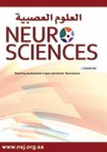
- Figure 1
Magnetic resonance imaging (MRI) of the brain with and without contrast. A) Axial fluid-attenuated inversion recovery (FLAIR) MRI image demonstrating periventricular and subcortical white matter lesions. B) Sagittal T2-weighted image showing corpus callosal white matter lesions. C) Axial T1-weighted image with contrast highlighting cerebellar leptomeningeal enhancement.
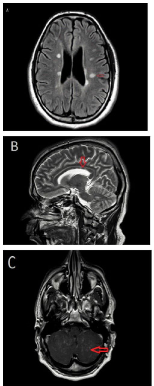
- Figure 2
Magnetic resonance imaging (MRI) of the spine with and without contrast. A) T2 sagittal image demonstrating a longitudinally extensive lesion from T1-T6, with cord swelling. B) Axial T1-weighted image, with contrast, showing multifocal punctate enhancement.
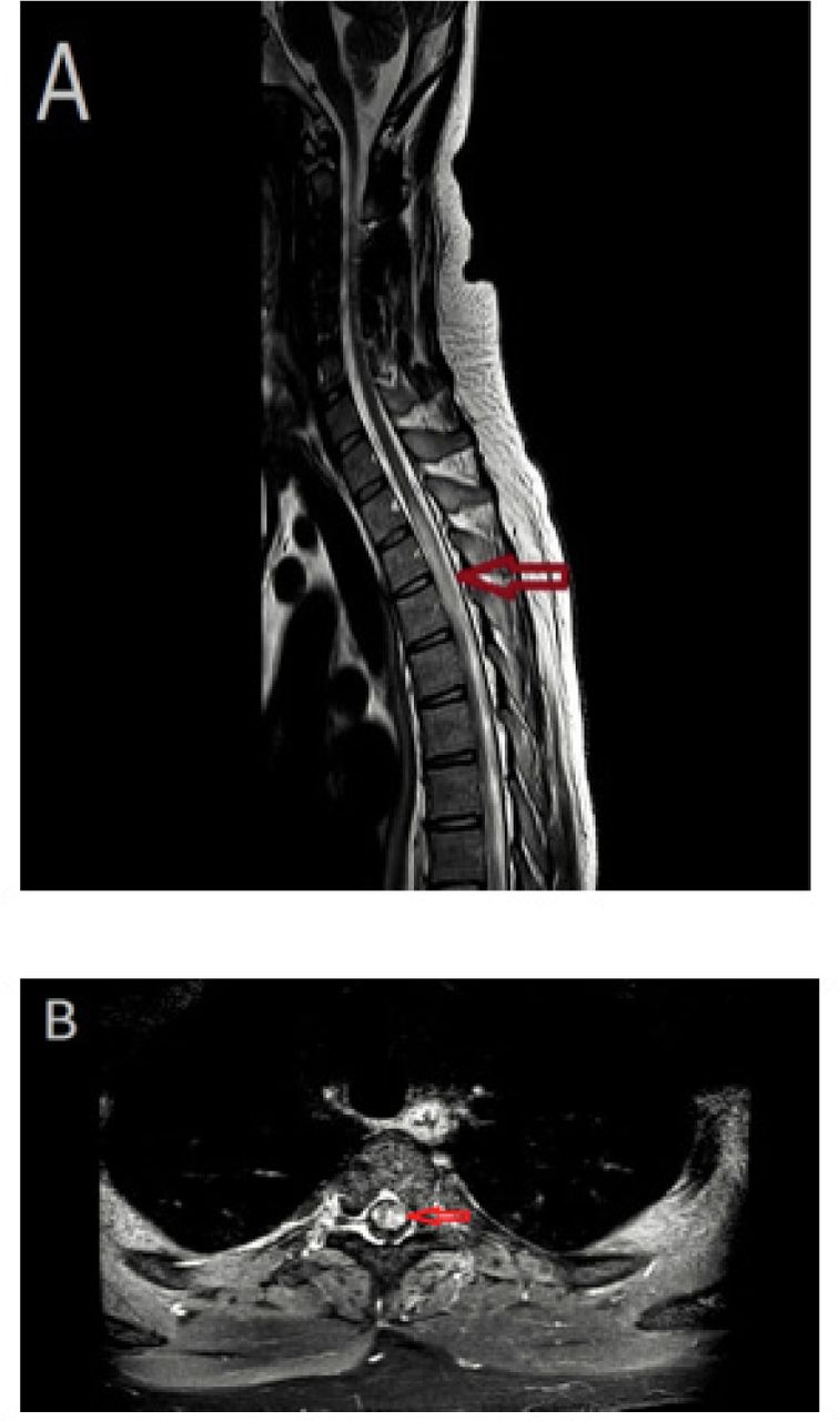
- Figure 3
Magnetic resonance imaging (MRI) of the brain and spine with contrast. A) Axial T1-weighted image, with contrast, showing complete resolution of cerebellar leptomeningeal enhancement. B) Sagittal T1-weighted MRI demonstrating an improvement of spinal cord abnormal enhancement.
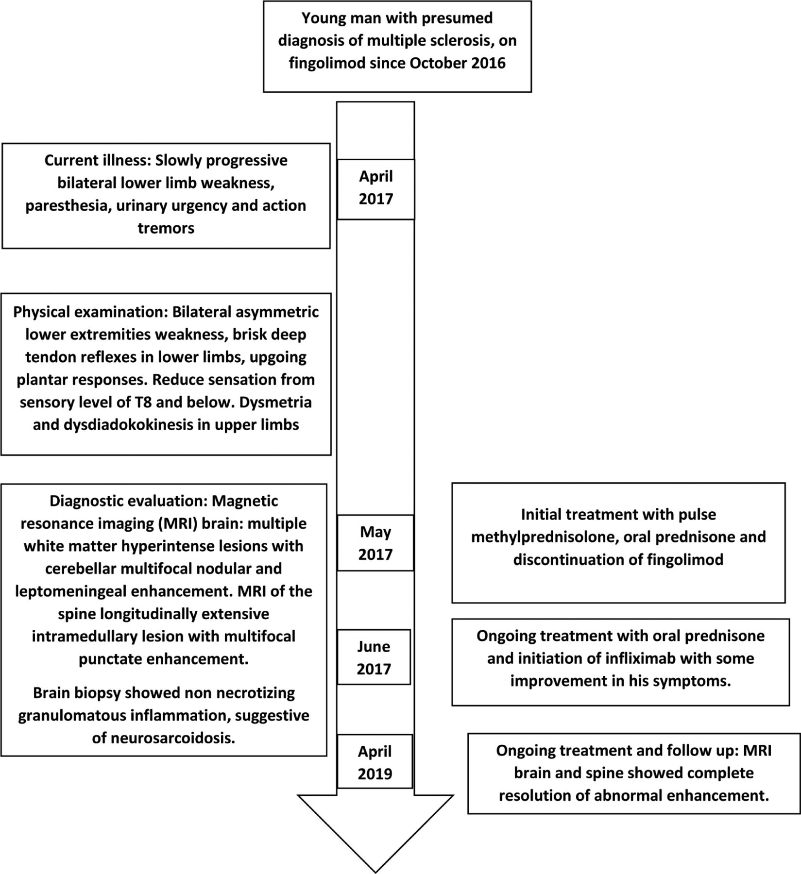
- Figure 4
Timeline of the presented case.


Jump to section
Related Articles
Cited By...
- No citing articles found.