


Article Figures & Data
Figures
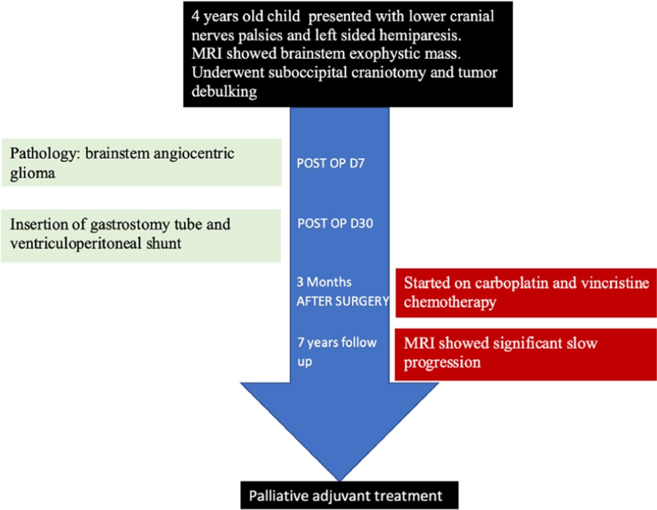
- Figure 1
Timeline showing the course of the patient during follow up and outcome.
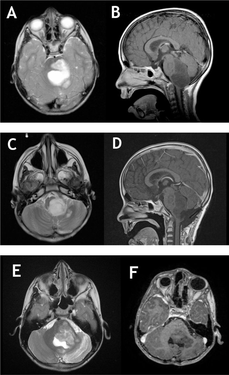
- Figure 2
Pre and post-operative MRI images of brainstem angiocentric glioma. a,b) T2 and T1 with gadolinium MRI showing a large brainstem none enhancing lesion with dorsal exophytic component compression the cerebellum, notice the significant compression on medulla. c,d) Postoperative images showing a large residual left intentionally on T2 and T1C+ images. e,f) After 7 years of follow up. Notice the progression of the lesion with further compression on cerebellum. No enhancement noticed or signs of transformation into a higher grade. The tumor is filling the entrapped CSF area on T2 weighted images.
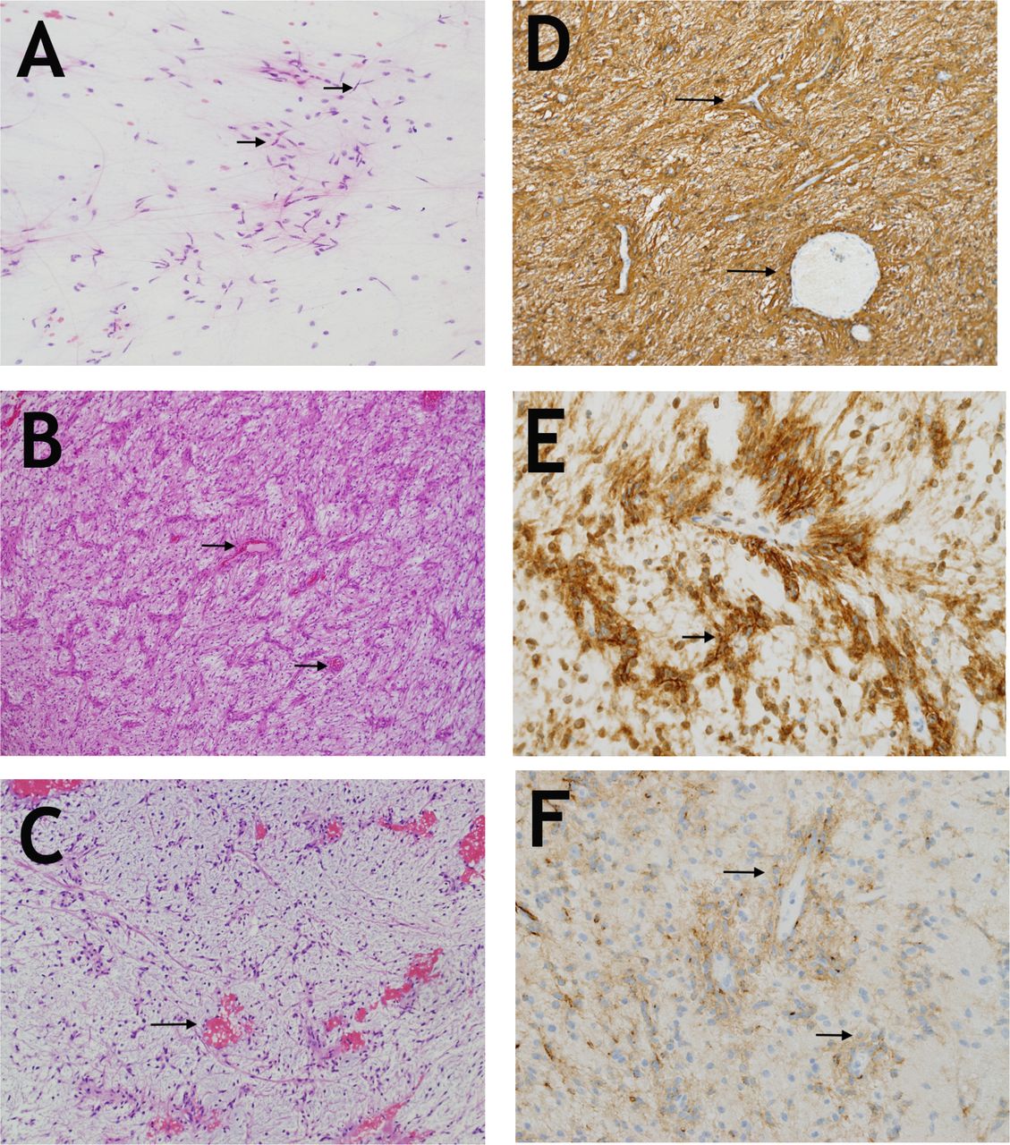
- Figure 3
Histopathological examination of the lesion a) Smear preparation showing bipolar cells (arrows) with long processes reminiscent of pilocytic astrocytoma (Hematoxylin and Eosin, x200). b) A low magnification image showing the neoplastic bipolar cells with their distinct angiocentric arrangement which is mostly parallel in this tumor (arrows). (Hematoxylin and Eosin, x100). c) The myxoid component is very hypocellular but retains the angiocentric pattern(arrows). (Hematoxylin and Eosin, x100). d) GFAP is diffusely expressed in tumor cells (arrows) and particularly highlights the angiocentric pattern (GFAP, polyclonal, Ventana, x200). e) D2-40 is strongly expressed (arrows), (clone D2-4, DakoCytomation, x400). f) EMA showing moderate staining of cells and their processes. Note the perinuclear accentuation of expression in some cells (arrows), (EMA, clone E29, Dako x400).
Tables
- Table 1
Summary of published cases of brainstem angiocentric gliomas. M: male, F:female, CN: cranial nerve, ETV: endoscopic third ventriculostomy, NTR: near total resection, VPS: ventriculoperitoneal shunt.
No. Author (year) Age (gender) Location Presentation/exam Surgery Chemotherapy Follow up Outcome MYB-QKI fusion 1 Covington et al (2009) 5 years (F) Right exophytic tectal lesion Unsteady gait, 4th and 7th CN palsies, hydrocephalus ETV, craniotomy and debulking No 2 years Stable lesion Yes 2 Weaver et al (2017) 5 years (F) Midline midbrain tegmentam Diplopia, 6th CN nerve palsy, hydrocephalus ETV, craniotomy and NTR NO 6 years Stable after resection No data 3 Weaver et al (2017) 6 years (M) Right pontine exophytic lesion Hemiparesis, facial palsy Stereotactic biopsy and ETV No 1.5 years Stable lesion No data 4 D’Aronco et al (2017) 7 years (M) Pontomedullary exophytic lesion respiratory failure, repated pneumonia Stereotactic biopsy Yes (carboplatin/vincristine) 10 months Progression Yes 5 D’Aronco et al (2017) 3 years (F) Pontomedullary exophytic lesion Facial palsy Stereotactic biopsy Yes (vinblastine and bevacizumab) 4 years Progression Yes 6 Chan et al (2017) 7 Years (M) Pons 6th CN palsy Stereotactic biopsy No data No data No data Yes 7 Current case (2019) 4 years (F) Left brainstem dorsal exophytic lesion Hydrocephalus, unsteady gait, hoarseness, choking with food, 6th,7th, and lower CN palsies Craniotomy and debulking, VPS Yes (carboplatin/vincristine) 8 years Progression Not done


Jump to section
Related Articles
Cited By...
- No citing articles found.