


Article Figures & Data
Figures
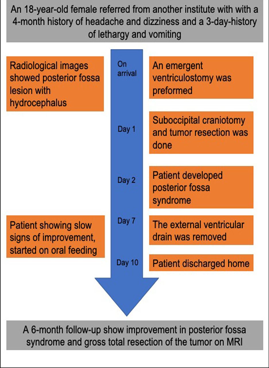
- Figure 1
- Timeline showing the clinical course of the patient and outcomes.
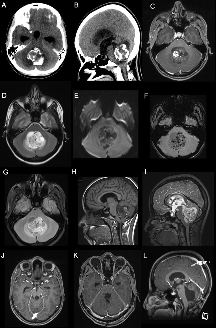
- Figure 2
- Pre-operative images. A and B) posterior fossa mass with coarse calcifications and a cystic component as well as hydrocephalus demonstrated on CT scan. C and H) T1-weighted post contrast MRI showing a hypointense lesion with heterogenous enhancement with gadolinium. D and I) Axial and sagittal T2-weighted MRI demonstrating heterogenous, hyperintense lesion in the fourth ventricle. E) Diffusion weighted images showing a large heterogeneous cystic lesion centered within the fourth ventricle compressing the adjacent structures with no significant restricted diffusion. F) Susceptibility-weighted images showing multiple areas of susceptibility effects in the lesion corresponding to calcification with some areas representing hemorrhage. G) The lesion again seen demonstrating heterogeneous signal intensity with adjacent small edema in the cerebellum on Fluid attenuated inversion recovery images (FLAIR). J-L) Postoperative MRI obtained at 6-month postoperatively showing no evidence of recurrence.
- Figure 3
- Histopathological and immunohistochemical findings of rosette-forming glioneuronal tumors, A) A smear shows true rosettes embedded in a loose fibrillary background, B) Hematoxylin and eosin (H-E) staining shows a ring like neurocytic rosette arranged around a neuropil core, C) Pilocytic astrocytoma like component exhibiting rosenthal fibers, D) Oligodendrocyte-like cells with perinuclear halo. (E) Neurocytic rosettes are positive for synaptophysin, f, g) GFAP and S100 showing positive staining in the pilocytic astrocytoma area, H) Prominent hyalinization in the glial area.


Tables
Tumor type Age Location Morphology Positive antibodies Ependymoma GII, GIII Children/ young adults Wall of the ventricles Spinal canal -Uniform round to oval cells with salt and pepper chromatin.- Perivascular pseudorosettes >true GFAP S100 EMA dot and ring Astroblastoma Children/ young adults Cerebrum - Astroblastic pseudorosettes- “stout” - (not fibrillar) processes - Prominent vascular hyalinization. GFAP S100EMA focal cytoplasmic or dotlike Rosettes forming glioneuronal tumor G1 Young adults 4th ventricle Cerebellum Biphasic:-- Neuronal component: small uniform cells forming neurocytic true or pseudorossettes.– Glial component: pilocytic astrocytoma or oligodendroglioma like Neurocytic rosettes: SynaptophysinNeuNGlial component:GFAP S100 Medulloblastoma GIV Children Cerebellum - Small round blue cell tumor- Brisk mitotic activity- Prominent karyorrhexis- Homer Wright rosettes Synaptophysin B-catenin CMYC P53 YAPGAB Embryonal tumor with multilayered rosettes / NOS GIV Children Cerebrum Brain stem Cerebellum 3 histological patterns in ETMR:- Embryonal tumor with abundant neuropil and true rosettes (ETANTR)– Medulloepithelioma– Ependymoblastoma Synaptophysin C19MC altered Pineoblastoma GIV Children Pineal gland - Small round blue cell tumor-Homer Wright rosettes- Flexner–Wintersteiner rosettes Synaptophysin Pituitary adenoma Adults Pituitary gland - Uniform nuclear morphology- Abundant cytoplasm- Perivascular rosettes - Papillae Synaptophysin Pit. Hormones Transcription factor
In this issue


Jump to section
Related Articles
Cited By...
- No citing articles found.