
Main menu

  • Home
  • Content
    • Latest
    • Ahead of print
    • Archive
  • Info for
    • Authors
    • Reviewers
    • Subscribers
    • Institutions
    • Advertisers
  • About Us
    • About Us
    • Editorial Office
    • Editorial Board
  • More
    • Alerts
    • Feedback
    • Folders
    • Help
  • Other Publications
    • Saudi Medical Journal

User menu

  • My alerts
  • Log in

Search

  • Advanced search
Neurosciences Journal
  • Other Publications
    • Saudi Medical Journal
  • My alerts
  • Log in
Neurosciences Journal

Advanced Search

  • Home
  • Content
    • Latest
    • Ahead of print
    • Archive
  • Info for
    • Authors
    • Reviewers
    • Subscribers
    • Institutions
    • Advertisers
  • About Us
    • About Us
    • Editorial Office
    • Editorial Board
  • More
    • Alerts
    • Feedback
    • Folders
    • Help
  • Follow psmmc on Twitter
  • Visit psmmc on Facebook
  • RSS
Case SeriesCase series
Open Access

Invasive aspergillosis of the central nervous system in immunocompetent patients in Saudi Arabia: Case series and review of the literature

Mahmoud S. Taha, Mahmoud I. Haddad, Ali A. Almomen, Marwa M Abdulkader and Rami A. Alhazmi
Neurosciences Journal October 2021, 26 (4) 379-384; DOI: https://doi.org/10.17712/nsj.2021.4.20210081
Mahmoud S. Taha
From the Department of Neurosurgery (Taha, Haddad), Department of Head and Neck Surgery (Almomen), Department of Pathology (Abdulkader), Department of Medical Imaging (Alhazmi), King Fahad Specialist Hospital, Dammam, Kingdom of Saudi Arabia
MD
  • Find this author on Google Scholar
  • Find this author on PubMed
  • Search for this author on this site
  • For correspondence: [email protected]
Mahmoud I. Haddad
From the Department of Neurosurgery (Taha, Haddad), Department of Head and Neck Surgery (Almomen), Department of Pathology (Abdulkader), Department of Medical Imaging (Alhazmi), King Fahad Specialist Hospital, Dammam, Kingdom of Saudi Arabia
MD
  • Find this author on Google Scholar
  • Find this author on PubMed
  • Search for this author on this site
Ali A. Almomen
From the Department of Neurosurgery (Taha, Haddad), Department of Head and Neck Surgery (Almomen), Department of Pathology (Abdulkader), Department of Medical Imaging (Alhazmi), King Fahad Specialist Hospital, Dammam, Kingdom of Saudi Arabia
MD
  • Find this author on Google Scholar
  • Find this author on PubMed
  • Search for this author on this site
Marwa M Abdulkader
From the Department of Neurosurgery (Taha, Haddad), Department of Head and Neck Surgery (Almomen), Department of Pathology (Abdulkader), Department of Medical Imaging (Alhazmi), King Fahad Specialist Hospital, Dammam, Kingdom of Saudi Arabia
MD
  • Find this author on Google Scholar
  • Find this author on PubMed
  • Search for this author on this site
Rami A. Alhazmi
From the Department of Neurosurgery (Taha, Haddad), Department of Head and Neck Surgery (Almomen), Department of Pathology (Abdulkader), Department of Medical Imaging (Alhazmi), King Fahad Specialist Hospital, Dammam, Kingdom of Saudi Arabia
MD
  • Find this author on Google Scholar
  • Find this author on PubMed
  • Search for this author on this site
  • Article
  • Figures & Data
  • eLetters
  • Info & Metrics
  • References
  • PDF
Loading

Article Figures & Data

Figures

  • Tables
  • Figure 1
    • Download figure
    • Open in new tab
    • Download powerpoint
    Figure 1

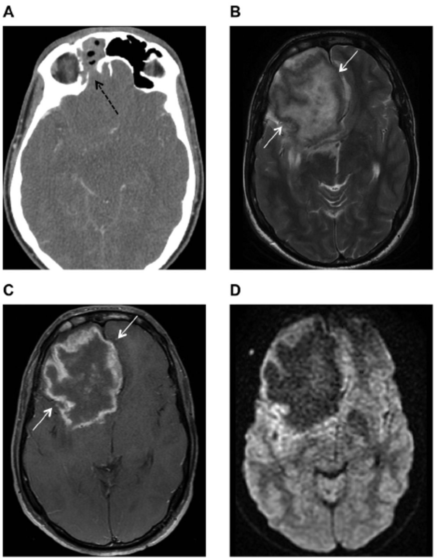
    - Axial enhanced brain computed tomography (CT) A) Axial enhanced brain computed tomography (CT) shows hyperdense opacification of the right frontal paranasal sinus with erosion of its posterior wall (black dashed arrow), B) Brain magnetic resonance imaging MRI scan Axial T2-weighted and, C) MRI T1-weighted post contrast show a large intra-axial lesion in the right frontal lobe with irregular thick peripheral enhancement (white arrows), D) MRI diffusion-weighted imaging shows no corresponding central diffusion restriction.

  • Figure 2
    • Download figure
    • Open in new tab
    • Download powerpoint
    Figure 2

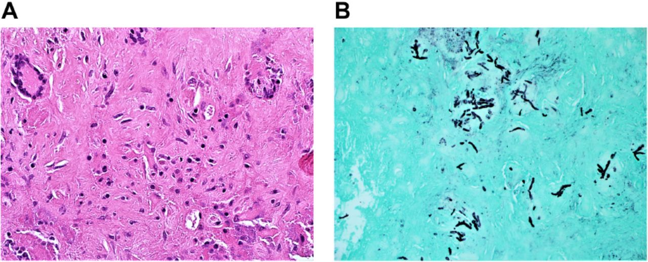
    - Hematoxylin- and eosin-stained sections (magnification, 40x) showing A) neuroglial tissue containing ill-defined granulomata with multinucleated giant cells. Thin hyphae with regular septation and branching at a 45° angle are noted, B) Grocott’s methenamine silver stain demonstrating thin hyphae and spores (magnification, 20x).

  • Figure 3
    • Download figure
    • Open in new tab
    • Download powerpoint
    Figure 3

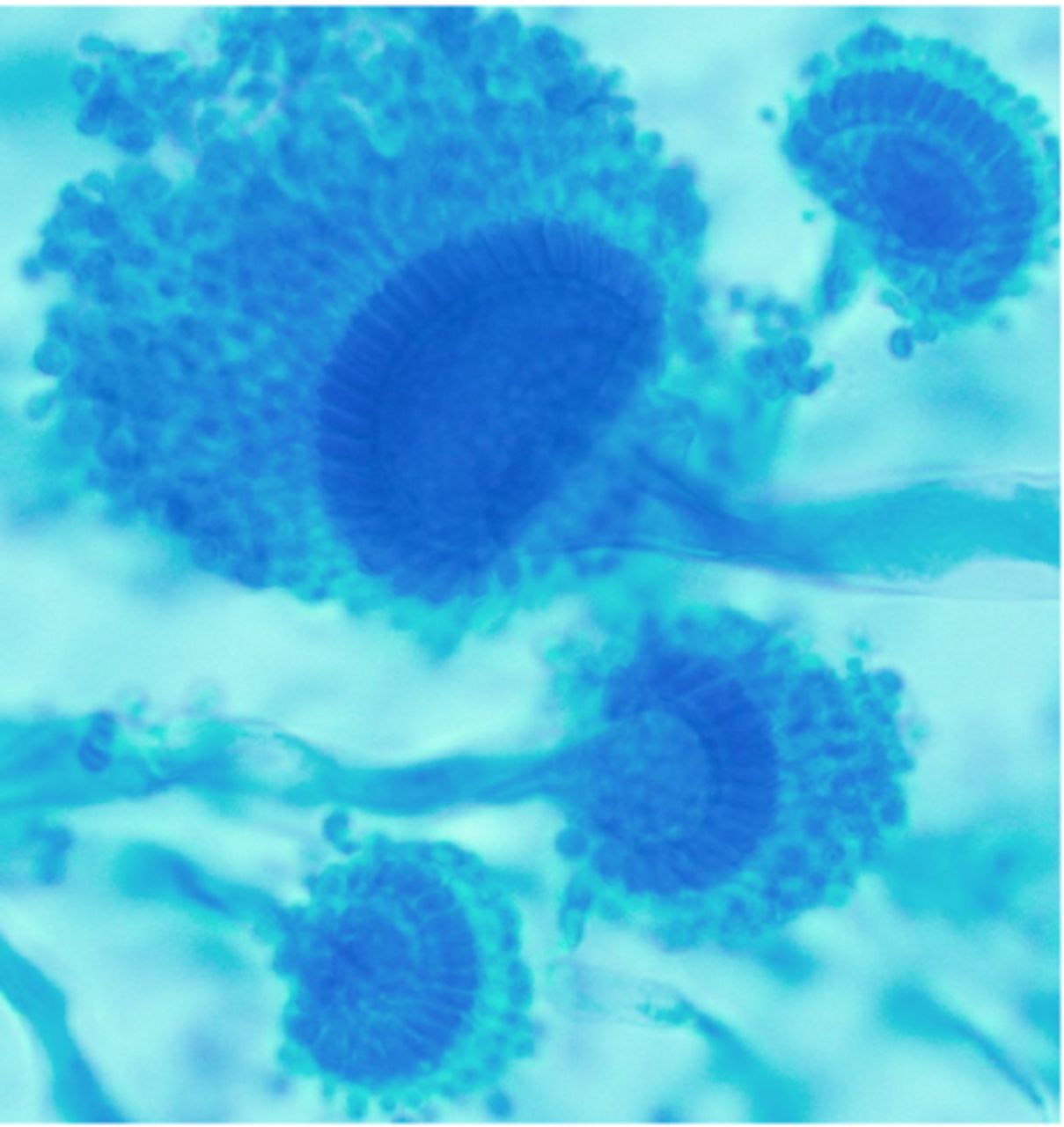
    - Detection methods for mycology: Specimens were inoculated on both selective fungal media (Mycosel, Becton, Dickenson and Company, MD, USA) and general fungal media Sabouraud Dextrose Agar (Titan Biotech Ltd, India) and incubated at 28°C–30°C for up to 4 weeks. Cultures with fungal growth were further worked up for mould identification using tease mount, scotch tape preparation and potato dextrose agar media for conidiation.The culture grew rapidly, producing a green–grey colony. The mould produced septate hyaline hyphae with smooth conidiophores supporting a dome-shaped vesicle. A row of phialides on the upper surface of the vesicle bore chains of round conidia in a columnar fashion characteristic of Aspergillus fumigatus.

  • Figure 4
    • Download figure
    • Open in new tab
    • Download powerpoint
    Figure 4

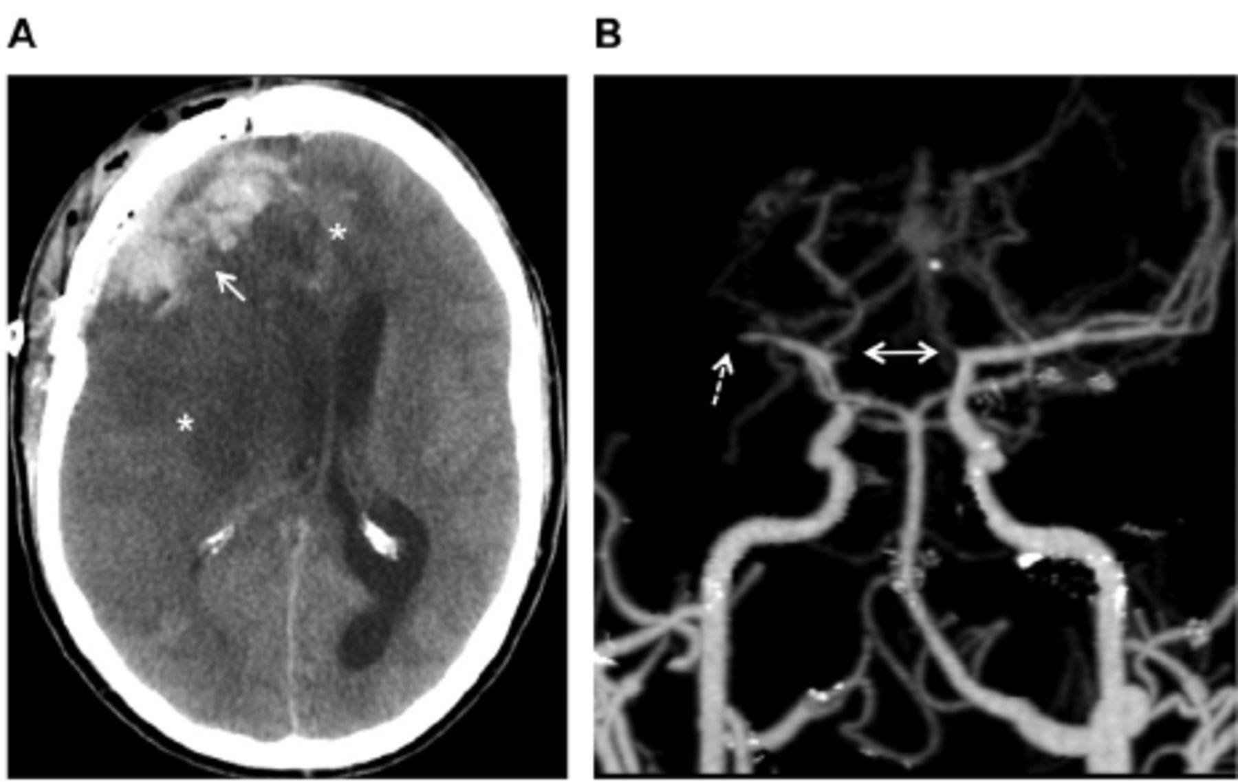
    - Axial plain brain CT shows (A) post-craniotomy changes in the right frontal aspect of the skull with haemorrhagic collection within the surgical bed (white arrow). Extensive ischemic low-attenuation tissue swelling in the right cerebral hemisphere and left frontal lobe is also observed (asterisks) along with a left-sided midline shift. (B) 3-dimensional reformatted MIP image from CTA shows occlusion of the right M1 segment (dashed arrow) and bilateral ACA (bidirectional arrow).

  • Figure 5
    • Download figure
    • Open in new tab
    • Download powerpoint
    Figure 5

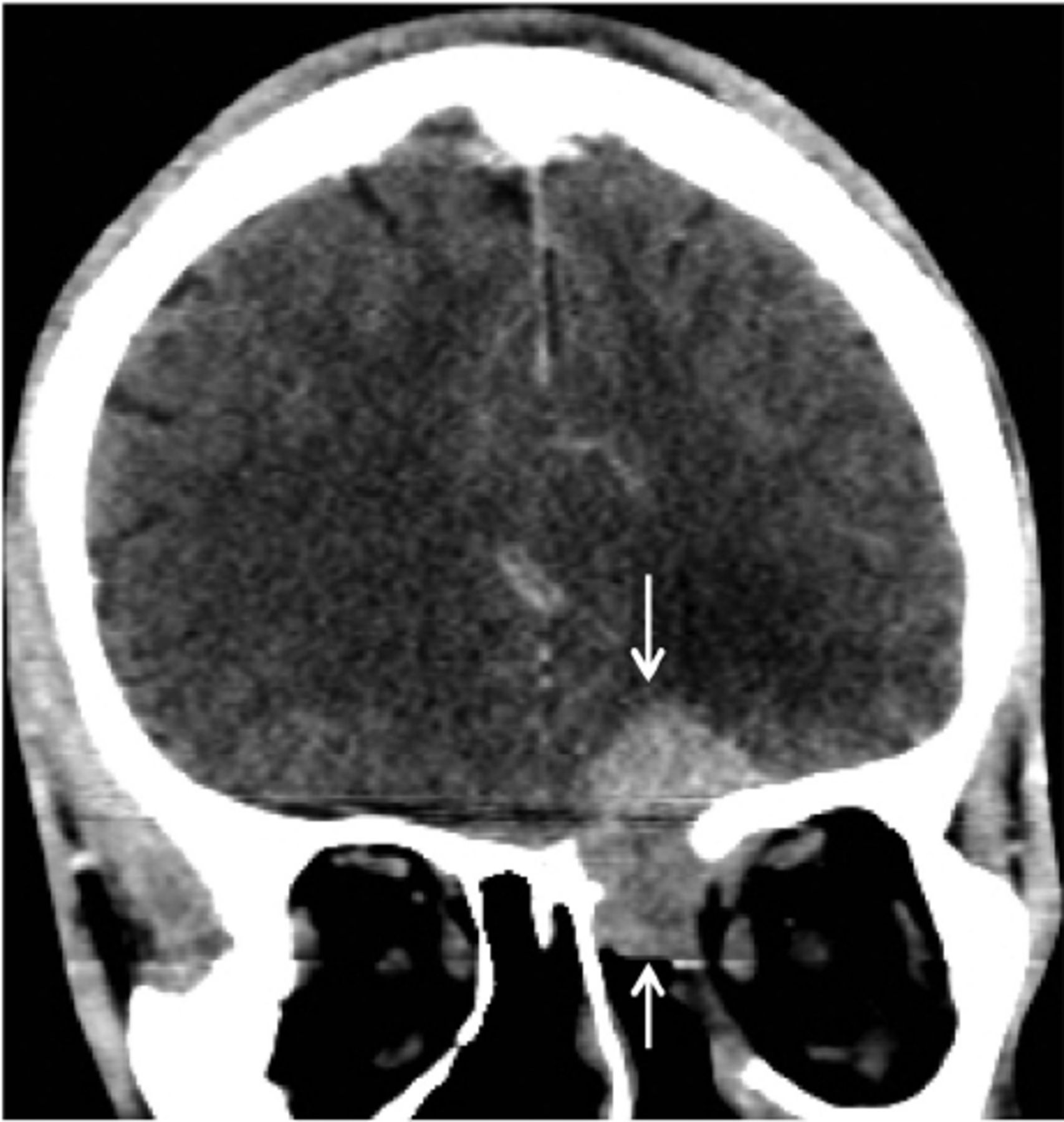
    - Enhanced coronal brain CT shows a growing mass centred in the anterior skull base with bone erosions (arrows) along with intracranial, extra-axial and ethmoid sinus soft tissue components.

  • Figure 6
    • Download figure
    • Open in new tab
    • Download powerpoint
    Figure 6

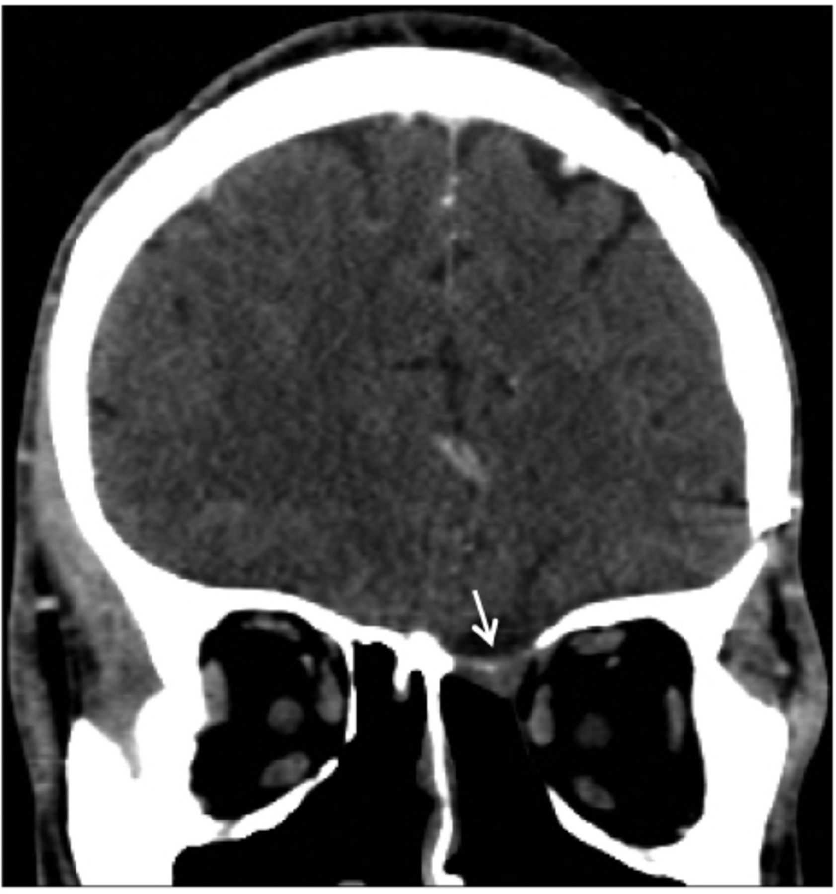
    - Enhanced coronal brain CT on post-operative follow up does not show any evidence of recurrence.

Tables

  • Figures
    • View popup
    Table 1

    - Summary of studies on IA of CNS in immunocompetent patients in Saudi Arabia including the current study.

    Study periodnAge: mean(range)Male/femaleAspergillus spp.Treatment anti-fungalClinical outcomeFollow up (months)
    Mohan et al7 (1987–92)442.2 (30–60)2/2A. flavus (4)Surgery/Amphotericin B3 no recurrence12-24 months
    1 death
    Ur-Rahman8 et al. (1983–94)947.3 (26–66)1/8A. flavus (6)Surgery/Amphotericin B plus flucytosine then itraconazole1 cured1-36 months
    6 residual or lost f/up
    A. fumigatus (3)
    2 death
    Baeesa et al9 (2000–12)1232 (17–50)4/8not definedSurgery/Amphotericin B plus itraconazole or variconazole8 cured12–50 months
    2 residual or recurrence
    2 deaths
    Taha et al (2012–18)333 (30–36)3/0not definedSurgery/Amphotericin B plus variconazole1 cured1-18 months
    2 deaths
PreviousNext
Back to top

In this issue

Neurosciences Journal: 26 (4)
Neurosciences Journal
Vol. 26, Issue 4
1 Oct 2021
  • Table of Contents
  • Cover (PDF)
  • Index by author
Print
Download PDF
Email Article

Thank you for your interest in spreading the word on Neurosciences Journal.

NOTE: We only request your email address so that the person you are recommending the page to knows that you wanted them to see it, and that it is not junk mail. We do not capture any email address.

Enter multiple addresses on separate lines or separate them with commas.
Invasive aspergillosis of the central nervous system in immunocompetent patients in Saudi Arabia: Case series and review of the literature
(Your Name) has sent you a message from Neurosciences Journal
(Your Name) thought you would like to see the Neurosciences Journal web site.
Citation Tools
Invasive aspergillosis of the central nervous system in immunocompetent patients in Saudi Arabia: Case series and review of the literature
Mahmoud S. Taha, Mahmoud I. Haddad, Ali A. Almomen, Marwa M Abdulkader, Rami A. Alhazmi
Neurosciences Journal Oct 2021, 26 (4) 379-384; DOI: 10.17712/nsj.2021.4.20210081

Citation Manager Formats

  • BibTeX
  • Bookends
  • EasyBib
  • EndNote (tagged)
  • EndNote 8 (xml)
  • Medlars
  • Mendeley
  • Papers
  • RefWorks Tagged
  • Ref Manager
  • RIS
  • Zotero
Share
Invasive aspergillosis of the central nervous system in immunocompetent patients in Saudi Arabia: Case series and review of the literature
Mahmoud S. Taha, Mahmoud I. Haddad, Ali A. Almomen, Marwa M Abdulkader, Rami A. Alhazmi
Neurosciences Journal Oct 2021, 26 (4) 379-384; DOI: 10.17712/nsj.2021.4.20210081
Twitter logo Facebook logo Mendeley logo
  • Tweet Widget
  • Facebook Like
  • Google Plus One
Bookmark this article

Jump to section

  • Article
    • Abstract
    • Methods
    • Results
    • Discussion
    • Acknowledgment
    • Footnotes
    • References
  • Figures & Data
  • eLetters
  • References
  • Info & Metrics
  • PDF

Related Articles

  • No related articles found.
  • PubMed
  • Google Scholar

Cited By...

  • No citing articles found.
  • Google Scholar

More in this TOC Section

  • Autoimmune epilepsy, retrospective case series of clinical features, management, and outcomes
  • Expanding the Allelic spectrum in ATP1A3-related disorders with 3 novel mutations and clinic features
  • Inherited CD59 deficiency, where neurology and genetics intertwine
Show more Case Series

Similar Articles

Navigate

  • home

More Information

  • Help

Additional journals

  • All Topics

Other Services

  • About

© 2025 Neurosciences Journal Neurosciences is copyright under the Berne Convention and the International Copyright Convention. All rights reserved. Neurosciences is an Open Access journal and articles published are distributed under the terms of the Creative Commons Attribution-NonCommercial License (CC BY-NC). Readers may copy, distribute, and display the work for non-commercial purposes with the proper citation of the original work. Electronic ISSN 1658-3183. Print ISSN 1319-6138.

Powered by HighWire