


Article Figures & Data
Figures
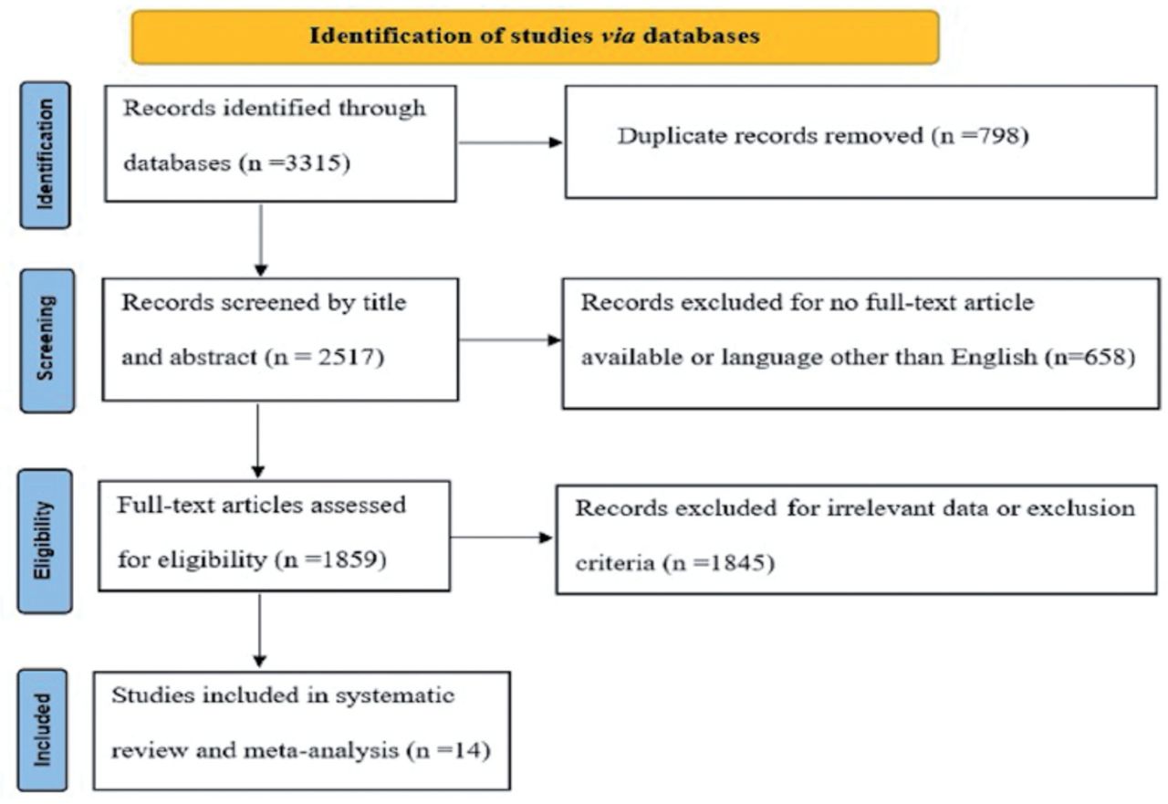
- Figure 1
- An outline of the PRISMA guidelines used to conduct this meta-analysis.
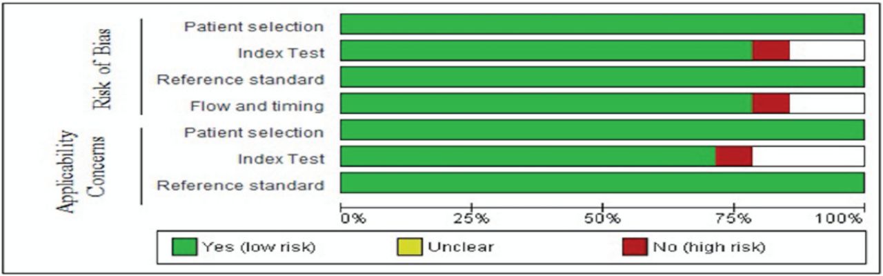
- Figure 2
- Risk of bias items presented as percentages across all articles. PET: positron emission tomography, PWI: perfusion-weighted imaging, CI: confidence interval
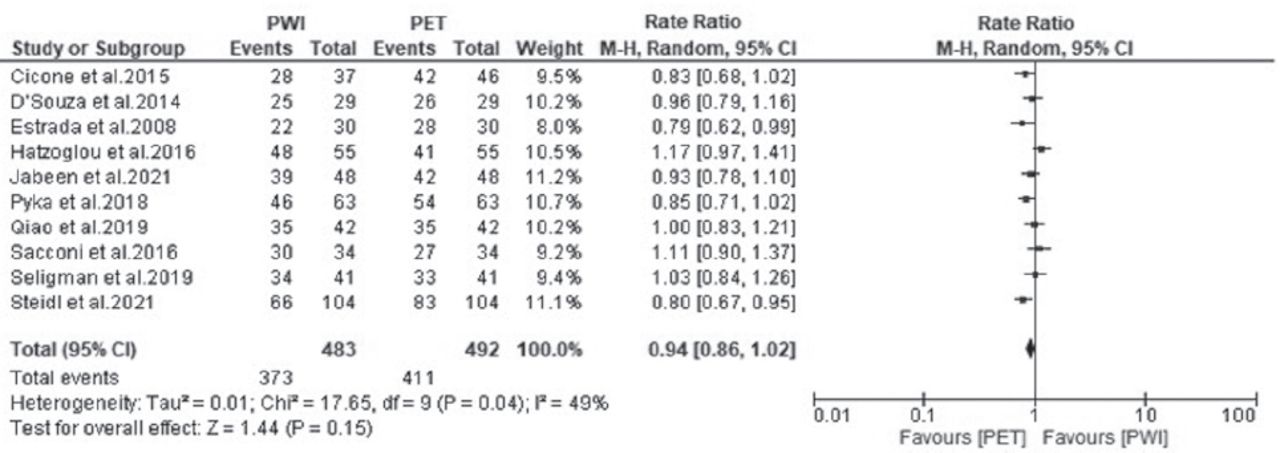
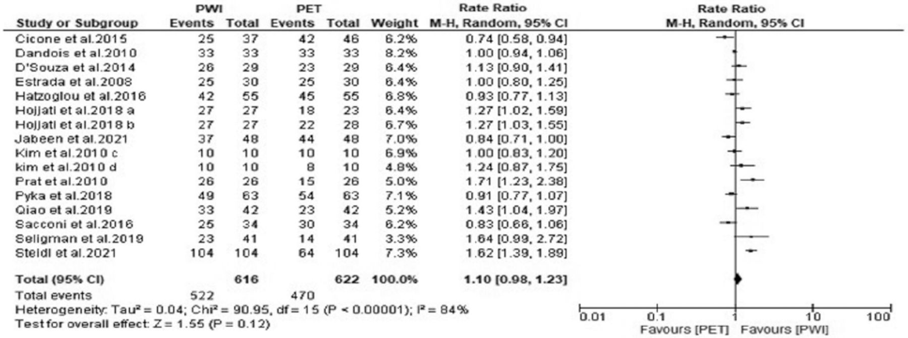
- Figure 3
- Forest plot of the rate ratio of accuracy between perfusion-weighted imaging (PWI) and positron emission tomography (PET). CI: confidence interval
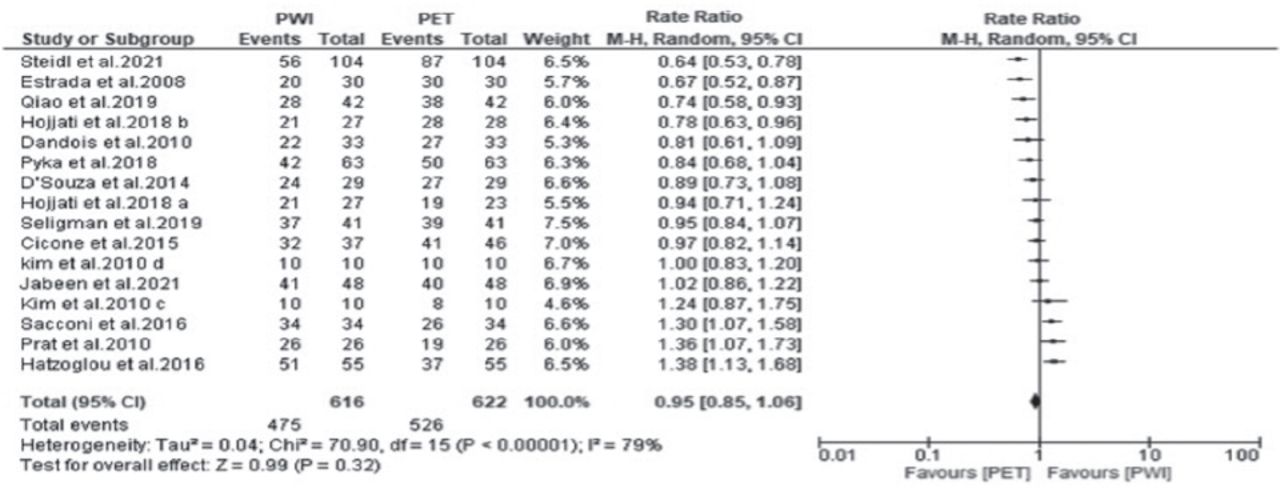
- Figure 4
- Forest plot of the rate ratio of sensitivity between perfusion-weighted imaging (PWI) and positron emission tomography (PET). CI: confidence interval
- Figure 5
- Forest plot of the rate ratio of sensitivity between perfusion-weighted imaging (PWI) and positron emission tomography (PET). CI: confidence interval
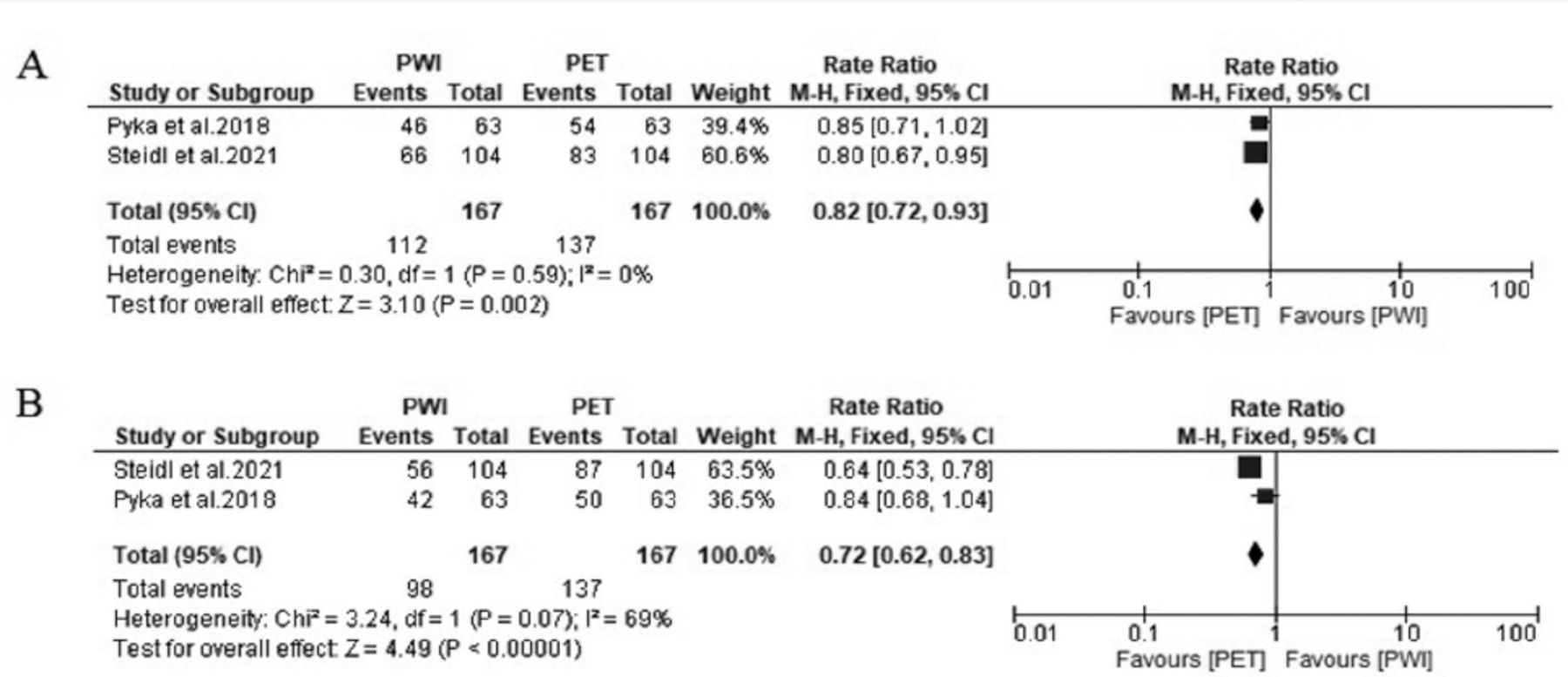
- Figure 6
- Forest plot showing the rate ratio of (A) accuracy and (B) sensitivity between PWI and positron emission tomography (PET).
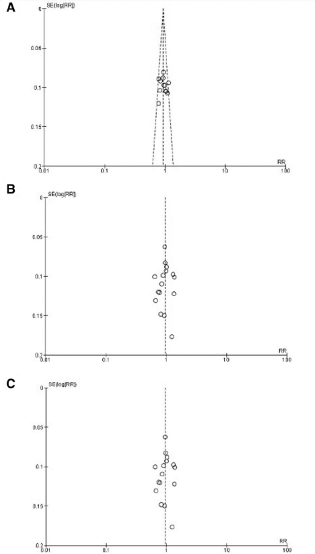
- Figure 7
- Funnel plot showing no publication bias in terms of (A) accuracy, (B) sensitivity, and (C) specificity among the studies.


Tables
Author name and year Study design Country Period since completion of radiation (month) Type of tumor Number of patients Cicone et al28 2015 Retrospective Italy 3 to 45 Brain metastases 42 Dandois et al29 2010 Retrospective Belgium 2 Astrocytic tumours with grades III Astrocytic tumours with grades IV grade III oligoastrocytomas 28 D’Souza et al30 2014 Prospective India 7 to 19 High-grade gliomas (III-IV) 29 Estrada et al31 2008 Prospective Mexico ND Choroid plexus carcinoma Anaplastic astrocytoma Glioblastoma multiforme Gliosarcoma Anaplastic oligoastrocytoma 30 Hatzoglou et al32 2016 Prospective USA ≤3 Gliomas (II to IV) Brain metastases 53 Hojjati et al33 2018 Retrospective USA 2 to 90 Glioblastoma multiforme 24 Jabeen et al34 2021 Retrospective India 3 to 12 Glioblastoma multiforme oligodendroglioma (grade II-IV) Anaplastic oligodendro glioma Anaplastic astrocytoma 48 Kim et al35 2010 Retrospective Korea 3 to 91 Anaplastic astrocytoma Glioblastoma Anaplastic oligodendroglioma 10 Prat et al36 2010 Retrospective Spain ND Anaplastic astrocytoma Astrocytoma Ependymoma Glioblastoma multiform 24 Pyka et al37 2018 Retrospective Germany 15 Glioblastoma Anaplastic Astrocytoma, Diffuse astrocytoma Oligodendroglioma Anaplastic oligodendroglioma 47 Qiao et al38 2019 Retrospective China >3 High-grade tumor (grade III-IV) 42 Sacconi et al39 2016 Retrospective USA ND Low- and high-grade tumor (grade II to IV) 20 Seligman et al40 2019 Retrospective Canada 2 to 156 Grade III Grade IV 41 Steidl et al41 2021 Retrospective Germany 6 to 12 Oligodendroglioma Astrocytoma Astrocytoma Glioblastoma 104 ND: not defined, USA: United States of America
Author name and year Age Number of lesions Modality and optimal cutoff The amino acid tracers of PET PET modality Follow-up period (month) Reference standard Cicone et al28 2015 38–84 46 (PET) 37 (PWI) *SUVLmax/Bkgrmax (rSUV), 1.59 *rCBV, 2.14 F-DOPA PET/CT 6 Histopathology+clinico-radiological follow-up Dandois et al29 2010 25-74 33 *rCBV, 182% *rCBV, 378% 11C-methionine PET 3 to 40 Histopathology D’Souza et al30 2014 15-61 29 *SUV, ND *rCBV, 182% 11C-methionine PET/CT 6 to 28 Histopathology+clinico-radiological follow-up Estrada et al31 2008 17-77 30 *SUV, ND *rCBV, 1.2 18F-FDG PET/MRI 6 to 21 Clinical and radiological criteria Hatzoglou et al322016 19–81 55 *SUVratio, 1.2 *Vpratio, 2.1 18F-FDG PET/CT 6 Histopathology+clinico-radiological follow-up Hojjati et al33 2018 34-81 28 (PET/MRI) 27 (PWI) 23 (PET/CT) *r-Mean, 1.47 *CBVmax, 3.32 18F-FDG PET/CT 8 to 18 Histopathology+clinico-radiological follow-up Jabeen et al34 2021 8-71 48 *TBRmax, 1.23 *rCBVratio, 1.38 11C-methionine PET/MRI 1 to 14 Histopathology+clinico-radiological follow-up Kim et al35 2010 31-66 10 *Lmax/Rmax, 2.64 *rCBV, 3.69 11C-methionine + 18F-FDG PET 6 to 50 Histopathology+clinical course Prat et al36 2010 18-70 26 ND 18F-FDG PET 5 Histopathology+clinico-radiological follow-up Pyka et al37 2018 42-64 63 *TBR, 2.07 *rCBV, 3.35 18F-FET PET/MRI 6 Histopathology+ imaging follow-up Qiao et al38 2019 ND 42 *TBRSUVmax, 1.85 *rCBVmean, 1.83 11C-methionine PET/CT 6 Histopathology+ Clinical follow up Sacconi et al39 2016 14–74 34 *SUVmean, <4.0 *rCBVmean, <1.74 18F-FDG PET/MRI 3 to 6 Histopathology Seligman et al40 2019 21-79 41 *Whole-tumor SUVmean divided by SUVmean of normal white matter >0.75 *Mean Ktrans of whole tumor divided by mean Ktrans of contralateral brain >4.5 18F-FDG PET/MRI 6 Histopathology+clinico-radiological follow-up Steidl et al41 2021 20–78 104 *Slope, <0.69 *rCBVmax, >2.85 18F-FET PET/MRI 6 Histopathology+clinico-radiological follow-up ND - not defined, PET - positron emission tomography, MRI - magnetic resonance imaging, CT - computed tomography, PWI - perfusion-weighted imaging, 18F-FDG - 18fluorine-fluorodeoxyglucose, 18F-FET - 18F-fluoro-ethyl-tyrosine, 18F-FLT- (18)Fluorothymidine, CBV - Cerebral Blood Volume, TBR - Tumor to Background Ratio SUV - Standardized Uptake Value, F-DOPA - fluorodopa
In this issue


Jump to section
Related Articles
Cited By...
- No citing articles found.