


Article Figures & Data
Figures
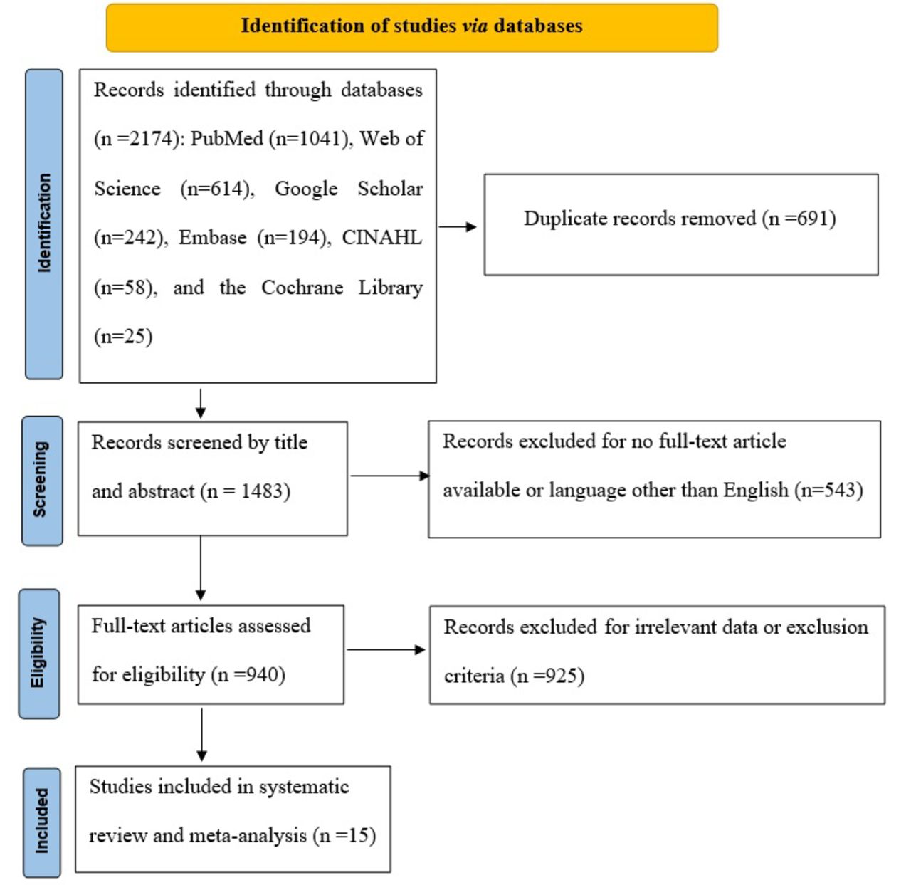
- Figure 1
- PRISMA study flowchart.
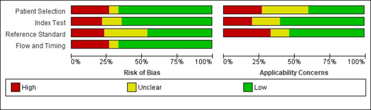
- Figure 2
- Risk of bias and applicability concerns graph: review authors’ judgements about each domain presented as percentages across included studies
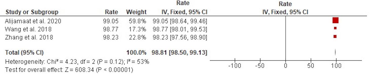
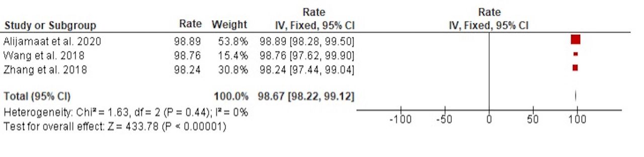
- Figure 3
- Pooled accuracy rates of 2D-3D CNN in the identification of MS lesions
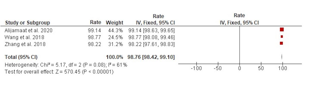
- Figure 4
- Pooled sensitivity rates of 2D-3D CNN in the identification of MS lesions.
- Figure 5
- Pooled specificity rates of 2D-3D CNN in the identification of MS lesions.
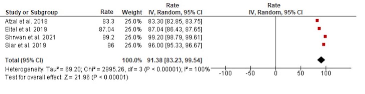
- Figure 6
- Pooled accuracy rates of 2D-3D CNN in the classification of MS lesions.
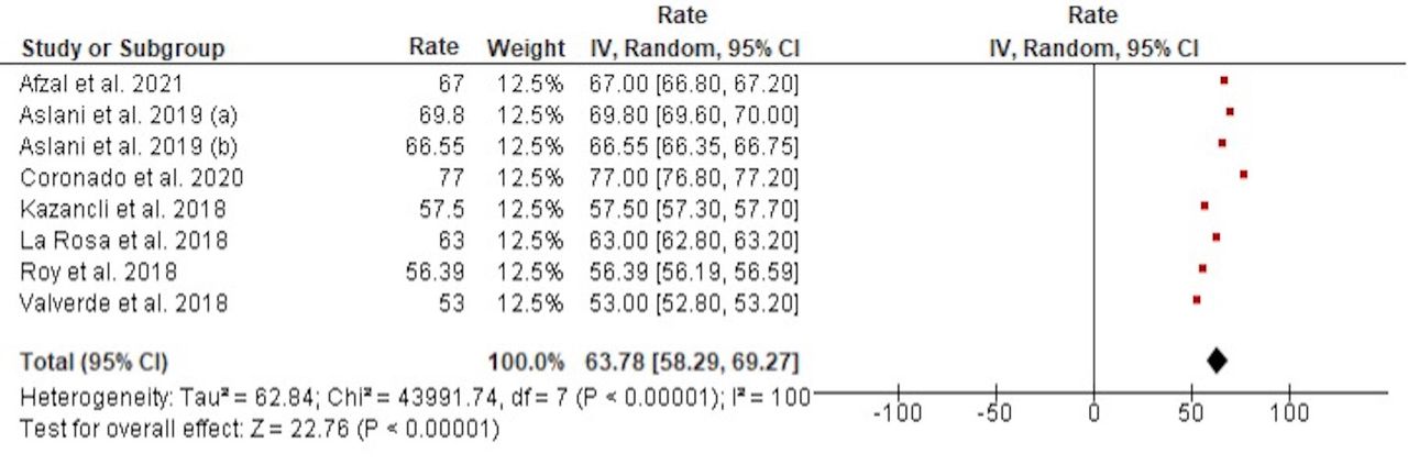
- Figure 7
- Pooled DSC of 2D-3D CNN in the segmentation of MS lesions.
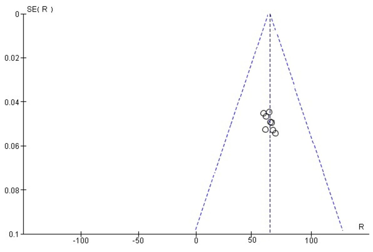
- Figure 8
- Funnel plot of DSC in studies investigating the segmentation of MS lesions


Tables
Article Country Dataset Sample size Diagnosis Application Deep learning architecture Performance Afzal el al, 201815 Australia John Hunter Hospital’s Dataset 21 -11 converted to MS-10 did not convert to MS Classification 2D-CNN Accuracy Afzal el al, 202116 Australia ISBI and MICCAI datasets 19 127 scans of MS Segmentation 2D-CNN -DSC-Sensitivity-Precision Alijamaat et al, 202017 Iran Laboratory of eHealth of the University of Cyprus 58 38 MS patients 20 healthy individuals Identification 2D-CNN -Accuracy-Precision-Sensitivity-Specificity Aslani et al., 201918 Italy -Private dataset-ISBI 2015 longitudinal dataset 51 -37 patients from private dataset -14 patients from ISBI 2015 longitudinal dataset Segmentation 2D-CNN DSC Aslani et al, 201919 Italy ISBI 2015 Longitudinal MS Lesion Segmentation 19 MS Segmentation 2D-CNN -DSC-Lesion-wise true-positive -Lesion-wise false-positive Coronado et al, 202020 USA CombiRx 1,006 Relapsing–remitting MS Segmentation 3D-CNN -DSC-Lesion-wise true-positive-Lesion-wise false-positive Eitel et al, 201921 Germany Clinical 147 76 MS patients 71 healthy patients Classification 3D-CNN Accuracy Kazancli et al, 201822 Spain Clinical 59 MS Segmentation 3D-CNN -DSC-True Positive Rate-False Discovery Rate-Volume Difference La Rosa et al, 201823 Switzerland Clinical 105 -Training dataset: 32 patients with EDSS scores ranged from 1 to 2
-Test dataset: 73 patients with EDSS scores ranged from 1 to 7.5Segmentation 3D-CNN -DSC-Lesion-wise false positive-Lesion-wise true positive-Volume difference Roy et al, 201824 USA ISBI 2015 19 -Training dataset: 5 patients with MS -Test dataset: 14 patients with MS Segmentation 2D-CNN DSC Shrwan et al, 202125 India Clinical 38 MS Classification 2D-CNN -Accuracy-Precision-Recall f_score Siar et al, 201926 Iran Clinical 1111 320 MS patients 791 healthy patients Classification 2D-CNN -Accuracy-Sensitivity-Specificity Valverde et al, 201827 Spain MICCAI 2008 MICCAI 2016 ISBI 2015 60 MS Segmentation 3D-CNN -DSC-Sensitivity-Precision Wang et al, 201828 China eHealth Laboratory and Private data 64 38 MS patients 26 healthy patients Identification 2D-CNN -Accuracy-Sensitivity-Specificity Zhang et al, 201829 China eHealth Laboratory and Private data 64 38 MS patients 26 healthy patients Identification 3D-CNN -Accuracy-Sensitivity-Specificity CNN: convolutional neural network, CombiRx: Combination Therapy in Patients with Relapsing-Remitting Multiple Sclerosis, DSC: Dice Similarity Coefficient, EDSS: Expanded Disability Status Scale, ISBI: International Symposium on Biomedical Imaging, MICCAI: Medical Image Computing and Computer Assisted Intervention, MS: Multiple sclerosis.
- Table 2
- Summary of CADS developed for MS using MRI neuroimaging modalities and details of deep learning architectures.
Article Preprocessing toolbox Others preprocessing Toolbox K Fold Details Classifier Loss function Optimizer Afzal el al., 201823 - data augmentation Keras - 6 convolutional layers + 6 Max Pooling - - Proposed Afzal el al., 202124 FMRIB Patch Extraction Keras, Tensor Flow - 2 convolutional layers + 2 Max Pooling + 1 fully connected Multinomial LR - - Alijamaat et al., 202025 - data augmentation, Histogram Stretching, discrete wavelet transform Keras, Tensor Flow - 15 convolutional layers + 1 Average Pooling + 1 fully connected + Dropout Sigmoid - Adam Aslani et al., 201926 FMRIB Decomposing 3D Data Into 2D Images Keras, Tensor Flow 4 3 Parallel ResNet50s + 5 MMFF Blocks + 4 MSFU Blocks + MPR Block Softmax Soft Dice Loss function Adam Aslani et al., 201927 - Data Augmentation Keras - ResNet50 + UFF Blocks - binary cross-entropy Adadelta Coronado et al., 202028 - Magnetic Resonance Imaging Automatic Processing Pipeline - - 5 convolutional + 4 Context Modules + 3 Up Sampling Modules + 2 Localization Modules + 2 Segmentation + 3 Strides + 3 De-Conv + 1 Upscaling Softmax Multiclass Weighted Dice Adam Eitel et al., 201929 FMRIB data augmentation Keras, Tensor Flow - 4 convolutional + 4 Max-Pooling + 4 Dropout Sigmoid - Adam Kazancli et al., 201830 Free Surfer Patch Extraction Tensor Flow - 2 convolutional + 2 Average Pooling + 2 batch normalization + 1 fully connected + 1 Dropout Softmax cross-entropy Adam La Rosa et al., 201831 FMRIB Manual Segmentation, LeMan-PV - - 4 convolutional + 2 Max Pooling + 4 batch normalization + 1 fully connected + 1 Dropout Softmax cross-entropy Adam Roy et al., 201832 - - Tensor Flow, Keras - 15 convolutional - - Adam Shrwan et al., 202133 - - Matlab R2020a - 3 convolutional + 3 batch normalization + 3 Max Pooling + 2 fully connected Softmax cross-entropy SGDM Siar et al., 201934 - - - - 25 Layers Softmax - - Valverde et al., 201835 FMRIB - Keras, Tensor Flow - 4 convolutional + 2 Max-Pooling + 4 batch normalization + 3 fully connected + 3 Dropout Softmax categorical cross-entropy ADADELTA Wang et al., 201836 - histogram stretching, data augmentation - - 11 convolutional + 11 batch normalization + 4 Pooling + 3 fully connected + 2 Dropout Softmax - - Zhang et al., 201837 - histogram stretching, data augmentation - - 7 convolutional +7 Pooling + 3 fully connected + 3 Dropout Softmax - - ADADELTA: adaptive learning rate method, Adam: A Method for Stochastic Optimization, CADS: Computer-aided detection software, CDMS: clinically defined multiple sclerosis, EDSS: Expanded Disability Status Scale, FMRIB: Functional Magnetic Resonance Imaging of the Brain, MMFF: multi-modal feature fusion block,, MRI: Magnetic resonance imaging, MRIAP: Magnetic Resonance Imaging Automatic Processing, MSFU: multi-scale feature upsampling block, MPR: multi-planes reconstruction, Matlab: matrix laboratory, SGDM: Stochastic Gradient Descent Momentum, UFF: upsampling fused featu
- Table 2
- Summary of CADS developed for MS using MRI neuroimaging modalities and details of deep learning architectures.
Article Clinical data about cases and controls Afzal el al., 201823 All patients included fulfilled the McDonald’s criteria. Out of these 21 patients, 10 converted to CDMS after one year, whereas 11 did not convert to CDMS after one year follow up. Afzal el al., 202124 21 scans of 5 subjects are available for training purposes and already preprocessed with several steps like skull stripping, denoising, bias correction, and co-registration. These 5 subjects have 4 time points and one subject having 5 time points with a gap of approximately 1 year. These 21 scans are provided for training purposes only. For testing purposes, 61 scans are provided from 14 subjects. Alijamaat et al., 202025 MRI images of 38 MS patients whose lesions are labeled by several neurologists and approved by radiologists. To increase the number of images, MRI images of 20 healthy individuals have been prepared by the authors and added to the existing data set. Aslani et al., 201926 19 subjects divided into two sets, 5 subjects for training and 14 subjects for testing.Each subject has MRI data with a different number of time-points, normally ranging between 4 to 6. Aslani et al., 201927 37 MS patients (22 females and 15 males) with mean age 44,6±12,2 years. The patient clinical phenotypes were 24 relapsing remitting MS, 3 primary progressive MS and 10 secondary progressive MS. The mean EDSS was 3,3±2, the mean disease duration was 13.1±8,7 years and the mean lesion load was 6.2±5.7 ml. Coronado et al., 202028 - Eitel et al., 201929 76 patients with relapsing-remitting MS according to the McDonald criteria 2010 and 71 healthy controls. Patients were excluded if they were outside the age range of 18 – 69 or did not have an MRI scan. All patients were examined under supervision of a board-certified neurologist at the NeuroCure Clinical Research Center (Charité – Universitätsmedizin Berlin) between January 2011 and July 2015. Kazancli et al., 201830 - La Rosa et al., 201831 -The training dataset was composed of 32 patients, 18 female / 14 male, mean age 34±10 years, with EDSS scores ranged from 1 to 2 (mean 1,6±0,3). Mean lesion volume is 0,11±0,40 ml (range 0.001-7.03 ml). Mean lesion load per case was 6,0±7,2 ml (range 0,3-37,2 ml).
-The test dataset was made up of 73 patients, 50 females and 23 males (mean age 38±10 years). EDSS scores ranged from 1 to 7.5 (mean 2,6±1,5). Mean lesion volume was 0,25±3,29 ml (range 0.002-159.827 ml). Mean lesion load per case was 14,3±27,9 ml (range 0.2-162.9 ml).Roy et al., 201832 128 patients enrolled in a natural history study of MS, 79 with relapsing-remitting, 30 with secondary progressive, and 19 with primary pro-gressive MS. Shrwan et al., 202133 - Siar et al., 201934 200 patients, including tumors and MS and healthy patients. Totally, the number of trench data for the brain tumor class was 461 images, 791 healthy patients, and 320 MS patients. The total number of data for the most 1286 images and test data was 384 images. Pictures were collected in the range of 6 to 80 years old and the average age was 43. Valverde et al., 201835 60 patients with a clinically isolated syndrome (Hospital Vall d’Hebron, Barcelona, Spain) were scanned on a 3 T Siemens with a 12-channel phased-array head coil (Trio Tim, Siemens, Germany) Wang et al., 201836 - Zhang et al., 201837 -There are 38 patients in the eHealth dataset. 676 slices associated with plaques were selected. All Brain lesions were identified and delineated by experienced MS neurologists and were confirmed by radiologists.
-Age-matched and gender-matched healthy controls (HC) of the eHealth dataset were included. The exclusion criteria for all volunteers were known neurological or psychiatric diseases, brain lesions, taking psychotropic medications, and contraindications to MR imaging.ADADELTA: adaptive learning rate method, Adam: A Method for Stochastic Optimization, CADS: Computer-aided detection software, CDMS: clinically defined multiple sclerosis, EDSS: Expanded Disability Status Scale, FMRIB: Functional Magnetic Resonance Imaging of the Brain, MMFF: multi-modal feature fusion block,, MRI: Magnetic resonance imaging, MRIAP: Magnetic Resonance Imaging Automatic Processing, MSFU: multi-scale feature upsampling block, MPR: multi-planes reconstruction, Matlab: matrix laboratory, SGDM: Stochastic Gradient Descent Momentum, UFF: upsampling fused featu
Parameter Number of studies Rate of DSC [95% CI] Heterogeneity Country Australia 1 67.00 [66.80-67.20] Chi2 =2121.51 Switzerland 1 63.00 [62.80-63.20] p<0.00001
I2 =100%USA 2 66.70 [66.56-66.83] Spain 2 55.25 [55.11-55.39] Italy 2 68.17 [68.04-68.31] DL architecture 2D CNN 4 64.94 [64.84-65.03] Chi2 =1067.22 3D CNN 4 62.63 [62.53-62.72] p<0.00001
I2 =99.9%Study excluded Rate of DSC (95% CI) Afzal et al, 2021 63.32 (57.06-69.58) Aslani et al, 2019 62.92 (56.88-68.96) Aslani et al, 2019 63.38 (57.10-69.66) Coronado et al, 2020 61.89 (57.20-66.58) Kazancli et al, 2018 64.68 (58.67-70.69) La Rosa et al., 2018 63.89 (57.55-70.23) Roy et al., 2018 64.84 (58.96-70.71) Valverde et al., 2018 65.32 (60.02-70.62)
In this issue


Jump to section
Related Articles
Cited By...
- No citing articles found.