
Main menu

  • Home
  • Content
    • Latest
    • Ahead of print
    • Archive
  • Info for
    • Authors
    • Reviewers
    • Subscribers
    • Institutions
    • Advertisers
  • About Us
    • About Us
    • Editorial Office
    • Editorial Board
  • More
    • Alerts
    • Feedback
    • Folders
    • Help
  • Other Publications
    • Saudi Medical Journal

User menu

  • My alerts
  • Log in

Search

  • Advanced search
Neurosciences Journal
  • Other Publications
    • Saudi Medical Journal
  • My alerts
  • Log in
Neurosciences Journal

Advanced Search

  • Home
  • Content
    • Latest
    • Ahead of print
    • Archive
  • Info for
    • Authors
    • Reviewers
    • Subscribers
    • Institutions
    • Advertisers
  • About Us
    • About Us
    • Editorial Office
    • Editorial Board
  • More
    • Alerts
    • Feedback
    • Folders
    • Help
  • Follow psmmc on Twitter
  • Visit psmmc on Facebook
  • RSS
Case ReportCase Report
Open Access

Thrombolytic therapy at systemic lupus onset with secondary antiphospholipid syndrome

A rare stroke experience

Juned J. Loharia, Junaid M. Alam, Hassan A. Abdelhadi and Tamer F. Marei
Neurosciences Journal January 2015, 20 (1) 55-60;
Juned J. Loharia
From the Department of Internal Medicine-ICU (Loharia, Alam), the Department of Internal Medicine-Neurology (Abdelhadi), and the Department of Medical Imaging (Marei), Imam Abdulrahman Binfaisal Hospital, National Guard Health Affairs, Dammam, Kingdom of Saudi Arabia
MD, MRCP(UK)
  • Find this author on Google Scholar
  • Find this author on PubMed
  • Search for this author on this site
Junaid M. Alam
From the Department of Internal Medicine-ICU (Loharia, Alam), the Department of Internal Medicine-Neurology (Abdelhadi), and the Department of Medical Imaging (Marei), Imam Abdulrahman Binfaisal Hospital, National Guard Health Affairs, Dammam, Kingdom of Saudi Arabia
DABIM
  • Find this author on Google Scholar
  • Find this author on PubMed
  • Search for this author on this site
  • For correspondence: [email protected] [email protected]
Hassan A. Abdelhadi
From the Department of Internal Medicine-ICU (Loharia, Alam), the Department of Internal Medicine-Neurology (Abdelhadi), and the Department of Medical Imaging (Marei), Imam Abdulrahman Binfaisal Hospital, National Guard Health Affairs, Dammam, Kingdom of Saudi Arabia
MRCP(UK), FRCP(Ed)
  • Find this author on Google Scholar
  • Find this author on PubMed
  • Search for this author on this site
Tamer F. Marei
From the Department of Internal Medicine-ICU (Loharia, Alam), the Department of Internal Medicine-Neurology (Abdelhadi), and the Department of Medical Imaging (Marei), Imam Abdulrahman Binfaisal Hospital, National Guard Health Affairs, Dammam, Kingdom of Saudi Arabia
MBBCh, FRCR
  • Find this author on Google Scholar
  • Find this author on PubMed
  • Search for this author on this site
  • Article
  • Figures & Data
  • eLetters
  • Info & Metrics
  • References
  • PDF
Loading

Article Figures & Data

Figures

  • Tables
  • Figure 1
    • Download figure
    • Open in new tab
    • Download powerpoint
    Figure 1

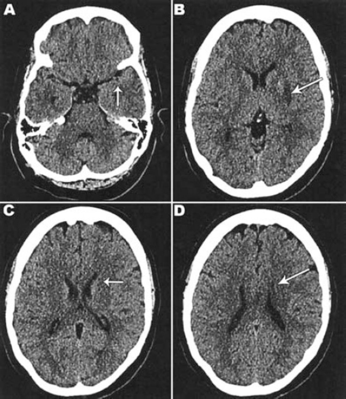
    Non-contrast CT scan brain showing A) and C) no CT signs of acute infarction or hemorrhage. B) Axial image and D) Coronal image showing focal hyper-density at the M2 segment of the left middle cerebral artery (MCA dot sign).

  • Figure 2
    • Download figure
    • Open in new tab
    • Download powerpoint
    Figure 2

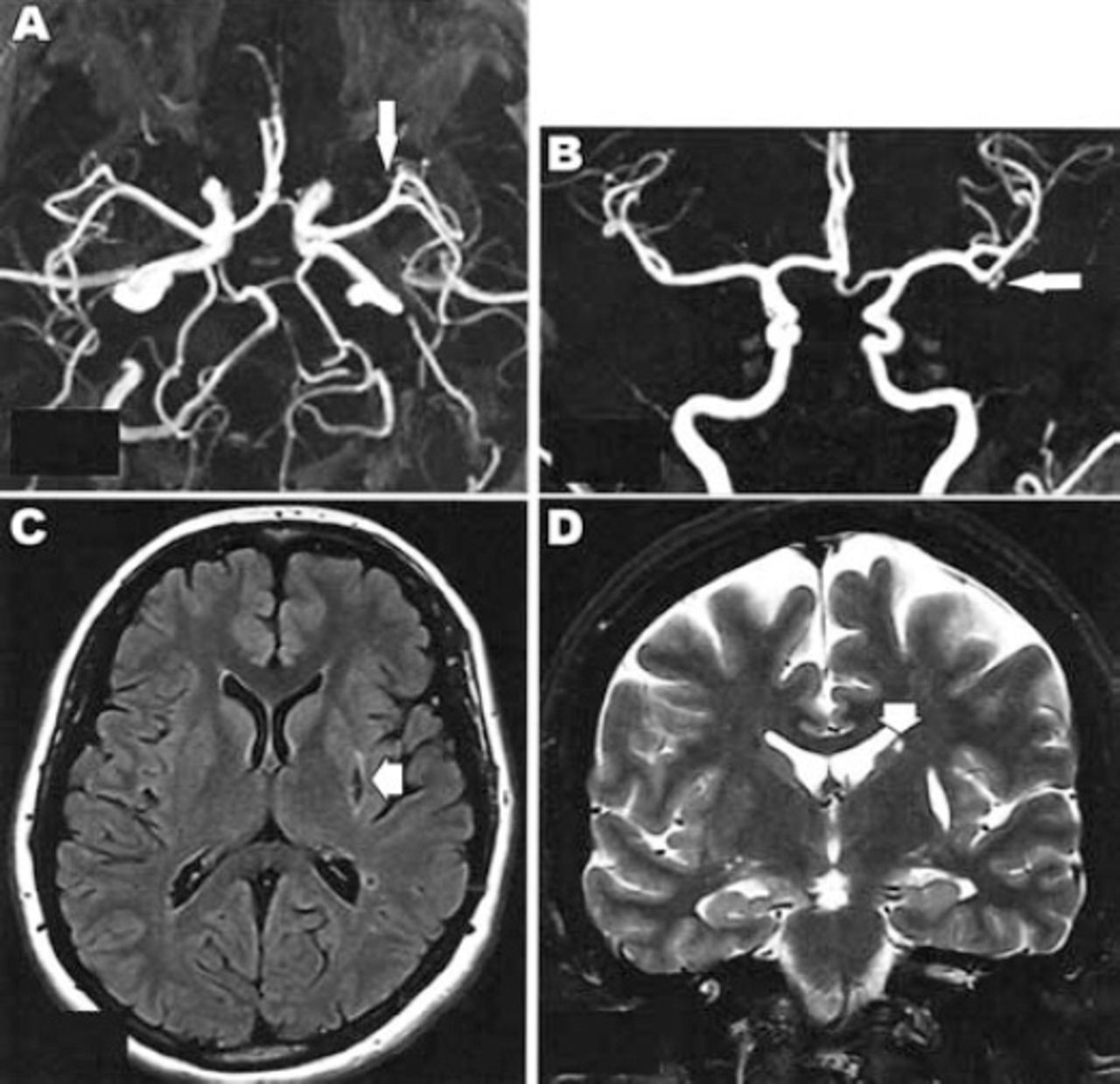
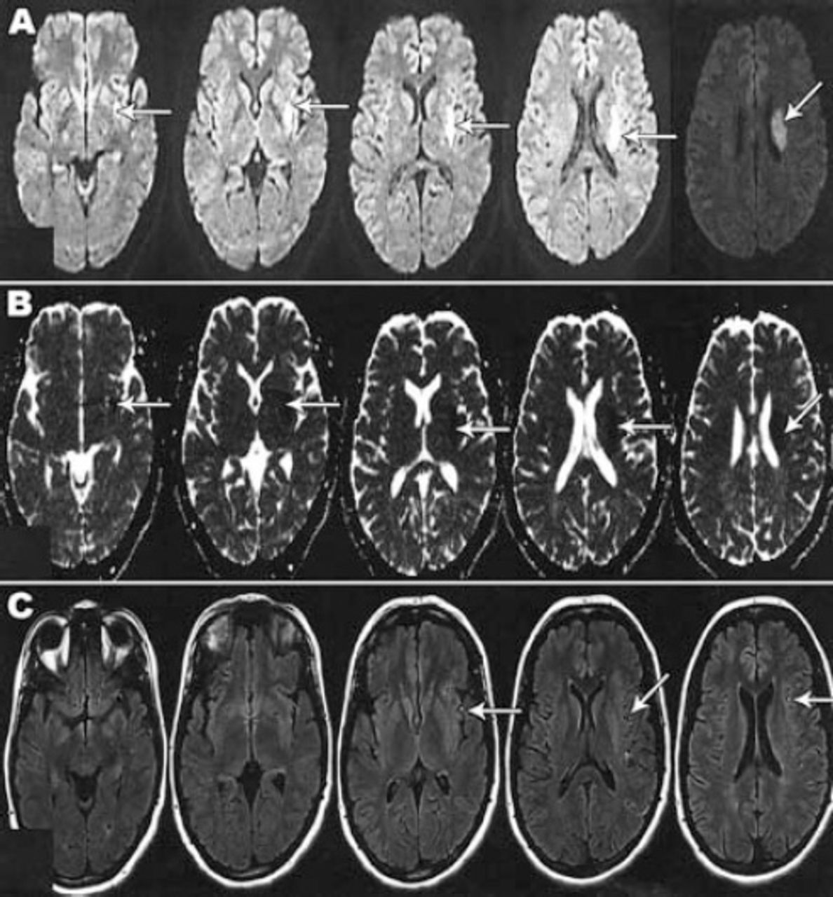
    Magnetic resonance imaging with diffusion-weighted image (DWI), apparent diffusion coefficient (ADC) and axial fluid attenuated inversion recovery (FLAIR) WI showing: A) Acute infarction of the left lentiform nucleus and corona radiata region with high signal on DWI (arrow in A); B) Low signal on ADC (arrow in B); C) FLAIR WI with loss of the normal signal void along the M2 and M3 branches of the left middle cerebral artery (arrow in C) implying slow blood flow.

  • Figure 3
    • Download figure
    • Open in new tab
    • Download powerpoint
    Figure 3

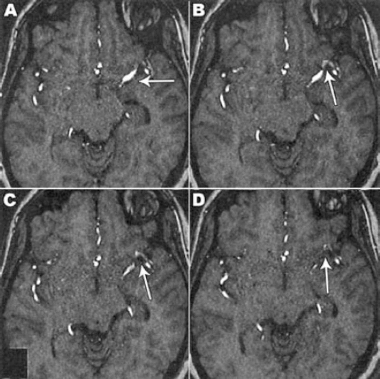
    3-D time-of-flight magnetic resonance angiogram (3 hours after stroke onset) showed A) left middle cerebral (MCA) segment with contrast enhancement, B), C), & D) signal attenuation from the M2 segment of the left MCA depicted with arrows graded as partial thrombo-embolic occlusion.

  • Figure 4
    • Download figure
    • Open in new tab
    • Download powerpoint
    Figure 4

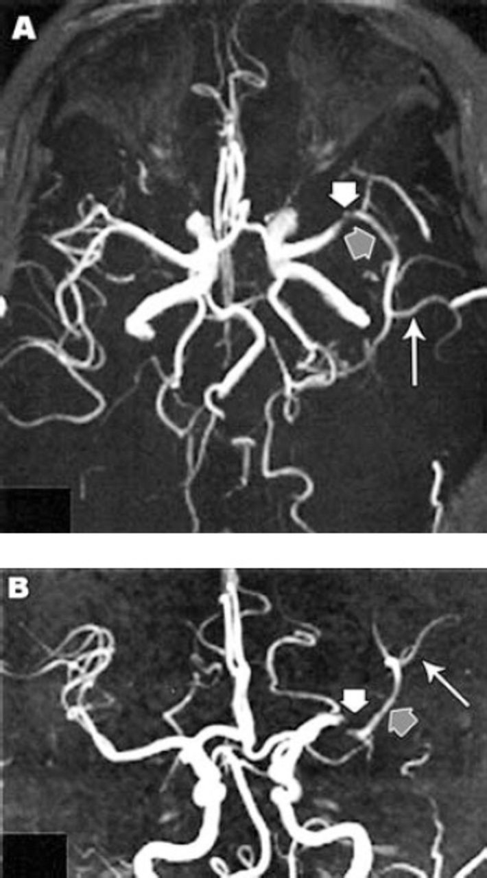
    Maximum intensity projection 3-D time-of-flight magnetic resonance angiogram of the circle of Willis (3 hours and 30 minutes after stroke onset) showing: A and B) thrombo-embolic filling defect at the M2 segment (short white arrows in A and B), narrowing distal M2 segment (short grey arrows in A and B) and pruning of M3 segment branches (long white arrow in A and B) of the left middle cerebral artery.

  • Figure 5
    • Download figure
    • Open in new tab
    • Download powerpoint
    Figure 5

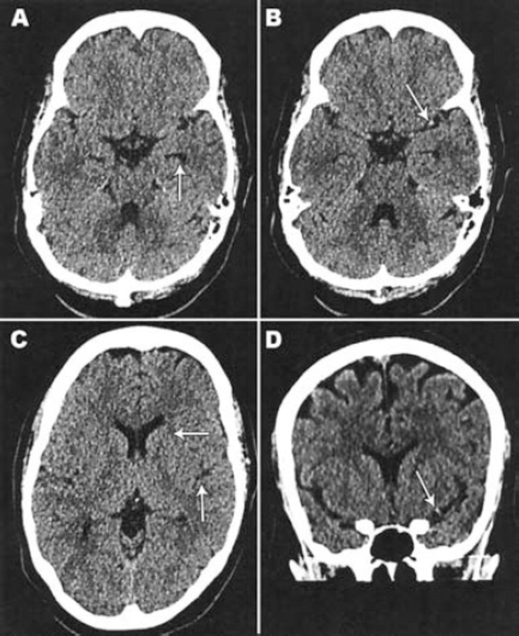
    Non-contrast CT scan brain (26 hours after stroke [22 hours after IV thrombolysis]) showed: A) previously seen hyperdense left middle cerebral artery dot sign in the M2 segment is less prominent. B), C) and D) showed acute linear infarction at the left lentiform and head of caudate nuclei.

  • Figure 6
    • Download figure
    • Open in new tab
    • Download powerpoint
    Figure 6

    Follow up coronal A) and axial B) maximum intensity projection 3-D time-of-flight magnetic resonance angiogram, axial fluid attenuated inversion recovery [FLAIR] C) and coronal D) T2 WI MRI showing: A) Patent M2 segment of left middle cerebral artery. B) Good filling of M3 segment branches. C) Chronic infarction at left lentiform nucleus. D) Chronic infarction at head of caudate nucleus.

Tables

  • Figures
    • View popup
    Table 1

    Complete laboratory test data (some test results were rechecked on follow-up as shown by values with / sign).

    Laboratory testReferencePatient value
    Hemoglobin (g/dl)12 - 1612
    White blood count (Ku/L)4 - 113.2
    Platelets (Ku/L)150 - 400180
    MCV (fl)76 - 9681.6
    MCH (pg)27 - 3225.3
    ESR (mm/hr)0 - 2044
    BUN (mmol/L)2.5 - 6.74.3
    Creatinine (umol/L)50 - 9859
    Blood sugar (mmol/L)2.9 - 7.85.6
    LDL (mmol/L)< 2.62.25
    PT (second/s)11 - 14.512.4
    PTT (seconds/s)26.1 - 37.336.1
    INR0.8 - 1.21
    TSH0.35 - 4.948.09 / 5.27
    Free T4 (pmol/L)9 - 1910.34 / 11.56
    25-OH Vit D (nmol/L)75 - 25078.4
    CRP (mg/L)< 56.7 / 4.1
    Fibrin degradation product (mg/L)0 - 0.52.9
    Fibrinogen (gm/L)1.5 - 4.11.6
    Anti-CCP IgG antibody (unit)< 200.84
    Sickle cell screeningNegative
    ANA index< 1.2> 12
    AntiDNA (IU/ml)< 2052.8
    Vitamin B12 (pmol/L)132 - 857175
    Homocysteine (umol/L)< 157.9
    Antithrombin III (%)75 - 125106
    Functional protein C (%)70 - 130104
    Protein S activity (%)65 - 14069.8
    APC resistance (%)0.9 - 1.30.9
    Factor 5 assay (unit/ml)0.5 - 1.50.74
    Lupus anticoagulant ratio0 - 1.22.1 / 2.5
     LA 1 (secs)30 - 4385.1 / 131
     LA 2 (secs)26.5 - 32.539.6 / 52.6
    Beta-2 glycoprotein
     IgG (U/ml)< 1035.68
     IgM (U/ml)< 123.59
     IgA (U/ml)< 36.43
    Anticardiolipin antibody
     IgG (GPL Unit/ml)< 1037.76 / 34.06
     IgM (MPL Unit/ml)< 76.02 / 6.30
     IgA (APL Unit/ml)< 106.23 / 8.57
     CANCA (U/ml)< 101.48
     PANCA (U/ml)< 64.78
     C3 (g/L)0.83 - 1.931.05
     C4 (g/L)0.150 - 0.5700.362
     Anti SM (U/ml)< 55.04
     Anti SS-A (U/ml)< 48.26
     Anti SS-B (U/ml)< 41.28
     24 hr urine protein (gm/day) - (g/L)0.05 - 0.14< 0.07
     24 hr urine creatinine (mmol/day)06.3 - 14.68.0
    PreviousNext
    Back to top

    In this issue

    Neurosciences Journal: 20 (1)
    Neurosciences Journal
    Vol. 20, Issue 1
    1 Jan 2015
    • Table of Contents
    • Cover (PDF)
    • Index by author
    Print
    Download PDF
    Email Article

    Thank you for your interest in spreading the word on Neurosciences Journal.

    NOTE: We only request your email address so that the person you are recommending the page to knows that you wanted them to see it, and that it is not junk mail. We do not capture any email address.

    Enter multiple addresses on separate lines or separate them with commas.
    Thrombolytic therapy at systemic lupus onset with secondary antiphospholipid syndrome
    (Your Name) has sent you a message from Neurosciences Journal
    (Your Name) thought you would like to see the Neurosciences Journal web site.
    Citation Tools
    Thrombolytic therapy at systemic lupus onset with secondary antiphospholipid syndrome
    Juned J. Loharia, Junaid M. Alam, Hassan A. Abdelhadi, Tamer F. Marei
    Neurosciences Journal Jan 2015, 20 (1) 55-60;

    Citation Manager Formats

    • BibTeX
    • Bookends
    • EasyBib
    • EndNote (tagged)
    • EndNote 8 (xml)
    • Medlars
    • Mendeley
    • Papers
    • RefWorks Tagged
    • Ref Manager
    • RIS
    • Zotero
    Share
    Thrombolytic therapy at systemic lupus onset with secondary antiphospholipid syndrome
    Juned J. Loharia, Junaid M. Alam, Hassan A. Abdelhadi, Tamer F. Marei
    Neurosciences Journal Jan 2015, 20 (1) 55-60;
    Twitter logo Facebook logo Mendeley logo
    • Tweet Widget
    • Facebook Like
    • Google Plus One
    Bookmark this article

    Jump to section

    • Article
      • Abstract
      • Case Report
      • Discussion
      • References
    • Figures & Data
    • eLetters
    • References
    • Info & Metrics
    • PDF

    Related Articles

    • No related articles found.
    • Google Scholar

    Cited By...

    • No citing articles found.
    • Google Scholar

    More in this TOC Section

    • Unmasking the mimic: Leprosy neuropathy misdiagnosed as chronic inflammatory demyelinating polyneuropathy: A case report from Saudi Arabia
    • Seminal vesicle schwannoma with chronic hemorrhage
    • Marchiafava-Bignami disease post-bariatric surgery: A case report and review of similar cases
    Show more Case Report

    Similar Articles

    Navigate

    • home

    More Information

    • Help

    Additional journals

    • All Topics

    Other Services

    • About

    © 2025 Neurosciences Journal Neurosciences is copyright under the Berne Convention and the International Copyright Convention. All rights reserved. Neurosciences is an Open Access journal and articles published are distributed under the terms of the Creative Commons Attribution-NonCommercial License (CC BY-NC). Readers may copy, distribute, and display the work for non-commercial purposes with the proper citation of the original work. Electronic ISSN 1658-3183. Print ISSN 1319-6138.

    Powered by HighWire