


Article Figures & Data
Figures
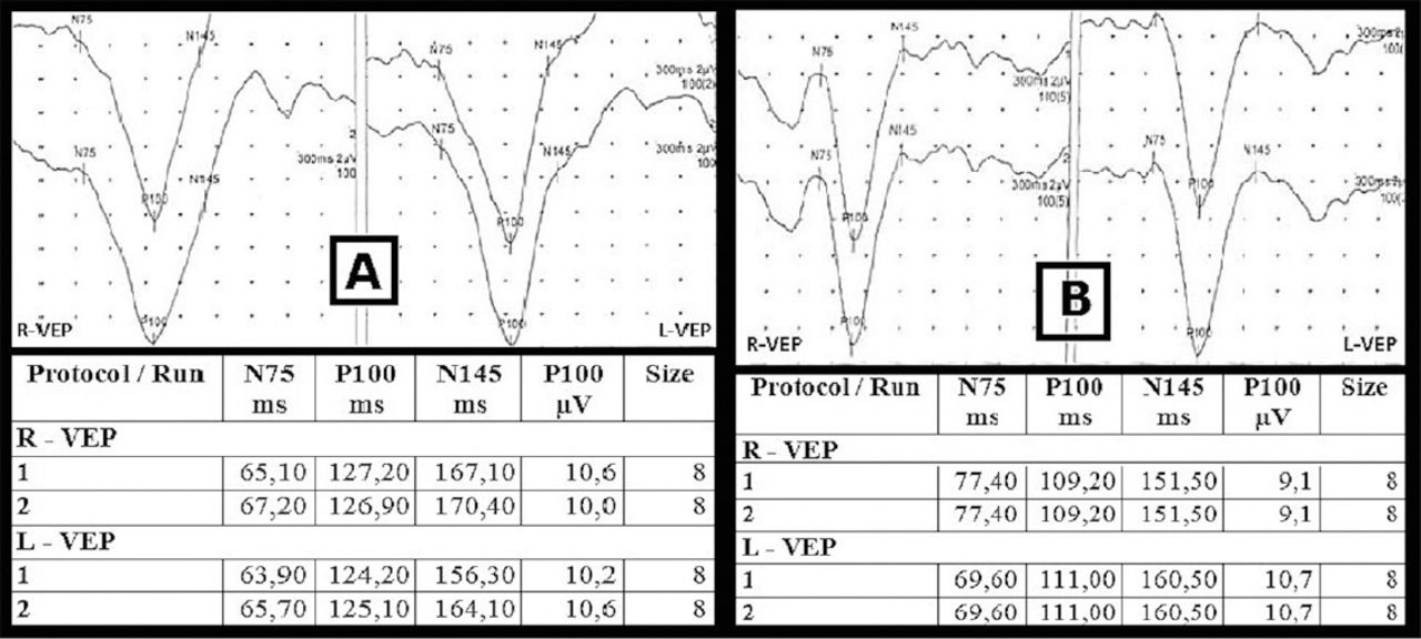
- Figure 1
Visual evoked potential (VEP) images of a patient who showed improvement in VEP values. Visual evoked potential images and values of a patient 3 hours before lumbar puncture (A). Visual evoked potential images and values of the same patients after one month of treatment (B). [X-axis: latency (ms), Y-axis: amplitude (µV)].

Tables
- Table 1
Demographic and clinical features of patients with idiopathic intracranial hypertension.
Demographic variables n (%) Age (mean±SD) Male 36.20±12.235 Female 38.11±11.872 Gender Male 27 (84) Female 5 (16) Obesity Yes 20 (63) No 12 (27) Papilledema Yes 28 (88) One-sided 1 (3) No 3 (9) Visual field Normal 7 (22) Pathological 25 (78) Visual acuity Normal 24 (75) Decreased 8 (25) CSF pressure (mm-water) (mean±SD) 386.88±82.753 Presenting complaint Headache 32 (100) Temporary loss of vision 21 (66) Photopsy 10 (31) Diplopia 9 (28) Eye pain 9 (28) Tinnitus 8 (25) Vertigo 5 (16) Time from the onset of the complaint to diagnosis (median) 8.50 (min: 3, max: 547) - Table 2
Comparison of the initial VEP values of IIH patients to the VEP values of the healthy control group.
Variables Initial VEP results of IIH patients (n=32) VEP results of the healthy control group (n=30) P-value Mean±SD Right P100, latency (ms) 115.10±10.12 101.48±2.47 <0.001 Left P100, latency (ms) 115.19±10.04 101.75±2.88 <0.001 Right amplitude (µV) 10.26±4.36 10.46±2.63 0.231 Left amplitude (µV) 9.75±4.20 10.36±2.91 0.122 IIH - idiopathic intracranial hypertension, VEP - visual evoked potential, SD - standard deviation
- Table 3
The relationship of pathological VEP results with gender, obesity, and visual acuity and visual field findings at admission.
Variables Initial VEP pathological (n=18) Initial VEP normal (n=14) P-value Gender Male 3 2 0.854 Female 15 12 Obesity No 5 7 0.198 Yes 13 7 Visual acuity Normal 11 13 0.040 Decreased 7 1 Visual field Normal 1 6 0.011 Pathological 17 8 VEP - visual evoked potential
Variables Initial VEP results of IIH patients Second VEP results of the IIH patients P-value Mean±SD Right P100, latency (ms) 115.10±10.12 118.84±12.536 0.045 Left P100, latency (ms) 115.19±10.04 118.69±11.047 0.049 Right amplitude (µV) 10.26±4.36 9.84±3.671 0.319 Left amplitude (µV) 9.75±4.20 9.69±3.693 0.887 IIH - idiopathic intracranial hypertension, VEP - visual evoked potential, SD - standard deviation
In this issue


Jump to section
Related Articles
Cited By...
- No citing articles found.