


Article Figures & Data
Figures
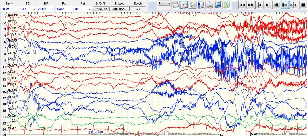
- Figure 1
Desynchronization (generalized electrodecremental ictal pattern) associated with epileptic spam.
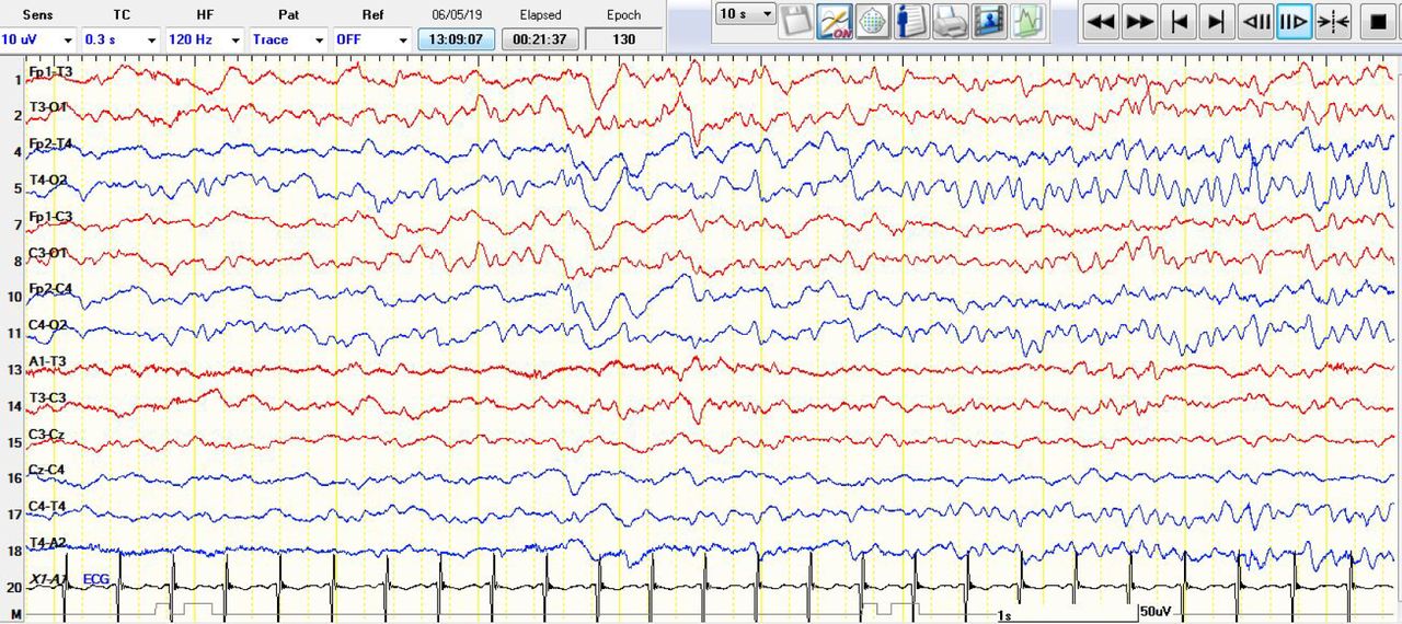
- Figure 2
There is a new rhythm medium amplitude alpha frequency 9Hz arising from right temporal T4 with evolution to higher amplitude and slower in frequencies which is representing an ictal discharge.
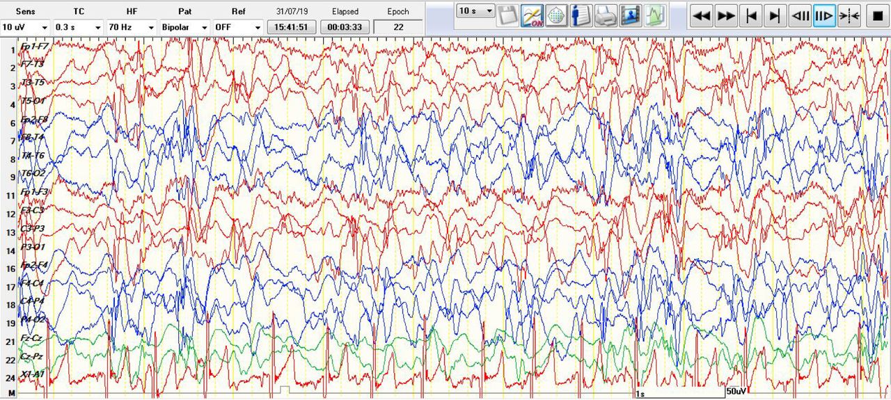
- Figure 3
Hypsarrhythmia, which is a continuous, high amplitude, asynchronous delta activities with interspersed independent multifocal spikes


Tables
- Table 1
Age, EEG and neuroimaging of patients with seizures ( IED= interictal discharges).
Patients Age EEG background &IED Ictal discharges Neuroimaging 1 Neonate Hypsarrhythmia with multifocal IED electrographic seizures seen over the left frontocentral Normal MRI brain 2 8 Months Normal for age background with multifocal IED Generalized electrodecremental ( epileptic spasm ) Normal MRI brain 3 Neonate Normal for age background with no IED An electrographic event 9Hz right temporal T4 ( Figure 2) Normal brain US 4 6 Months Hypsarrhythmia with multifocal IED Generalized electrodecremental (epileptic spasm ) MRI brain: bilateral symmetrical T2 hyperintensity involving the globuspallidi bilaterally and the lateral thalamic nuclei 5 10 Months Generalized slow background with Left temporal IED Three electrographic seizures were recorded all focal arising from left hemisphere MRI brain: Diffuse cerebral atrophy 6 3 Months Generalized voltage attenuation of background with no IED Generalized electrodecremental (epileptic spasm ) MRI brain: Interval progression of the diffuse cerebral atrophy. 7 7 Months Hypsarrhythmia with Multifocal IED Generalized electrodecremental (epileptic spasm ) MRI brain: bilateral symmetrical T2 hyperintensity involving the globus pallidi bilaterally and the lateral thalamic nuclei . 8 4 Months Normal for age background with Left temporal IED Ictal discharges at left temporal region associated with clinical bradycardia Normal MRI brain 9 2 Months Normal for age background with Bilateral temporal IED Build up sharp waves on left temporal region mostly electrographic seizure CT head : brain atrophy and craniosynostosis 10 Neonate Normal for age background with multifocal IED Generalized electrodecremental (epileptic spasm) Normal brain US 11 4 Months Hypsarrhythmia withMultifocal IED Generalized electrodecremental (epileptic spasm) Normal MRI brain EEG data n (%) Seizures(Electrographic & Electroclinical) 11 (6.4) from total EEG Electrographic seizures 3 (36.3) Electroclinical seizures 8 (63.6) History of ICU admission or hospitalization 2 (18.2) Abnormal neuroimaging 5 (45.45) Focal seizures 5 (45.45) Generalized seizures 6 (45.45)


Jump to section
Related Articles
Cited By...
- No citing articles found.