
Main menu

  • Home
  • Content
    • Latest
    • Ahead of print
    • Archive
  • Info for
    • Authors
    • Reviewers
    • Subscribers
    • Institutions
    • Advertisers
  • About Us
    • About Us
    • Editorial Office
    • Editorial Board
  • More
    • Alerts
    • Feedback
    • Folders
    • Help
  • Other Publications
    • Saudi Medical Journal

User menu

  • My alerts
  • Log in

Search

  • Advanced search
Neurosciences Journal
  • Other Publications
    • Saudi Medical Journal
  • My alerts
  • Log in
Neurosciences Journal

Advanced Search

  • Home
  • Content
    • Latest
    • Ahead of print
    • Archive
  • Info for
    • Authors
    • Reviewers
    • Subscribers
    • Institutions
    • Advertisers
  • About Us
    • About Us
    • Editorial Office
    • Editorial Board
  • More
    • Alerts
    • Feedback
    • Folders
    • Help
  • Follow psmmc on Twitter
  • Visit psmmc on Facebook
  • RSS
Case ReportCase Report
Open Access

Kearns-Sayre syndrome with rare imaging finding of SLC25A4 Mutation

Huan Zhao, Min Shi, Fang Yang and Xuhong Yang
Neurosciences Journal April 2022, 27 (2) 111-115; DOI: https://doi.org/10.17712/nsj.2022.2.20210123
Huan Zhao
From the Department of Neurology, Hospital of Chengdu University of Traditional Chinese Medicine, Chengdu, Sichuan Province, People’s Republic of China.
  • Find this author on Google Scholar
  • Find this author on PubMed
  • Search for this author on this site
Min Shi
From the Department of Neurology, Hospital of Chengdu University of Traditional Chinese Medicine, Chengdu, Sichuan Province, People’s Republic of China.
  • Find this author on Google Scholar
  • Find this author on PubMed
  • Search for this author on this site
Fang Yang
From the Department of Neurology, Hospital of Chengdu University of Traditional Chinese Medicine, Chengdu, Sichuan Province, People’s Republic of China.
  • Find this author on Google Scholar
  • Find this author on PubMed
  • Search for this author on this site
Xuhong Yang
From the Department of Neurology, Hospital of Chengdu University of Traditional Chinese Medicine, Chengdu, Sichuan Province, People’s Republic of China.
  • Find this author on Google Scholar
  • Find this author on PubMed
  • Search for this author on this site
  • For correspondence: [email protected]
  • Article
  • Figures & Data
  • eLetters
  • Info & Metrics
  • References
  • PDF
Loading

Article Figures & Data

Figures

  • Tables
  • Figure 1
    • Download figure
    • Open in new tab
    • Download powerpoint
    Figure 1

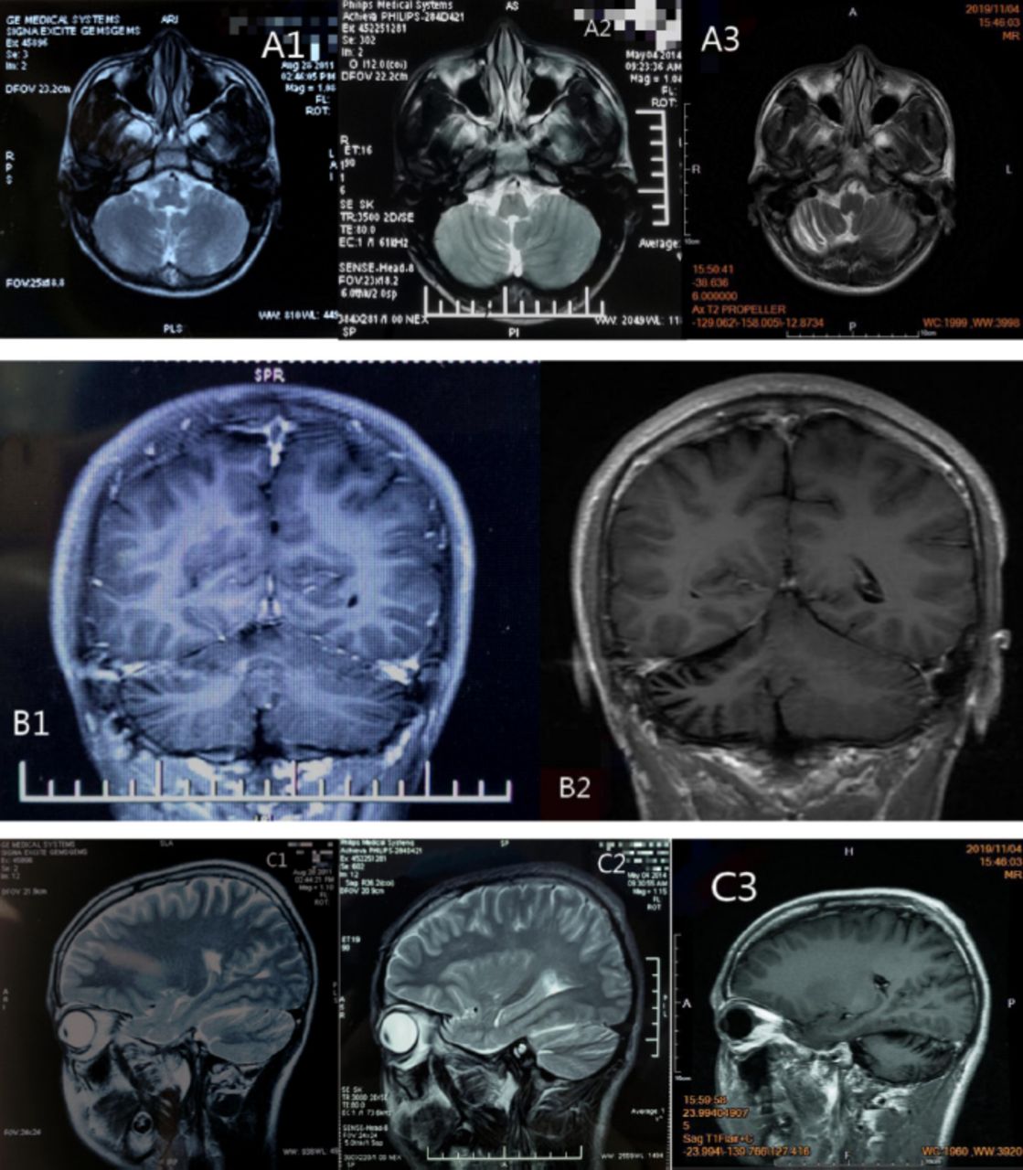
    - MRI images A1 and C1) are the cranial MRI images on May 14, 2011. They reveal obvious sulcus of vermis cerebelli; A2, B1 and C2) are the cranial MRI images on August 4, 2014. They reveal slight narrowing of the gyrus of the right cerebellar hemisphere; A3, B2 and C3) are the cranial MRI images on November 4, 2019. They reveal narrowing of the gyrus of the right cerebellar hemisphere and widening of the sulcus.

  • Figure 2
    • Download figure
    • Open in new tab
    • Download powerpoint
    Figure 2

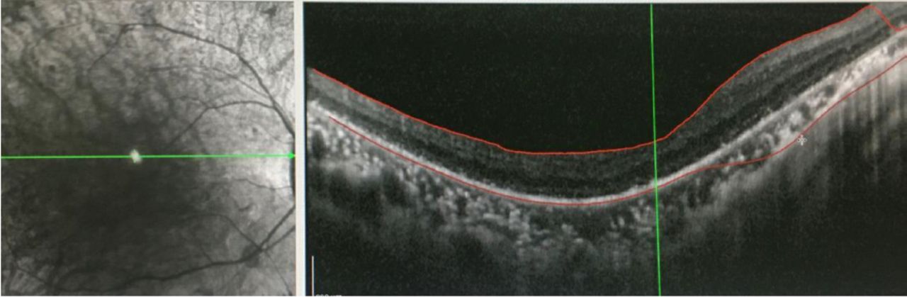
    - Outer retinal atrophy in the right eye revealed by OCT of both eyes in the Department of Ophthalmology of our hospital in November 2019.

  • Figure 3
    • Download figure
    • Open in new tab
    • Download powerpoint
    Figure 3

    - The physical examination image A) November 1, 2019: both pupils are of the same size and roundness, with a diameter of about 3mm, sensitive to light reflex, blepharoptosis for the right, and mild restriction for movement in the downward and lower right directions; B) April 20, 2020: both pupils are of the same size and roundness, with a diameter of about 3 mm, sensitive to light reflex, blepharoptosis for the right, unable to move in the downward and lower right directions.

Tables

  • Figures
    • View popup
    Table 1

    - Patient timeline.

    DatesRelevant past medical history and interventions
    Before 2011No relevant past medical history or interventions
    DatesSummaries from initial and follow-up visitsDiagnostic testing (including dates)Interventions
    May, 2011At the age of 13 years, the patient’s parents complained that the child’s language skills were lower than normal children1. On May 14, 2011 no abnormality was found on the children’s intelligence screening test.
    2. A1 and C1 are the Cranial MRI images on May 14, 2011 (Figure 1)
    Undetermined diagnosis; No clinical intervention
    August, 2014At the age of 16 years, symptoms slowly and significantly worsened, specifically with elongated speech, heterogeneous pitch, and often fulminant.Results revealed obvious sulcus of vermis cerebelli; A2, B1 and C2 are the Cranial MRI images on August 4, 2014 (Figure 1)Undetermined diagnosis; No clinical intervention
    November, 2017At the age of 19 years, the patient gradually developed right blepharoptosis, the inability to move in the lower and right lower direction (Figure 1), and exhibited double vision, as well as significantly elongated speech, heterogeneous pitch, and often fulminantNo diagnostic testingUndetermined diagnosis; No clinical intervention
    November, 2019At the age of 21 years, aggravation of the above symptoms occurred1. In November, 2019 laboratory test, electrophysiological examination, cerebrospinal fluid examination, ECG and other related examinations were performed. The results are provided in Table 2.
    2. In November, 2019 OCT showed outer retinal atrophy in the right eye (see Figure 2).
    3. In November, 2019 a cranial MRI showed significant atrophy of the right cerebellar hemisphere (see A3, B2 and C3 in Figure 1).
    4. In November, 2019 a quantitative polymerase chain reaction revealed that the patient had mitochondrial DNA deletion (mitochondrial/nuclear DNA: 18.7%) (mitochondria DNA depletion [mitochondria/nuclear DNA: 18.7%]), and that the patient’s mother had mitochondria DNA depletion (mitochondria/nuclear DNA: 2.7%).
    Kearns-Sayre syndrome was diagnosed. The patient received trophic nerve therapy during hospitalization
    • View popup
    Table 2

    - Laboratory examination, electrophysiological examination, cerebrospinal fluid (CSF) examination and other examinations.

    ExaminationsResults
    Hematology
    White blood cell5.05*109/L
    Percentage of Neutrophils37.5%
    Whole blood C-reactive protein<0.50mg/L
    Cholesterol(-)
    Bile acid(-)
    Liver enzyme(-)
    Tumor markers(-)
    Autoimmune antibody profile(-)
    Fasting blood glucose6.3mmol/l
    Glycosylated hemoglobin5.2
    T31.61 nmol/L
    T4102.5 nmol/L
    Parathyroid gland hormone(-)
    Sex hormone(-)
    VitB12680 pmol/L
    Folate11.3 nmol/L
    Coenzyme Q10/
    Vitamin E(-)
    Anti-GAD antibody/
    Cerebrospinal fluid
    Total white blood cells4.1
    Red blood cell10.3
    Protein254 mg/dL
    IgG1.87 mg/dL
    EEG(-)
    Electromyography
    F-wave(-)
    H-reflex(-)
    Repetitive stim(-)
    Fundus Optical Tomography (OCT)Outer retinal atrophy
    Abdominal ultrasonography(-)
    Chest CT(-)
    ECGExtension of PR interval

    ECG - electrocardiogram, IgG - Immunoglobulin G, EEG - electroencephalogram, T4 - thyroxine

      • View popup
      Table 3

      - Negative test result of mitochondrial gene hotspot mutation in MELAS (Mitochondrial Encephalomyopathy) syndrome using PCR-Sanger.

      Test methodTest SitesTest resultsReference values
      PCR-sanger sequencingm.3093CC
      m.3243AA
      m.3244GG
      m.3252AA
      m.3256CC
      m.3258TT
      m.3260AA
      m.3271TT
      m.3291TT
      m.13513GG
    PreviousNext
    Back to top

    In this issue

    Neurosciences Journal: 27 (2)
    Neurosciences Journal
    Vol. 27, Issue 2
    1 Apr 2022
    • Table of Contents
    • Cover (PDF)
    • Index by author
    Print
    Download PDF
    Email Article

    Thank you for your interest in spreading the word on Neurosciences Journal.

    NOTE: We only request your email address so that the person you are recommending the page to knows that you wanted them to see it, and that it is not junk mail. We do not capture any email address.

    Enter multiple addresses on separate lines or separate them with commas.
    Kearns-Sayre syndrome with rare imaging finding of SLC25A4 Mutation
    (Your Name) has sent you a message from Neurosciences Journal
    (Your Name) thought you would like to see the Neurosciences Journal web site.
    Citation Tools
    Kearns-Sayre syndrome with rare imaging finding of SLC25A4 Mutation
    Huan Zhao, Min Shi, Fang Yang, Xuhong Yang
    Neurosciences Journal Apr 2022, 27 (2) 111-115; DOI: 10.17712/nsj.2022.2.20210123

    Citation Manager Formats

    • BibTeX
    • Bookends
    • EasyBib
    • EndNote (tagged)
    • EndNote 8 (xml)
    • Medlars
    • Mendeley
    • Papers
    • RefWorks Tagged
    • Ref Manager
    • RIS
    • Zotero
    Share
    Kearns-Sayre syndrome with rare imaging finding of SLC25A4 Mutation
    Huan Zhao, Min Shi, Fang Yang, Xuhong Yang
    Neurosciences Journal Apr 2022, 27 (2) 111-115; DOI: 10.17712/nsj.2022.2.20210123
    Reddit logo Twitter logo Facebook logo Mendeley logo
    • Tweet Widget
    • Facebook Like
    • Google Plus One
    Bookmark this article

    Jump to section

    • Article
      • Abstract
      • Case Report
      • Discussion
      • References
    • Figures & Data
    • eLetters
    • References
    • Info & Metrics
    • PDF

    Related Articles

    • No related articles found.
    • PubMed
    • Google Scholar

    Cited By...

    • No citing articles found.
    • Google Scholar

    More in this TOC Section

    • Cauda equina syndrome by extrusion of lumbar disc after lumbar sympathetic neurolysis
    • Post COVID-19 Guillain–Barre syndrome presents as sensory dominant neuropathy with reversible conduction failure
    • Thrombosis of intraorbital arterio-venous fistula
    Show more Case Report

    Similar Articles

    Navigate

    • home

    More Information

    • Help

    Additional journals

    • All Topics

    Other Services

    • About

    © 2023 Neurosciences Journal Neurosciences is copyright under the Berne Convention and the International Copyright Convention. All rights reserved. Neurosciences is an Open Access journal and articles published are distributed under the terms of the Creative Commons Attribution-NonCommercial License (CC BY-NC). Readers may copy, distribute, and display the work for non-commercial purposes with the proper citation of the original work. Electronic ISSN 1658-3183. Print ISSN 1319-6138.

    Powered by HighWire