
Main menu

  • Home
  • Content
    • Latest
    • Ahead of print
    • Archive
  • Info for
    • Authors
    • Reviewers
    • Subscribers
    • Institutions
    • Advertisers
  • About Us
    • About Us
    • Editorial Office
    • Editorial Board
  • More
    • Alerts
    • Feedback
    • Folders
    • Help
  • Other Publications
    • Saudi Medical Journal

User menu

  • My alerts
  • Log in

Search

  • Advanced search
Neurosciences Journal
  • Other Publications
    • Saudi Medical Journal
  • My alerts
  • Log in
Neurosciences Journal

Advanced Search

  • Home
  • Content
    • Latest
    • Ahead of print
    • Archive
  • Info for
    • Authors
    • Reviewers
    • Subscribers
    • Institutions
    • Advertisers
  • About Us
    • About Us
    • Editorial Office
    • Editorial Board
  • More
    • Alerts
    • Feedback
    • Folders
    • Help
  • Follow psmmc on Twitter
  • Visit psmmc on Facebook
  • RSS
Case ReportCase Report
Open Access

Sporadic subependymal giant cell astrocytoma with somatic TSC2 mutation: A case report

Ali H. Alassiri, Turki M. Alfayea, Tariq I. Aljared and Khaled R. Alenezi
Neurosciences Journal April 2024, 29 (2) 139-143; DOI: https://doi.org/10.17712/nsj.2024.2.20230089
Ali H. Alassiri
From the College of Medicine (Alassiri, Alfayea), King Saud bin Abdulaziz University for Health Sciences, from the Department of Pathology and Laboratory Medicine (Alassiri), Department of Oncology (Alfayea), Department of Surgery (Aljared), Department of Medical Imaging (Alenezi), King Abdulaziz Medical City, and from the King Abdullah International Medical Research Center (Alassiri, Alfayea), Riyadh, Kingdom of Saudi Arabia
MD
  • Find this author on Google Scholar
  • Find this author on PubMed
  • Search for this author on this site
  • ORCID record for Ali H. Alassiri
  • For correspondence: [email protected]
Turki M. Alfayea
From the College of Medicine (Alassiri, Alfayea), King Saud bin Abdulaziz University for Health Sciences, from the Department of Pathology and Laboratory Medicine (Alassiri), Department of Oncology (Alfayea), Department of Surgery (Aljared), Department of Medical Imaging (Alenezi), King Abdulaziz Medical City, and from the King Abdullah International Medical Research Center (Alassiri, Alfayea), Riyadh, Kingdom of Saudi Arabia
MD
  • Find this author on Google Scholar
  • Find this author on PubMed
  • Search for this author on this site
Tariq I. Aljared
From the College of Medicine (Alassiri, Alfayea), King Saud bin Abdulaziz University for Health Sciences, from the Department of Pathology and Laboratory Medicine (Alassiri), Department of Oncology (Alfayea), Department of Surgery (Aljared), Department of Medical Imaging (Alenezi), King Abdulaziz Medical City, and from the King Abdullah International Medical Research Center (Alassiri, Alfayea), Riyadh, Kingdom of Saudi Arabia
MD
  • Find this author on Google Scholar
  • Find this author on PubMed
  • Search for this author on this site
Khaled R. Alenezi
From the College of Medicine (Alassiri, Alfayea), King Saud bin Abdulaziz University for Health Sciences, from the Department of Pathology and Laboratory Medicine (Alassiri), Department of Oncology (Alfayea), Department of Surgery (Aljared), Department of Medical Imaging (Alenezi), King Abdulaziz Medical City, and from the King Abdullah International Medical Research Center (Alassiri, Alfayea), Riyadh, Kingdom of Saudi Arabia
MD
  • Find this author on Google Scholar
  • Find this author on PubMed
  • Search for this author on this site
  • Article
  • Figures & Data
  • eLetters
  • Info & Metrics
  • References
  • PDF
Loading

Article Figures & Data

Figures

  • Tables
  • Figure 1
    • Download figure
    • Open in new tab
    • Download powerpoint
    Figure 1

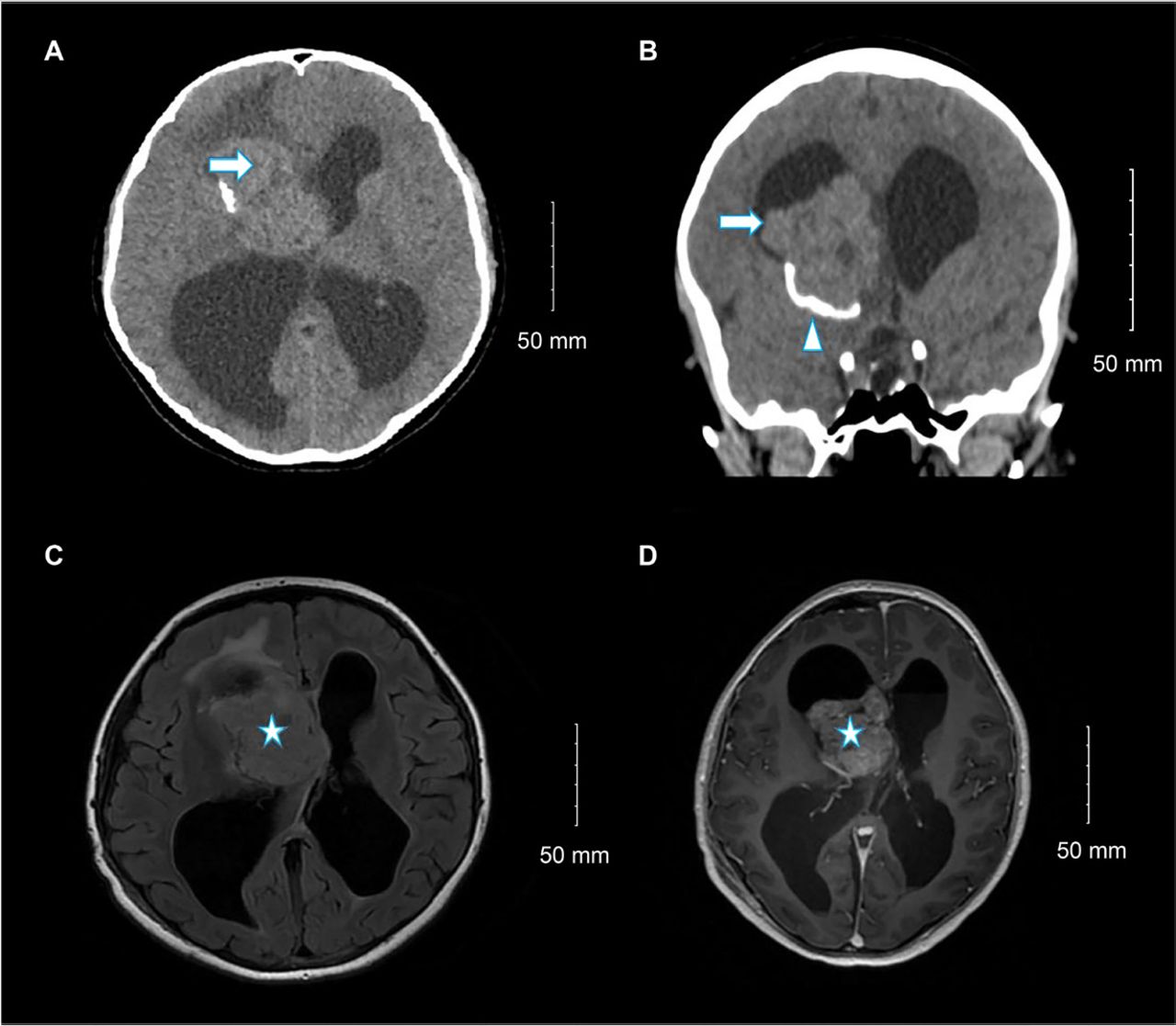
    - Selected (A) axial and (B) coronal images of unenhanced brain computed tomography show a large right lateral ventricle frontal horn dense mass (arrow) associated with peripheral calcification (arrowhead) causing marked hydrocephalus. Selected (C) axial fluid-attenuated inversion recovery and (D) axial T1 post-contrast brain magnetic resonance imaging show large, well-defined, lobulated intraventricular mass lesions within the right lateral ventricle at the region of foramen of Monro with heterogeneous moderate enhancement (star) and hydrocephalus.

  • Figure 2
    • Download figure
    • Open in new tab
    • Download powerpoint
    Figure 2

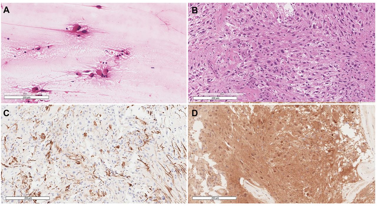
    - Selected views of A) Smear slide at 20× showing the large eosinophilic tumor cells with processes emanating from them. B) Hematoxylin and eosin-stained section at 20× power reveals the classic histomorphology of subependymal giant cell astrocytoma with some of the cells showing well-defined borders while others show elongated cytoplasmic processes. C) Glial fibrillary acidic protein stain highlighting scattered positive tumor cells. D) Positive immunohistochemical staining for S100.

  • Figure 3
    • Download figure
    • Open in new tab
    • Download powerpoint
    Figure 3

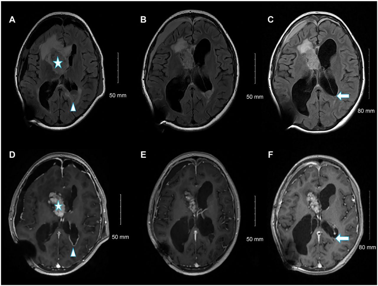
    - From left to right, selected fluid-attenuated inversion recovery magnetic resonance images (A, C, and E), and selected axial T1 post-contrast images at the same level (B, D, and F). Upon admission (A, B), the residual heterogeneously enhanced the right lateral ventricle frontal horn component (star). A new left occipital ependymal/subependymal nodule with peripheral enhancement (arrowhead) is visible. Follow-up examination at 3 months (C, D) revealed an interval reduction in the size of the left occipital ependymal/subependymal nodule, with peripheral enhancement. Follow-up examinations at 6 months (E, F) showed a further reduction in the size of the left occipital ependymal/subependymal nodule (arrow).

Tables

  • Figures
    • View popup
    Table 1

    - Timeline table.

    DatesRelevant past medical history and interventions
    6 months earlierIntermittent morning headaches with on/off nausea and vomiting over six months diagnosed as sinusitis in a private clinic
    DateSummaries from initial and follow-up visitsDiagnostic testing (including dates)Interventions
    23 Mar 2022Presented to emergency department with increasing early morning headaches with nausea and vomiting over last one month. Physical examination revealed decreased sensation in the left V2 maxillary branch of the trigeminal nerve. Also bilateral papilledema and optic disc pallor. Otherwise, intact power, other sensations, and no cerebellar signs.Eye ultrasound showed 10 mm optic sheath diameter Non-contrast brain computed tomography (CT) demonstrated active hydrocephalus and right lateral ventricular mass with associated calcificationDexamethasone 8 mg STAT then 4 mg q6hrs
    24 Mar 2022Patient was admitted to pediatric intensive care unit (PICU) in stable conditionMRI revealed a heterogeneously enhancing well-defined right lateral intraventricular mass measuring 6×5.3×3.3 cm.Emergency right septostomy with insertion of external ventricular drain (EVD)
    30 Mar 2022Patient intubated and ventilated after surgery. The preoperative symptoms markedly improved after tumor debulking, however, she developed three episodes of tonic-clonic seizures.Pathological diagnosis is subependymal giant cell astrocytoma Tumor genomic analysis showed pathogenic TSC2 mutation No clinical or radiological TSC stigmataRight frontal craniotomy and maximum safety resection of about 70% of the intraventricular tumor Levetiracetam 500 mg BID
    12 Apr 2022Patient extubated successfully but found to have buccofascial apraxia and mild left sided weakness which improved a lot with PT and OT White blood cell count in the cerebrospinal fluid normalized after 2 weeks of broad spectrum antibioticsHigh white blood cell count in the cerebrospinal fluid without clinical signs of infectionBroad spectrum antibiotics for two weeks Removal of EVD and insertion of right occipital ventriculoperitoneal (VP) shunt
    21 Apr 2022Discharged in good condition with no deficits  
    19 May 2022Re-admitted for evacuation of post-operative subdural hemorrhage. Patient complained of headache and facial asymmetryMRI brain revealed stable residual tumor but there was a new left occipital subependymal 2.7 cm nodule with peripheral enhancementEverolimus (mTOR inhibitor) was administered at a starting dose of 4.5 mg/m2 per day then increased to 5 mg daily for two weeks, followed by a further increase to 7.5 mg daily
    4 Sep 2022Outpatient visit The patient was asymptomatic and reported no adverse effectsFollow-up brain MRIs at three and six months intervals showed a reduction in the residual tumor size and occipital noduleContinue Everolimus treatement until it fails or the side effects become intolerable
PreviousNext
Back to top

In this issue

Neurosciences Journal: 29 (2)
Neurosciences Journal
Vol. 29, Issue 2
1 Apr 2024
  • Table of Contents
  • Cover (PDF)
  • Index by author
Print
Download PDF
Email Article

Thank you for your interest in spreading the word on Neurosciences Journal.

NOTE: We only request your email address so that the person you are recommending the page to knows that you wanted them to see it, and that it is not junk mail. We do not capture any email address.

Enter multiple addresses on separate lines or separate them with commas.
Sporadic subependymal giant cell astrocytoma with somatic TSC2 mutation: A case report
(Your Name) has sent you a message from Neurosciences Journal
(Your Name) thought you would like to see the Neurosciences Journal web site.
Citation Tools
Sporadic subependymal giant cell astrocytoma with somatic TSC2 mutation: A case report
Ali H. Alassiri, Turki M. Alfayea, Tariq I. Aljared, Khaled R. Alenezi
Neurosciences Journal Apr 2024, 29 (2) 139-143; DOI: 10.17712/nsj.2024.2.20230089

Citation Manager Formats

  • BibTeX
  • Bookends
  • EasyBib
  • EndNote (tagged)
  • EndNote 8 (xml)
  • Medlars
  • Mendeley
  • Papers
  • RefWorks Tagged
  • Ref Manager
  • RIS
  • Zotero
Share
Sporadic subependymal giant cell astrocytoma with somatic TSC2 mutation: A case report
Ali H. Alassiri, Turki M. Alfayea, Tariq I. Aljared, Khaled R. Alenezi
Neurosciences Journal Apr 2024, 29 (2) 139-143; DOI: 10.17712/nsj.2024.2.20230089
Twitter logo Facebook logo Mendeley logo
  • Tweet Widget
  • Facebook Like
  • Google Plus One
Bookmark this article

Jump to section

  • Article
    • Abstract
    • CASE Report
    • Discussion
    • Acknowledgments
    • Footnotes
    • References
  • Figures & Data
  • eLetters
  • References
  • Info & Metrics
  • PDF

Related Articles

  • No related articles found.
  • PubMed
  • Google Scholar

Cited By...

  • No citing articles found.
  • Google Scholar

More in this TOC Section

  • Unmasking the mimic: Leprosy neuropathy misdiagnosed as chronic inflammatory demyelinating polyneuropathy: A case report from Saudi Arabia
  • Seminal vesicle schwannoma with chronic hemorrhage
  • Marchiafava-Bignami disease post-bariatric surgery: A case report and review of similar cases
Show more Case Report

Similar Articles

Navigate

  • home

More Information

  • Help

Additional journals

  • All Topics

Other Services

  • About

© 2025 Neurosciences Journal Neurosciences is copyright under the Berne Convention and the International Copyright Convention. All rights reserved. Neurosciences is an Open Access journal and articles published are distributed under the terms of the Creative Commons Attribution-NonCommercial License (CC BY-NC). Readers may copy, distribute, and display the work for non-commercial purposes with the proper citation of the original work. Electronic ISSN 1658-3183. Print ISSN 1319-6138.

Powered by HighWire