


Article Figures & Data
Figures
- Figure 1
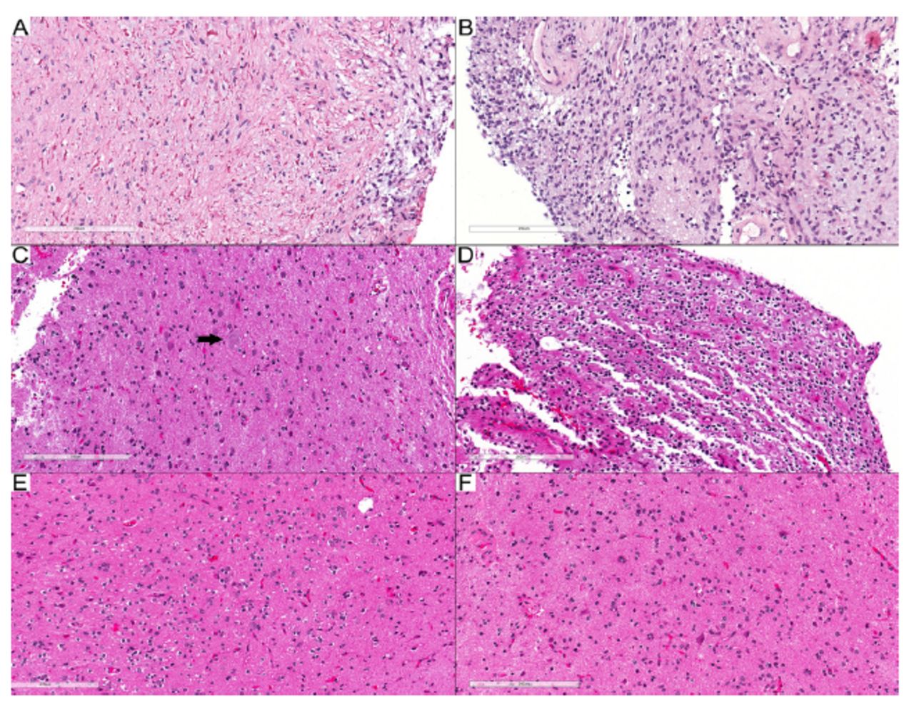
- Tumors with modified diagnoses. A, B) Case 24: DLGNT. A) Biphasic tumor with piloid glial cells and numerous Rosenthal fibers. B) Glial component with round to oval nuclei. C–F) Case 25: PLNTY. C) Dysmorphic ganglion cell (arrow). D) Oligodendroglioma-like component. (E, F) Infiltrative component. Scale bars: 200 μm (A–F).
- Figure 2
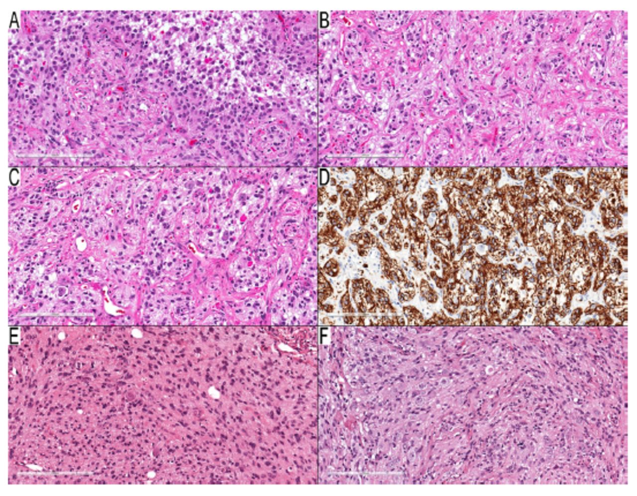
- Tumors with debatable diagnoses. A–D) Case 29: temporal lobe mass in a three-year-old boy. A) Neoplastic cells with round and elongated nuclei and many EGBs. B, C) Many dysmorphic ganglion cells with binucleated form in (C). D) Diffusely positive GFAP in the background, but negative in dysmorphic ganglion cells. E–F) Case 31: cerebellar tumor in a 3-year-old boy. E) Infiltrative components consist of elongated neoplastic cells with entrapped neurons. F) Many dysmorphic ganglion cells. Scale bars: 200 μm (A–F).
- Figure 3
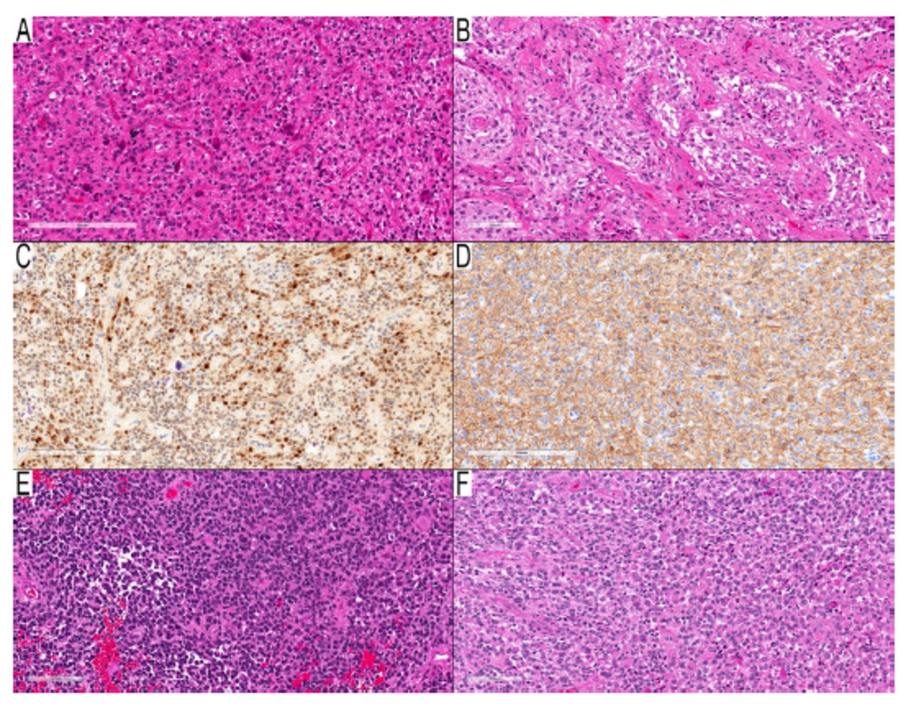
- Two cases with morphology of high-grade astrocytoma and MC of PXA. A–D) Case 34: parieto-temporal mass in a nine-year-old girl. A) Cellular component with round neoplastic cells infiltrative around entrapped nuclei. B) Two granular mitoses in the center of the field. (C) NeuN immunostain, an immunopositive subset of neoplastic nuclei, which raised the possibility of a neuronal component, but there was no clear neuronal differentiation. (D) GFAP immunostain shows a cytoplasmic rim in many neoplastic cells and the background. (E–F) Case 35: temporal lobe mass in a two-year-old boy. (E) High-cellular area with round-oval hyperchromic neoplastic cells and significant mitotic activity. (F) Many neoplastic cells with eccentric round eosinophilic cytoplasm resembling rhabdoid or epithelioid neoplastic cells. Scale bars: 200 μm (A, C, D); 100 μm (B, E, F).
- Figure 4
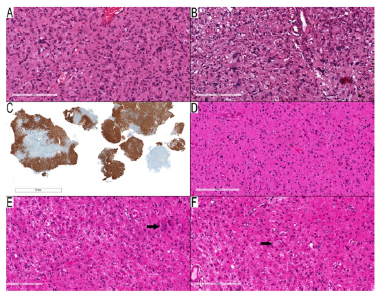
- Examples of cases in the non-contributary category. A–C) Case 46: parietal lobe mass in a thirteen-year-old woman. A) Spindled neoplastic cells infiltrate around entrapped neurons with scattered pleomorphic neoplastic cells. B) Scattered dysmorphic ganglion cells, but no EGBs present. C) CD34 immunostaining shows diffuse staining in the neoplastic cells. D–F) Case 47: occipital/temporal lobe mass in an eleven-year-old girl. D) Infiltrative glial component with round to slightly elongated nuclei. E) Area with many EGBs and scattered pleomorphic ganglion cells. (F) A rare example of a binucleated dysmorphic ganglion cell (arrow). Scale bars: 200 μm (A, B, C–F); 5 mm (C).


Tables
No. Age (Y) Sex Site PD NGS AF MC V12.5 MC V12.8 Final diagnosis 1 1.7 M OP PA BRAF V600E 0.29 PA-M (0.99) PA-M (0.99) PA 2 7 F Ce PA BRAF A598_T599insV 0.31 PA-I (0.99) PA-I (0.99) PA 3 17 M TL PA BRAF V600Dfs*47 0.08 PA-H (0.89) PA-H (0.79) PA 4 11 M FL;PL PA BRAF V600E 0.19 PA-H (0.93) PA-H (0.96) PA 5 12 F OP PA BRAF V600E 0.13 PA-M (0.99) PA-M (0.99) PA 6 15 M Ce PA BRAF L410Q 0.22 PA-I (0.99) PA-I (0.99) PA 7 1.6 M TL PA BRAF V600E 0.28 PA-H (0.98) PA-H (0.81) PA 8 15 M OP PA BRAF V600E 0.15 PA-M (0.99) PA-M (0.99) PA 9 15 M Ce PA BRAF T599dup, Ad1 0.48 PA-I (0.89) PA-M (0.59) PA 10 1.2 M OP PA BRAF V600E 0.2 PA-M (0.99) PA-M (0.98) PA 11 14 F BS PA-R BRAF T599dup 0.59 PA-I (0.98) PA-I (0.90) PA 12 16 M TL PA BRAF V600E 0.26 PA-H (0.94) PA-H (0.98) PA 13 14 F TL GG BRAF V600E 0.24 GG (0.99) GG (0.99) GG 14 0.75 F TL GG BRAF V600E 0.18 GG (0.94) GG (0.98) GG 15 2 M TL GG BRAF V600E 0.18 GG (0.75) GG (0.85) GG 16 3 M TL GG BRAF V600E 0.41 GG (0.99) GG (0.99) GG 17 14 F TL PXA BRAF V600E 0.36 PXA (0.99) PXA (0.99) PXA 18 13 M TL PXA BRAF V600E 0.48 PXA (0.99) PXA (0.99) PXA 19 9 F FL PXA BRAF V600E 0.51 PXA (0.99) PXA (0.99) PXA 20 14 F FL PXA BRAF V600E 0.29 PXA (0.99) PXA (0.99) PXA 21 14 M TL PXA BRAF V600E 0.41 PXA (0.99) PXA (0.99) PXA 22 10 M PL PXA BRAF V600E 0.3 PXA (0.99) PXA (0.99) PXA 23 12.3 F TL As- Inf BRAF V600E 0.3 GG (0.50) GG (0.80) LG GNT vs As 23 16.3 F Hip PXA BRAF V600E 0.24 PXA (0.99) PXA (0.99) PXA 24 3.6 F SC PA BRAF V600E 0.49 DLGNT-1 (0.99) DLGNT-1 (0.99) DLGNT 25 13.9 F TL GG BRAF V600E 0.69 PLNTY (0.73) PLNTY (0.48) PLNTY 26 18 M TL GG BRAF V600E 0.12 PA-H (0.83) PA-H (0.97) GG 27 19 F OL;TL GG BRAF V600E 0.19 PA-H (0.94) PA-H (0.81) GG 28 10 M TL GG BRAF V600E 0.14 PA-H (0.82) GG (0.32) GG 29 3 M TL GG BRAF V600E 0.38 PA-H (0.99) PA-H (0.99) GG 30 15 F TL GG BRAF V600E 0.19 PA-H (0.85) PA-H (0.99) GG 31 3 M Ce GG BRAF V600E 0.29 PA-I (0.99) PA-I (0.95) GG 32 0.25 F OP DIA BRAF V600E 0.28 PA-M (0.99) PA-M (0.99) DIA vs PA 33 6 M OP DIG BRAF V600E 0.34 PA-M (0.99) PA-M (0.99) PA vs DIG 34 9 F PL;TL HG As BRAF V600E 0.46 PXA (0.99) PXA (0.99) pHGG, NEC 35 2 M TL HG As BRAF V600E 0.89 PXA (0.99) PXA (0.98) pHGG, NEC 36 11 F TL GG BRAF V600E 0.07 C-RM (0.56) C-IM (0.43) GG 37 14 M FL GG BRAF T599dup IF ins 0.24 C-RM (0.64) C-RM (0.46) GG 38 4 M BS GG BRAF V600E 0.15 C-RM (0.38) C-RM (0.44) GG 39 4 F FL GG BRAF V600E, Ad2 0.09 C-RM (0.74) C-RM (0.54) GG 40 2 M TL GG BRAF V600E 0.18 PA-H (0.45) PA-H (0.57) GG 41 16 M TL GG BRAF V600E 0.08 C-RM (0.98) C-RM (0.86) GG 42 20 F BS LG As BRAF V600E 0.11 C-RM (0.98) C-RM (0.96) LG As 43 11 F PL;TL PA/PMA BRAF V600E 0.16 C-RM (0.99) C-RM (0.97) PA/PMA 44 9 M TL HG As, NEC BRAF V600E, Ad3 0.53 pHGG, RTK1 (0.38) DPHGG, RTK1 (0.47) pHGG, NEC 45 11 F Th DMG, K27M BRAF V600E, Ad4 0.16 G-IDHw-M (0.43) C-IM (0.88) DMG, K27M 46 13 F PL LG G/GN BRAF V600E 0.33 GG (0.52) PA-H (0.24) GG 47 11 F OL;TL LG G/GN BRAF V600E 0.26 PXA (0.52) PXA (0.36) PXA Ad: additional mutations [Ad1: P53 X307_splice and CDH1 X177_splice; Ad2: NF1 C1792*; Ad3: TP53 R248L; Ad4: H3F3A K28M, TERT promotor mutation (not specified)]. AF: allelic frequency; BS: brainstem; C-IM: control tissue, inflammatory microenvironment; C-RM: control tissue, reactive tumor microenvironment; Ce: cerebellum; DLGNT-1: diffuse leptomeningeal glioneuronal tumor, subtype 1; F: female; FL: frontal lobe; G-IDHw-M: glioblastoma, IDH-wildtype, mesenchymal type; Hip: hippocampus; LV: lateral ventricle; M: male; ND: not done; NEC: not elsewhere classified; OL: occipital lobe; OP: optic pathway; PA-H: pilocytic astrocytoma, hemispheric; PA-I: pilocytic astrocytoma, infratentorial; PA-M: pilocytic astrocytoma, midline; pHGG: diffuse pediatric-type high-grade glioma; PL: parietal lobe; PLNTY: polymorphous low-grade neuroepithelial tumor of the young; PXA: pleomorphic xanthoastrocytoma; SC: spinal cord; Th: thalamus; TL: temporal lobe; Y: years
In this issue


Jump to section
Related Articles
Cited By...
- No citing articles found.