


Article Figures & Data
Figures
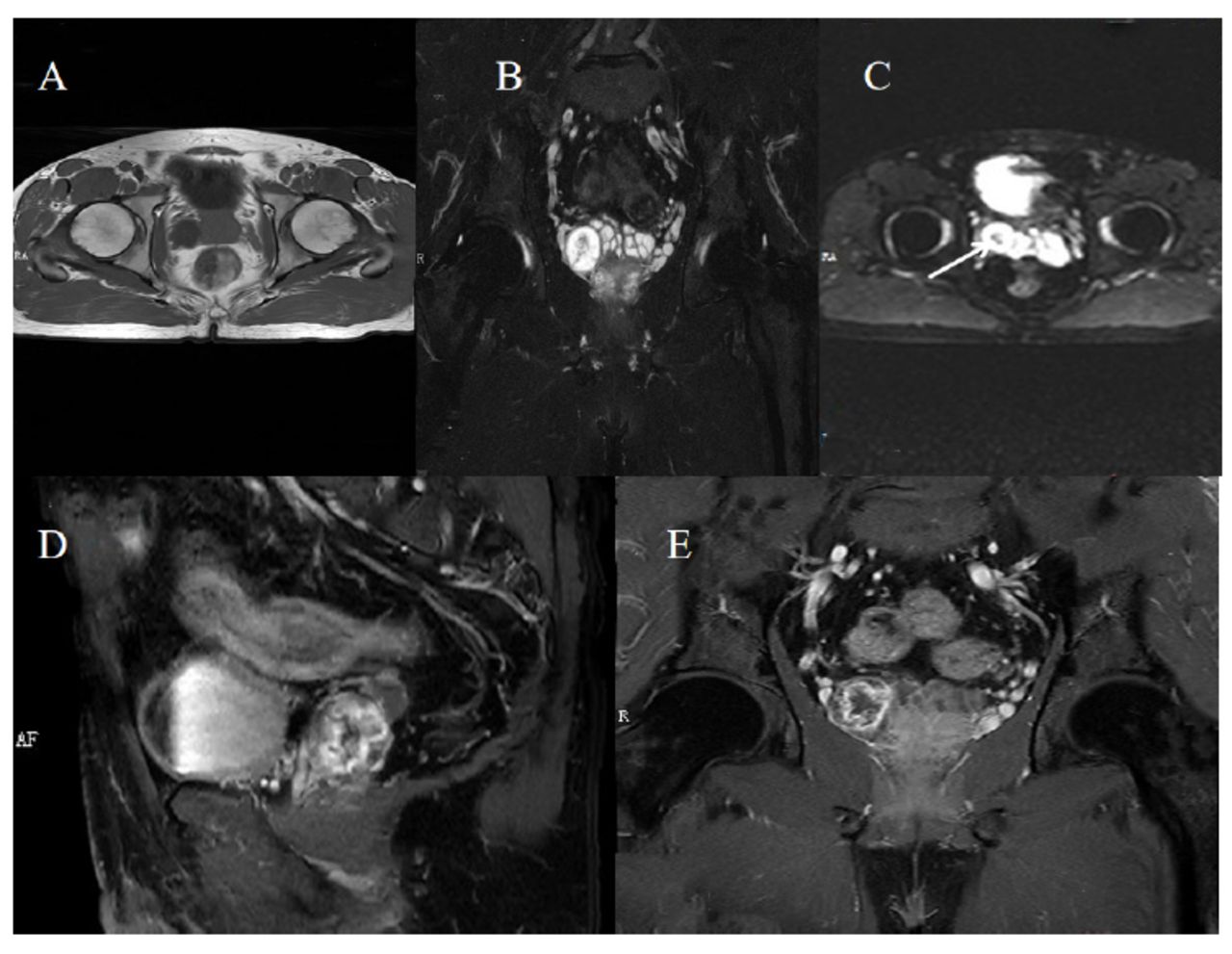
- Figure 1
- Contrast-enhanced MRI scan. A) Pelvic MRI transverse section T1 sequence. B) Coronal T2 sequence shows the linear, sheet-like low signal intensity of nerve fibers. C) The B50 sequence shows low signal intensity in the central area, suggesting hemorrhage. D and E) Contrast-enhanced scans of the T1 sequence show heterogeneous enhancement with intact capsule structure and punctate vascular-like enhancement.
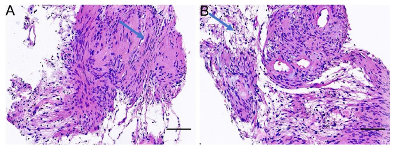
- Figure 2
- The HE-stained image of the lesion tissues. Arrows indicate the dense area A) and the sparse area (B). In the dense area, tumor cells were spindle-shaped with indistinct cell boundaries. The nuclei of tumor cells were elongated and arranged in a palisade pattern. The tumor cell nuclei were round and deeply stained in the sparse area, with no significant atypia, and mucinous degeneration was observed in small areas. Additionally, a few fibers and striated muscle tissues were visible in the puncture specimen. Scale bars are 100μm.
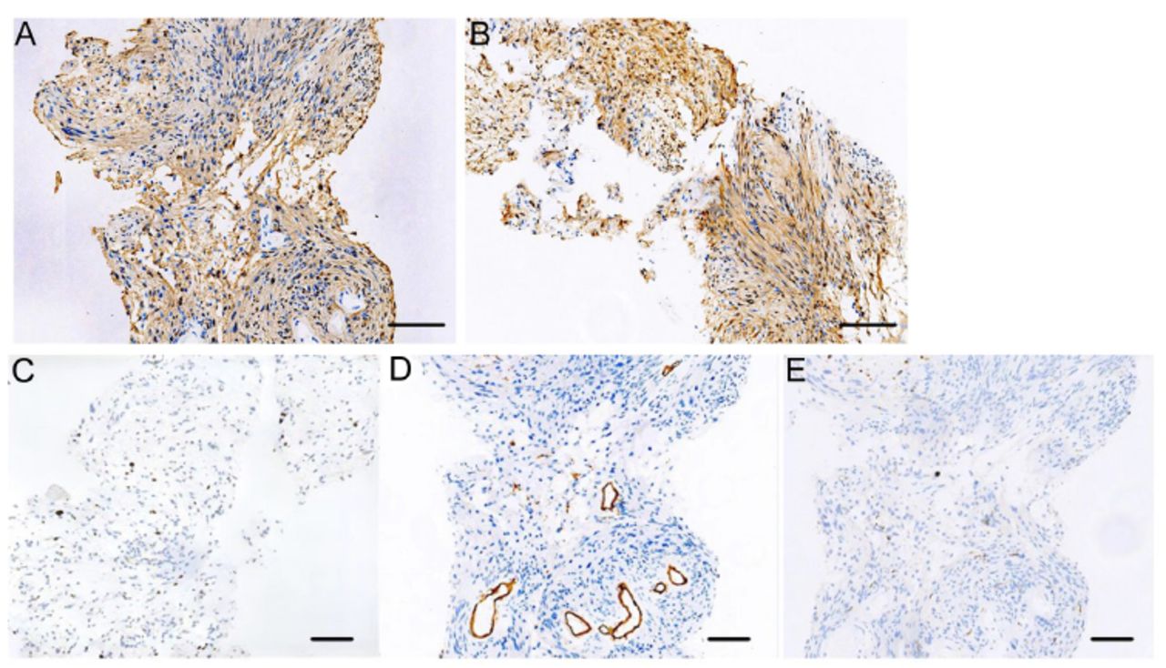
- Figure 3
- Immunohistochemical staining of the lesion tissues. The images demonstrate strong positive expression of S-100 A) and NSE B), a Ki67 index of 5% C) and a negative expression for CD34 D) and Desmin E) Scale bars are 100 μm.


Tables
Dates Relevant past medical history and interventions 2023.3.23 A 54-year-old man with no significant medical history, including drug allergies, genetic diseases, or psychiatric disorders. Date Summaries from initial and follow-up visits Diagnostic testing (including dates) Interventions 2023.3.23 Patient was incidentally found to have a right-sided seminal vesicle mass during a routine physical examination. A right-sided seminal vesicle mass was initially found by ultrasound. Subsequent contrast-enhanced CT scan revealed a lesion at the anterior margin of the right seminal vesicle gland. No special instructions. 2023.3.26 The patient reported no urinary symptoms. Concerned about disease progression, he sought care at our hospital MRI enhancement scan on 3.27 showed right seminal vesicle occupancy with hemorrhage; Puncture biopsy under ultrasound was performed on 3.28; Pathological examination on 3.30 diagnosed spindle cell soft tissue tumor in the seminal vesicle department, and Schwannoma was considered. No special medications are used, and surgery was recommended. 2023.4.1 The patient was subsequently referred to a Higher-level hospital for laparoscopic tumor resection. The diagnosis was confirmed postoperatively. He was discharged a week later. 2023.4.3 Laparoscopic resection of pelvic mass; 2023.4.7 postoperative pathologic examination confirmed the diagnosis of Seminal Vesicle Schwannoma. The patient underwent laparoscopic resection of the tumor with no complications.
In this issue


Jump to section
Related Articles
Cited By...
- No citing articles found.