


Article Figures & Data
Figures
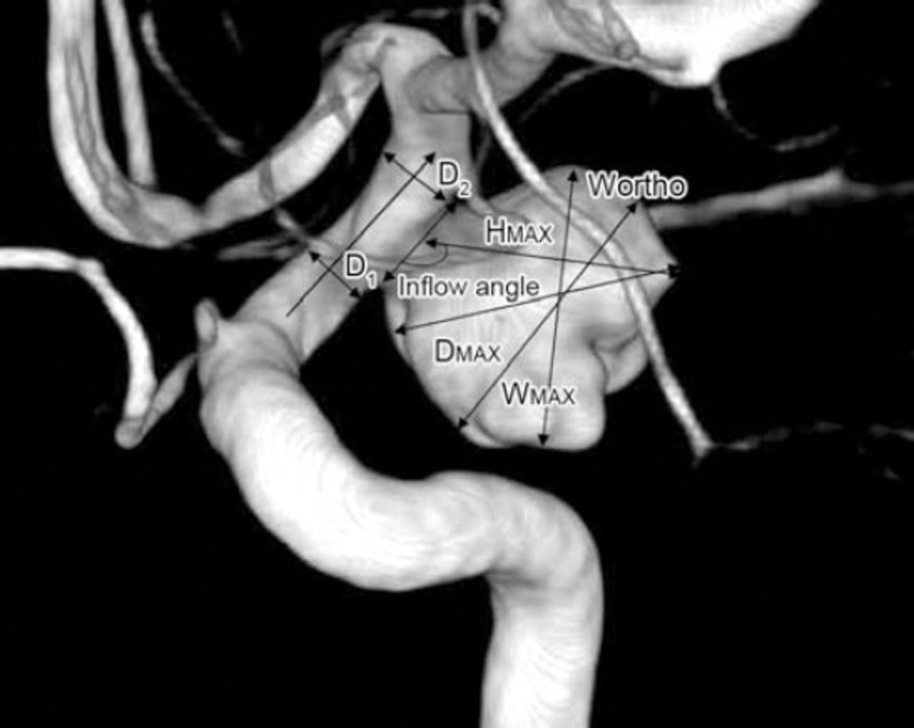
- Figure 1
Sidewall aneurysm measurements. Patient (male, 46 years) was hospitalized because of acute headache for one day. Head CT showed subarachnoid hemorrhage. Two days later 2 aneurysms in each carotid were found on digital subtraction angiography. The left carotid aneurysm is shown: maximal diameter (Dmax), longest dimension from the center of the neck to the dome tip (Hmax or height), maximal longitudinal diameter parallel with the neck plane (Wortho), dome width perpendicular to height (Wmax), the angle between axis of flow in the parent vessel at the level of the aneurysm neck and the aneurysm’s main axis from the center of the neck to the tip of the dome (inflow angle). The average value of both sides of the aneurysm neck=D1+D2/2 (average parent artery diameter).
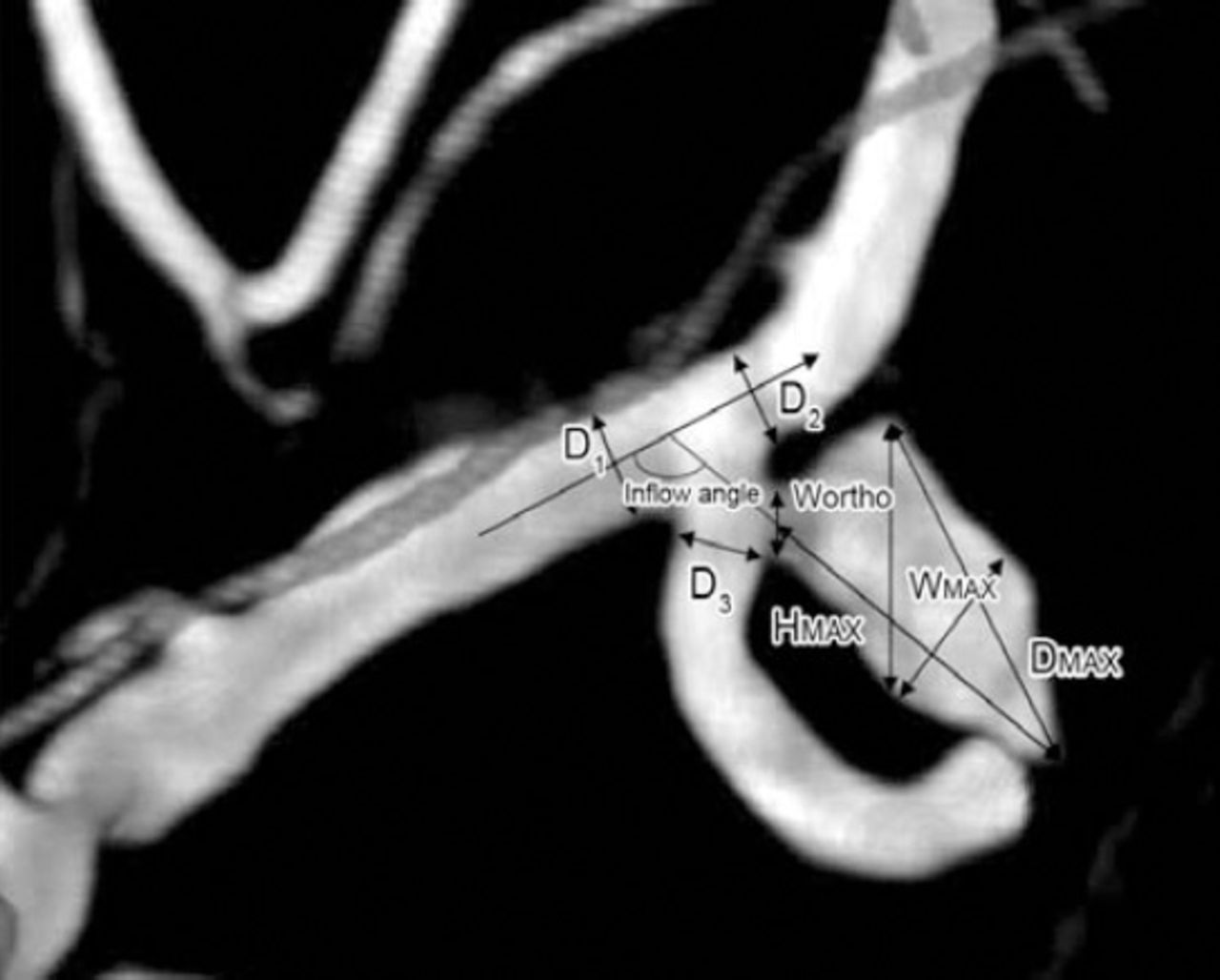
- Figure 2
Bifurcation aneurysm assessments. Patient (male, 66 years) was hospitalized because of acute headache for one day. Head CT showed subarachnoid hemorrhage. Two days later 2 aneurysms in the left middle cerebral artery and right posterior communicating artery respectively were found on digital subtraction angiography. The left middle cerebral aneurysm is shown: Maximal diameter (Dmax), longest dimension from the center of the neck to the dome tip (Hmax or Height), maximal longitudinal diameter parallel with the neck plane (Wortho), dome width perpendicular to height (Wmax), the angle between axis of flow in the parent vessel and the aneurysm’s main axis from the center of the neck to the tip of the dome (inflow angle). The average value of artery diameter=D1+D2+D3/3.


Tables
Variable Number (%) and type of aneurysms 1R:1UR 1R:2UR 1R:3UR Total Patients 27/34 (79.4) 6/34 (17.7) 1/34 (2.9) 34/34 (100) Age (mean±SD) 55.9±11.1 56.2±9.45 48 55.79±10.64 Male:Female 13:14 1:5 1:0 15:19 Hypertension 9/27 (33.3) 3/6 (50.0) 1/1 (100) 13 (38.2) Smoking 5/27 (18.5) 2/6 (33.3) 1/1 (100) 8 (23.5) ↵* Data presented as number of ruptured aneurysms (R) and unruptured aneurysms (U)
- Table 2
Comparisons of morphological aneurysm parameters between ruptured and unruptured aneurysms.*
Parameter Ruptured (34 aneurysms) Unruptured (42 aneurysms) Mann-Whitney U Z P-value Dmax (mm) 4.50 (4.14) 2.10 (2.38) 318.500 -4.136 0.000 Hmax (mm) 4.16 (3.07) 2.00 (1.55) 294.000 -4.396 0.000 Aspect ratio 1.95 (1.31) 1.05 (0.39) 293.500 -4.394 0.000 Bottleneck factor 1.72 (0.99) 1.03 (0.27) 413.500 -3.190 0.001 Bulge location 0.35 (0.50) 0.10 (0.40) 515.500 -2.163 0.031 Size ratio 2.12 (2.04) 0.83 (0.47) 157.500 -5.814 0.000 Height-width ratio 1.12 (0.75) 0.95 (0.13) 265.000 -4.695 0.000 Inflow angle (°) 129.50 (56.75) 90.00 (39.00) 326.500 -4.057 0.000 ↵* Data presented as median and (interquartile range)
- Table 3
Evaluation of the risk factors of size ratio and height-width ratio with aneurysm rupture by binary logistic regression analyses.
Variable B S.E. Wald P OR 95% CI Constant -5.346 1.264 17.887 0.001 0.005 Size ratio <1 13.983 0.001 ≥1 ≤1.6 2.392 0.872 7.520 0.006 10.931 1.98-60.39 >1.6 3.066 0.843 13.236 0.001 21.462 4.11-111.96 Height-width ratio ≤1 >1 2.192 0.690 10.105 0.001 8.954 2.32-34.60 - Table 4
Spearman correlation coefficients of morphological risk factors by bivariate correlation analysis among multiple cerebral aneurysm patients.
Bivariate correlations Hmax Aspect ratio Bottleneck factor Height-width ratio Bulge location Size ratio Inflow angle Dmax 0.942† (0.958†) 0.627† (0.226) 0.438† (0.408†) 0.354* (-0.140) 0.157 (0.328*) 0.726† (0.761†) -0.002 (0.271) Hmax - 0.574† (0.256) 0.520† (0.466†) 0.359* (-0.150) 0.111 (0.325*) 0.728† (0.718†) 0.072 (0.314*) Aspect ratio - - 0.546† (0.634†) 0.643† (0.479†) 0.308 (0.538†) 0.684† (0.309*) 0.044 (-0.001) Bottleneck factor - - - 0.507† (0.007) 0.065 (0.722†) 0.540† (0.429†) 0.035 (0.148) Height-width ratio - - - - 0.017 (0.155) 0.542† (-0.019) -0.034 (-0.010) Bulge location - - - - - 0.186 (0.491†) -0.153 (0.124) Size ratio - - - - - - 0.261 (0.200) ↵* p<0.05,
↵† p<0.01. Data with and without brackets represents spearman correlation coefficients of morphological risk factors in unruptured and ruptured aneurysms; correlation coefficient (R) ≥0.7 indicates strong correlation; 0.7-0.4 indicates moderate correlation, and <0.4 indicates weak correlation
In this issue


Jump to section
Related Articles
Cited By...
- No citing articles found.