
Main menu

  • Home
  • Content
    • Latest
    • Ahead of print
    • Archive
  • Info for
    • Authors
    • Reviewers
    • Subscribers
    • Institutions
    • Advertisers
  • About Us
    • About Us
    • Editorial Office
    • Editorial Board
  • More
    • Alerts
    • Feedback
    • Folders
    • Help
  • Other Publications
    • Saudi Medical Journal

User menu

  • My alerts
  • Log in

Search

  • Advanced search
Neurosciences Journal
  • Other Publications
    • Saudi Medical Journal
  • My alerts
  • Log in
Neurosciences Journal

Advanced Search

  • Home
  • Content
    • Latest
    • Ahead of print
    • Archive
  • Info for
    • Authors
    • Reviewers
    • Subscribers
    • Institutions
    • Advertisers
  • About Us
    • About Us
    • Editorial Office
    • Editorial Board
  • More
    • Alerts
    • Feedback
    • Folders
    • Help
  • Follow psmmc on Twitter
  • Visit psmmc on Facebook
  • RSS
Case ReportCase Report
Open Access

Incidentally diagnosed giant invasive sacral schwannoma

Its clinical features and surgical management without stability

Guray Togral, Murat Arikan, Askin E. Hasturk and Safak Gungor
Neurosciences Journal July 2014, 19 (3) 224-228;
Guray Togral
From the Departments of Orthopedics and Traumatology (Togral, Arikan, Gungor), and Neurosurgery (Hasturk), Oncology Training and Research Hospital, Ankara, Turkey
MD
  • Find this author on Google Scholar
  • Find this author on PubMed
  • Search for this author on this site
Murat Arikan
From the Departments of Orthopedics and Traumatology (Togral, Arikan, Gungor), and Neurosurgery (Hasturk), Oncology Training and Research Hospital, Ankara, Turkey
MD
  • Find this author on Google Scholar
  • Find this author on PubMed
  • Search for this author on this site
Askin E. Hasturk
From the Departments of Orthopedics and Traumatology (Togral, Arikan, Gungor), and Neurosurgery (Hasturk), Oncology Training and Research Hospital, Ankara, Turkey
MD
  • Find this author on Google Scholar
  • Find this author on PubMed
  • Search for this author on this site
  • For correspondence: [email protected]
Safak Gungor
From the Departments of Orthopedics and Traumatology (Togral, Arikan, Gungor), and Neurosurgery (Hasturk), Oncology Training and Research Hospital, Ankara, Turkey
MD
  • Find this author on Google Scholar
  • Find this author on PubMed
  • Search for this author on this site
  • Article
  • Figures & Data
  • eLetters
  • Info & Metrics
  • References
  • PDF
Loading

Article Figures & Data

Figures

  • Tables
  • Figure 1
    • Download figure
    • Open in new tab
    • Download powerpoint
    Figure 1

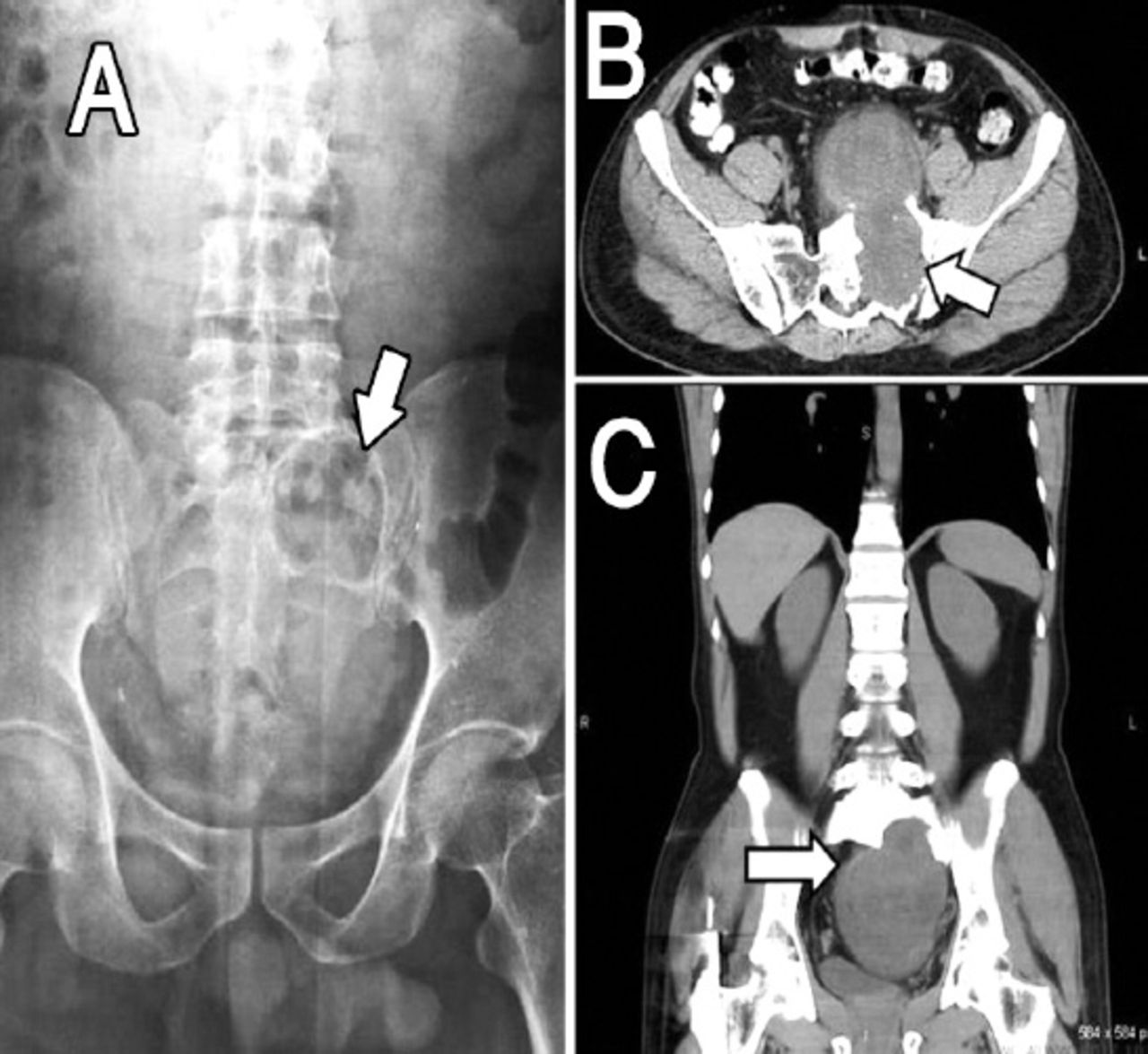
    Preoperative images showing: A) Patient x-ray showing a sclerotic margined, smooth contoured cavity lesion invading the left half of the sacrum. B) Axial CT showing lobulated contoured hypo dense heterogeneous mass that has destructed the neural foramens, filling the pelvis and displacing the bladder anteriorly. C) Coronal CT cross section showing a hyperdense sclerotic lobulated contoured mass, from the left half of the sacrum and filling the pelvis.

  • Figure 2
    • Download figure
    • Open in new tab
    • Download powerpoint
    Figure 2

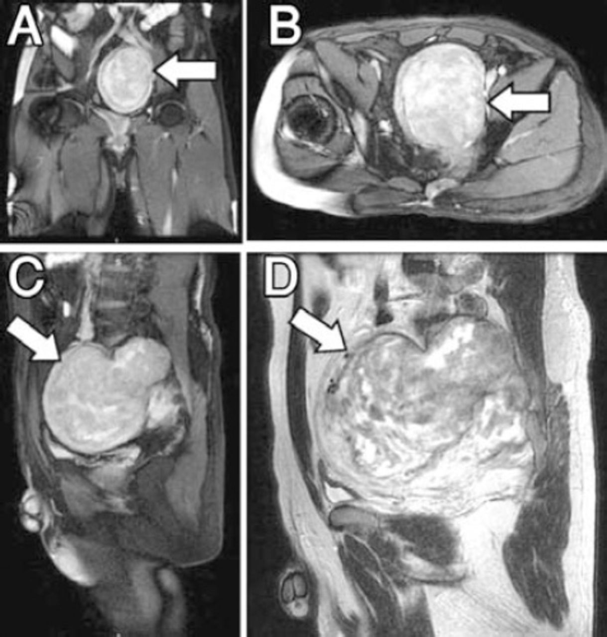
    Preoperative MRI sections showing: A) T2 weighted coronal MRI showing hyperintense lobulated mass filling the pelvis. B) T2 weighted axial MRI showing hyperintense mass with its largest diameter 138 × 91 mm. C) T2 weighted sagittal MRI showing a dense heterogeneous opaque mass with agent uptake, sagittal 146 × 114 mm sized mass invading the left sacral spinal canal. D) T1 weighted sagittal MRI showing opaque involvement belonging to calcified and necrotic areas inside the lesion.

  • Figure 3
    • Download figure
    • Open in new tab
    • Download powerpoint
    Figure 3

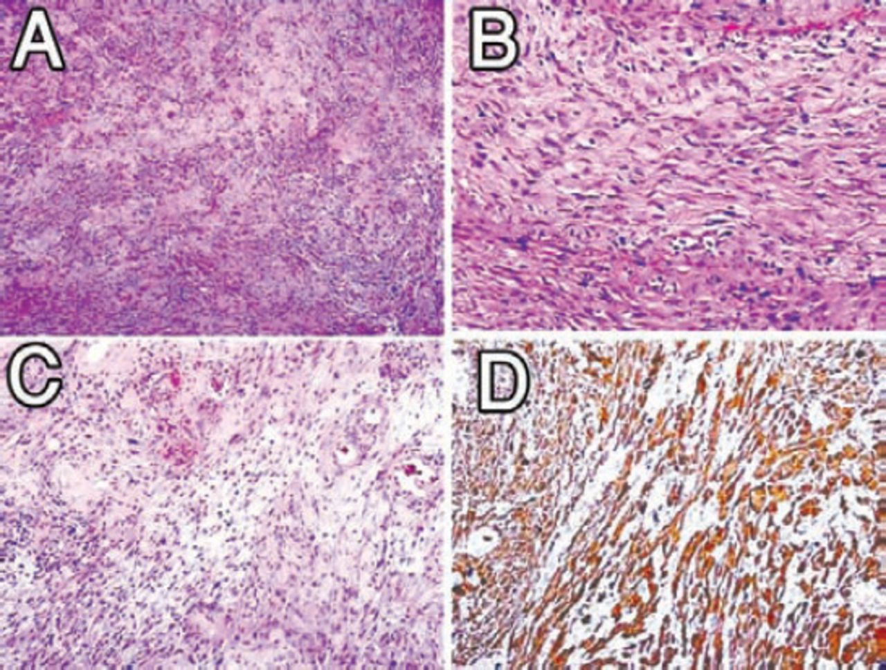
    Histopathological examination of specimens: A) Irregular fascicles of spindle cells, nuclear palisading, Hematoxylin & Eosin (HE) × 100. B) Cellular detail. Oval blunt-ended or elongated nuclei, HE × 400. C) Hyalinized blood vessels and relatively myxoid stroma, HE × 200. D) Diffuse S100 protein positivity, HE × 400.

  • Figure 4
    • Download figure
    • Open in new tab
    • Download powerpoint
    Figure 4

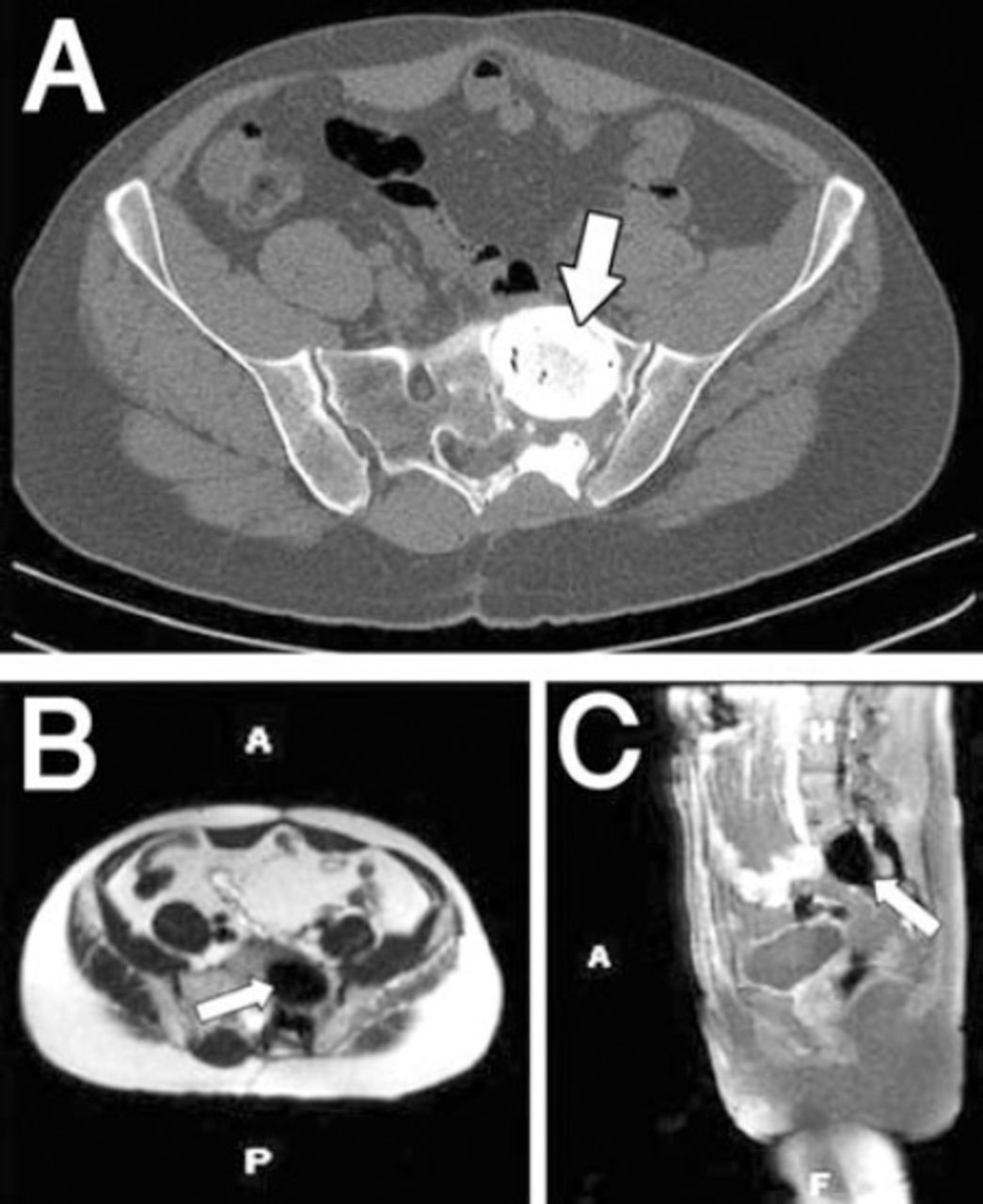
    There was no recurrence detected on MRI in the postoperative first year. A) Axial CT, B) Axial MRI, C) Sagittal MRI. A - anterior, P - posterior, H - head, F - foot

Tables

  • Figures
    • View popup
    Table 1

    Modified Sridhar classification of benign nerve sheath tumors.5

    Original types
    Type IIntraspinal tumor <2 vertebral segments in length; a: intradural; b: extradural
    Type IIIntraspinal tumor >2 vertebral segments in length (giant tumor)
    Type IIIIntraspinal tumor with extension into nerve root foramen
    Type IVIntraspinal tumor with extraspinal extension (dumbbell tumors); a: extraspinal component <2.5 cm; b: extraspinal component >2.5 cm (giant tumor)
    Type VTumor with erosion into vertebral bodies (giant invasive tumor), lateral and posterior extensions into myofascial planes
    Additional types for spinal intraosseous schwannoma
    Type VITumor in entirely intravertebral location without intraspinal portion
    Type VIIIntraspinal tumor with erosion into vertebral bodies (invasive tumor) and extension into nerve root foramen
PreviousNext
Back to top

In this issue

Neurosciences Journal: 19 (3)
Neurosciences Journal
Vol. 19, Issue 3
1 Jul 2014
  • Table of Contents
  • Cover (PDF)
  • Index by author
Print
Download PDF
Email Article

Thank you for your interest in spreading the word on Neurosciences Journal.

NOTE: We only request your email address so that the person you are recommending the page to knows that you wanted them to see it, and that it is not junk mail. We do not capture any email address.

Enter multiple addresses on separate lines or separate them with commas.
Incidentally diagnosed giant invasive sacral schwannoma
(Your Name) has sent you a message from Neurosciences Journal
(Your Name) thought you would like to see the Neurosciences Journal web site.
Citation Tools
Incidentally diagnosed giant invasive sacral schwannoma
Guray Togral, Murat Arikan, Askin E. Hasturk, Safak Gungor
Neurosciences Journal Jul 2014, 19 (3) 224-228;

Citation Manager Formats

  • BibTeX
  • Bookends
  • EasyBib
  • EndNote (tagged)
  • EndNote 8 (xml)
  • Medlars
  • Mendeley
  • Papers
  • RefWorks Tagged
  • Ref Manager
  • RIS
  • Zotero
Share
Incidentally diagnosed giant invasive sacral schwannoma
Guray Togral, Murat Arikan, Askin E. Hasturk, Safak Gungor
Neurosciences Journal Jul 2014, 19 (3) 224-228;
Twitter logo Facebook logo Mendeley logo
  • Tweet Widget
  • Facebook Like
  • Google Plus One
Bookmark this article

Jump to section

  • Article
    • Abstract
    • Case Report
    • Discussion
    • Acknowledgments
    • Footnotes
    • References
  • Figures & Data
  • eLetters
  • References
  • Info & Metrics
  • PDF

Related Articles

  • Intradural lumbar cystic schwannoma
  • How often is low back pain or sciatica not due to lumbar disc disease?
  • Google Scholar

Cited By...

  • No citing articles found.
  • Google Scholar

More in this TOC Section

  • Unmasking the mimic: Leprosy neuropathy misdiagnosed as chronic inflammatory demyelinating polyneuropathy: A case report from Saudi Arabia
  • Seminal vesicle schwannoma with chronic hemorrhage
  • Marchiafava-Bignami disease post-bariatric surgery: A case report and review of similar cases
Show more Case Report

Similar Articles

Navigate

  • home

More Information

  • Help

Additional journals

  • All Topics

Other Services

  • About

© 2025 Neurosciences Journal Neurosciences is copyright under the Berne Convention and the International Copyright Convention. All rights reserved. Neurosciences is an Open Access journal and articles published are distributed under the terms of the Creative Commons Attribution-NonCommercial License (CC BY-NC). Readers may copy, distribute, and display the work for non-commercial purposes with the proper citation of the original work. Electronic ISSN 1658-3183. Print ISSN 1319-6138.

Powered by HighWire