


Article Figures & Data
Figures
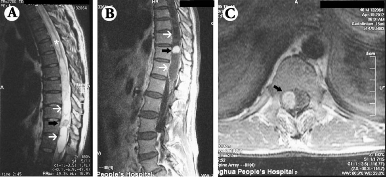
- Figure 1
Patient MRI showing: A) T2WI showed 2 hyperintense lesions (white arrows) separated by a hypointense nodule (black arrow) from T9 to T12 levels. The syrinx extending the whole thoracic segments was shown (asterisk). B) T1WI enhancement showed homogeneous enhancement of the middle nodule, while its bipolar lesions were not enhanced. C) Axial enhanced scan showed an enhanced lesion in the spinal canal (black arrow).
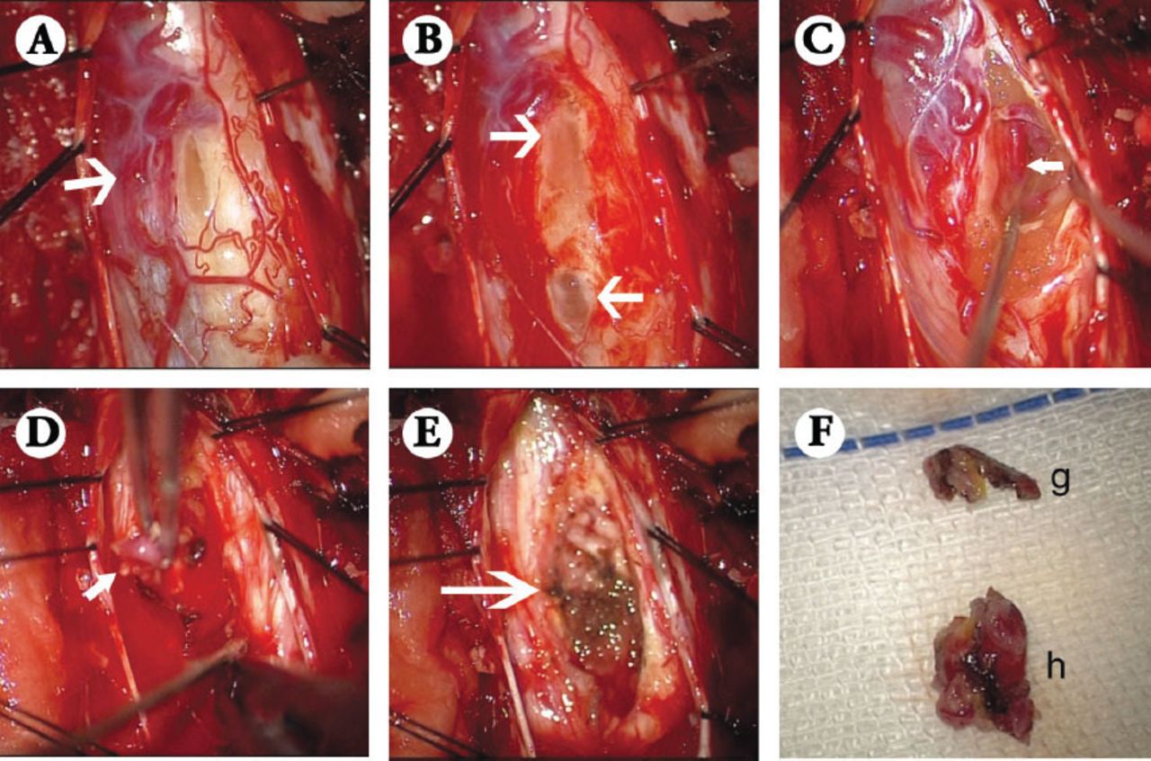
- Figure 2
Microsurgical view of the lesions during operation showing: A) Large draining veins on the dorsal and dorsolateral spinal cord surface were seen with the opening cyst after dural opening. B) Two separated cysts were opened, and light yellow fluid flowed out. C) A large vascular anomaly was exposed deeper inside the spinal cord (white arrow). D) The vascular conglomeration was resected in the spinal cord (white arrow). E) Cauterized and Gelfoam placement for hemostasis after successful resection of the tumors. F) Resected specimens for pathological studies, one was part of the cystic capsules (g), and the other was the vascular conglomeration that measured approximately 0.5×0.5×0.5cm in size (h).
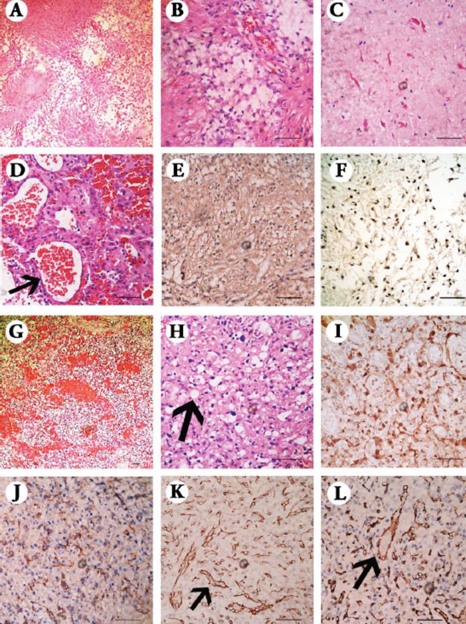
- Figure 3
Microscopic view of the tumor with Hematoxylin and Eosin and immunohistochemical stainings showing: A) Biphasic pattern with varying proportions of compacted areas and loose-textured areas (×100). B) Biphasic pattern with compact portions yielding bipolar piloid cells, while the loose-textured areas possessed round to oval, cytologically bland nuclei and relatively short, cobweb-like processes, which are fibril-poor (×400). C) The Rosenthal fibers were observed as tapered corkscrew-shaped, brightly eosinophilic, hyaline masses (×400). D) The abundant glomeruloid vasculature (×400). E) The glial fibrillary acidic protein was positive in the compact region while negative in the loose-textured area (×400). F) The p53 was positive in the area of pilocytic astrocytoma (×400). G) Two components of stromal cells and abundant vascular cells (×100). H) Stromal cells that were characterized by large lipid-containing vacuoles (×400). I) S-100 was positive mainly in the area of stromal cells (×400). J) CD56 was positive mainly in the area of stromal cells (×400). K) the VIII factor was mainly positive in the area rich in endothelial cells (×400). L) CD31 was mainly positive in the area rich in endothelial cells (×400).
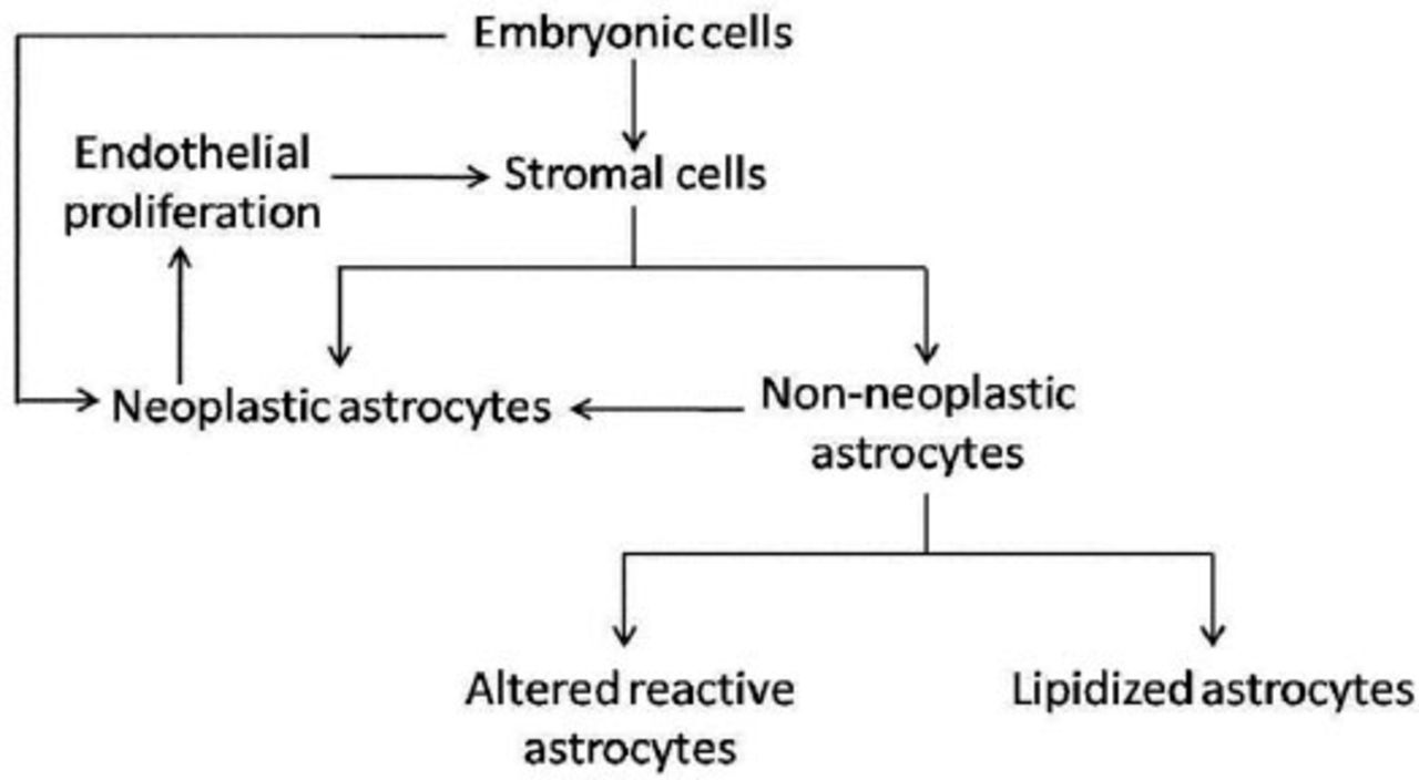
- Figure 4
The illustration of hypotheses on the association of hemangioblastoma and pilocytic astrocytoma.


In this issue


Jump to section
Related Articles
Cited By...
- No citing articles found.