


Article Figures & Data
Figures
- Figure 1
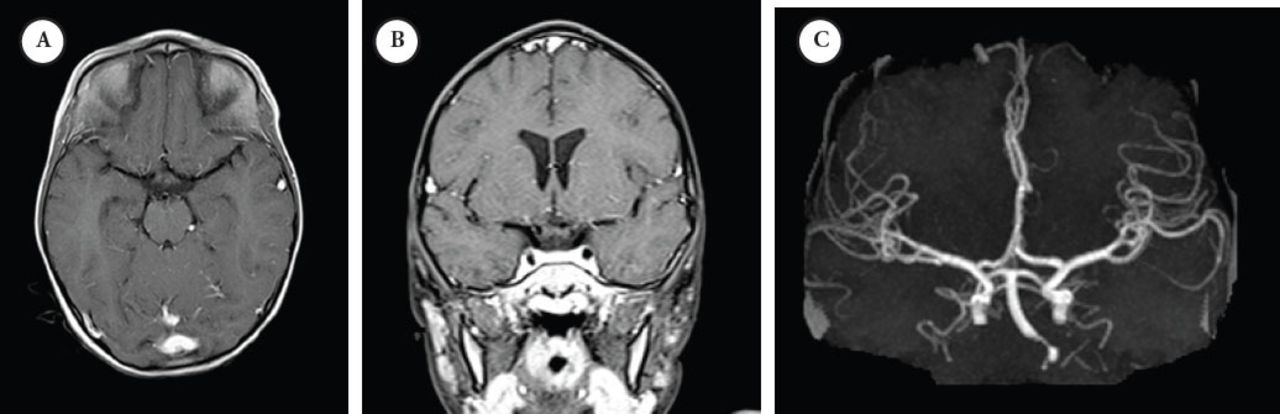
Initial presentation study A) post-contrast T1-wighted MRI and B) at the level of the Circle of Willis and maximum-intensity projection reconstruction MRI image C) demonstrating normal flow void and vascular anatomy at the circle of Willis, particularly at the left ICA and MCA. MCA - middle cerebral arteries, ICA - internal carotid artery
- Figure 2
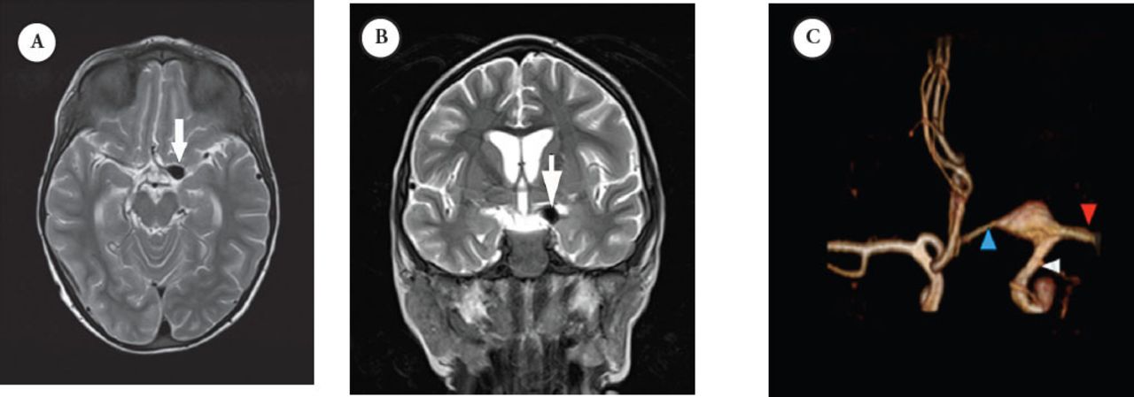
Second presentation MRI study: selected axial A) and coronal B) T2-weighted MRI at the region of circle of Willis showing an aneurysm at the junction between the distal ICA, M1-segment of MCA and the A1-segment of ACA, demonstrated as a flow void. This was well demonstrated with a C) 3D-reconstruction of the MRA. ICA - internal carotid artery, MCA -middle cerebral arteries, ACA - anterior and middle cerebral arteries
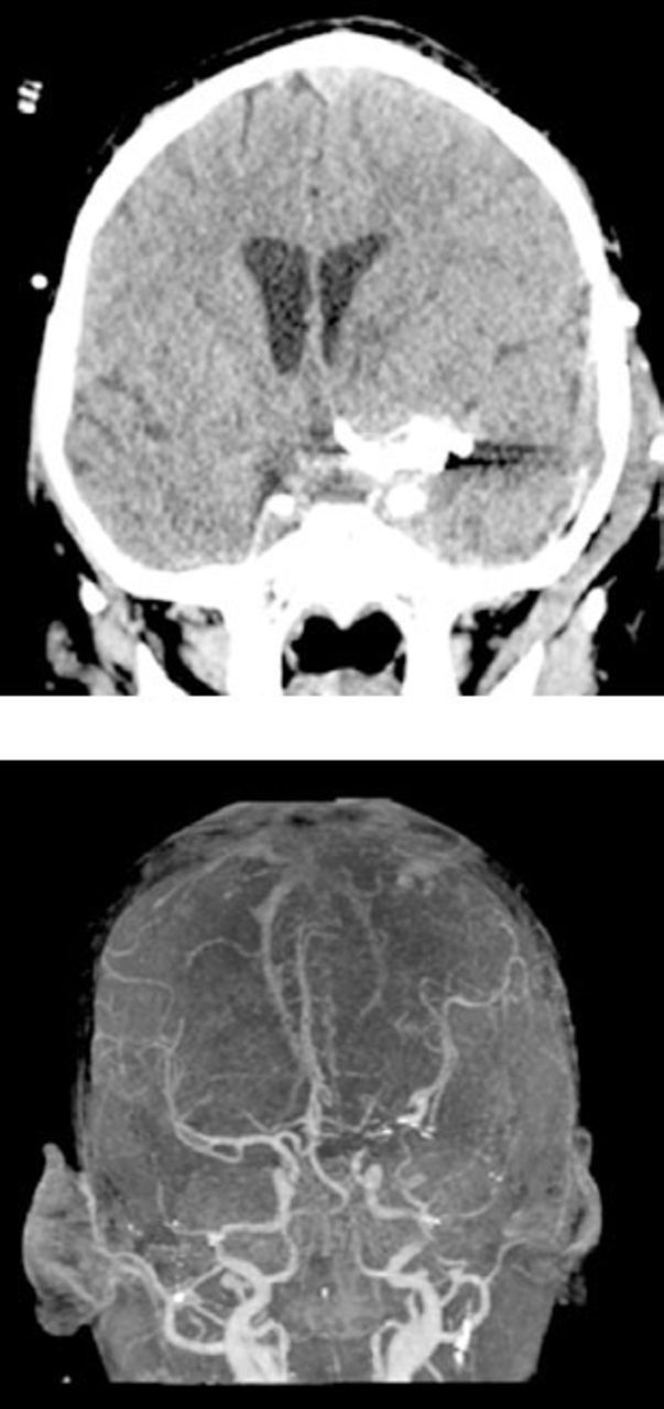
- Figure 3
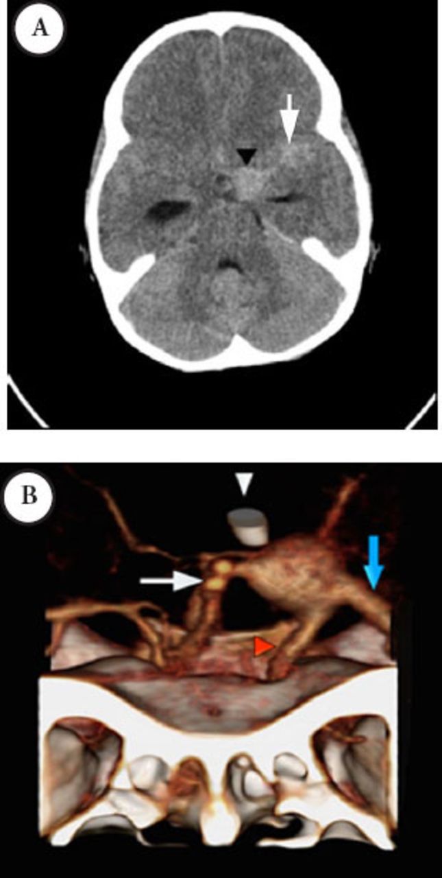
Third presentation CT study selected axial non-contrast enhanced CT of the brain A) showing interval increase in the dimensions of the aneurysm and SAH due rupture of the aneurysm. There was hydrocephalus seen as marked dilatation of the temporal horns of the temporal horns of the lateral ventricles. 3D-reconstruction of a contrast-enhanced CTA B) after insertion of EVD, demonstrating the large fusiform aneurysm at the junction between the distal ICA, M1-segment of MCA and the A1- segment of ACA. The bony landmarks of the cranial fossa and the anterior cloned process are also shown. MCA - middle cerebral arteries, ICA - internal carotid artery, EVD - external ventricular drain, ACA- anterior and middle cerebral arteries, CTA - CT angiography, SAH - subarachnoid hemorrhage
- Figure 4
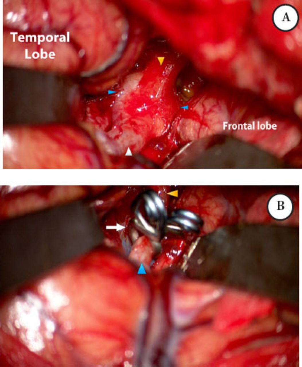
Intraoperative microscopic photograph through A) left frontotemporal craniotomy and transsylvian approach demonstrating the extent of the aneurysm, B) Repair of the fusiform ICA/MCA aneurysm was reconstructed with 2 fenestrated aneurysm clips. MCA - middle cerebral arteries, ICA - internal carotid artery.
- Figure 5
Selected coronal non-contrast CT image of the brain A) showing the clip applied across the aneurysm. Maximum-intensity projection reconstruction B) and 3D reconstruction of the postoperative contrast-enhanced MRA, demonstrates the reconstruction of the aneurysm and the patent distal circulation.


Tables
Year /No. of cases Age at aneurysm diagnosis (years)/gender/HIV source Presentation Radiological modality/Radiological findings/vessels involved Management Outcome Kure et al, 198914 6 yrs/male/congenital Aphasia and quadriplegia Fusiform dilatation of circle of Willis NS Death after 25 days in the hospital Lang et al, 199119 8 yrs/male/congenital NS Fusiform dilatations of left ICA, MCA, and ACA AZT 5 years before CNS event Died after one month secondary to cardiomyopathy Husson et al, 199213 1 11 yrs/male/blood transfusion Asymptomatic Multiple saccular aneurysms of right MCA and fusiform aneurysm of left ACA and MCA DDI one year prior to the presentation changed to AZT after aneurysm diagnosis CNS symptoms after 16 months 2 12 yrs/male/blood transfusion Asymptomatic multiple aneurysmal dilatations of the right MCA and ACA DDI was continued after the diagnosis Remained asymptomatic 3 12 yrs/female/congenital Fever, lethargy, seizure and upper limbs weakness Multiple fusiform ICA, ACA, MCA and right PCA aneurysms NS Death within 2 months of presentation Dubrovsky et al, 19982 1 13 yrs/male/uncertain Acute right hemiparesis Fusiform dilatations of left supraclinoid ICA and proximal ACA and MCA AZT 18 months prior to the presentation, continued after diagnosis of the aneurysm Death after 3 months secondary to cerebral infarction 2 11 yrs/female/congenital Acute change in mental status Fusiform aneurysms of left ICA and proximal MCA and bilateral PCA AZT started one year prior to the presentation Death after 6 months secondary to infection 3 6 yrs/female/Congenital Seizure Multiple fusiform aneurysms of the right ICA and proximal right MCA AZT since 2-year-old. Radiotherapy for brain lymphoma Neurological status worsened gradually. Died later secondary to HIV-enteropathy 4 12 yrs/male/blood transfusion Headache and left-sided weakness Fusiform aneurysms of left ICA, MCA, and ACA ART (DDI 2 years before the presentation Multiple opportunistic infections after the presentation died after 36 months 5 10 yrs/male/perinatal Collapsed at school Not carried out DDI started 2 years prior to the presentation Death at presentation Fulmer et al, 19985 11 yrs/female/congenital Asymptomatic Multiple ICA, MCA, and ACA fusiform aneurysms Observation Death after 3 years was secondary to other AIDs complications Mazzoni et al, 200015 8 yrs/female/congenital Sudden loss of consciousness followed by seizure and mild hemiparesis multiple saccular and fusiform aneurysms in the proximal arteries, predominantly, in the left MCA AZT was replaced with DDI at the age of 3 years. HAART (AZT, 3TC, RTV) was initiated after aneurysm diagnosis Patient recovered after one month. After 4 months, repeated MRA showed no changes in the aneurysms Carvalho neto et al, 20016 6 yrs/male/congenital Seizure Aneurysmal dilatation of circle of Willis, more in the right. AZT and DDI were started at the age of 2 years. Supportive management for the CNS event The seizures stopped, and the patient was discharged with follow up in the clinic Nunes et al, 20013 2 months old/female/congenitally-acquired Right hemiparesis and coma Saccular aneurysm of left basilar artery. IV AZT at birth. She was on AZT and DDI at presentation. Conservative management for SAH Death after 12 days of SAH Patsalides et al, 200220 7 cases, (7yrs – 15 yrs), 5 males and 2 females, 4 perinatal and 3 through blood transfusion NS Aneurysms involved ICA, MCA, ACA, PCA, basilar and vertebral arteries All were taking ART before the diagnosis of aneurysmal arteriopathy 2 cases were alive through out the observation period; 4 cases died secondary to other AIDS-related complications and a single case died secondary to CNS complications Petropolous et al, 200317 12 yrs/male/NS Asymptomatic Multiple saccular aneurysms of proximal right ICA and proximal basilar artery Continued the earlier started HAART (AZT, 3TC, NVP) with follow-up of the aneurysms After 2 years follow-up, no new CNS symptoms, and MRI showed no changes Martinez-Longoria et al, 20041 12 yrs/female/congenital Headache, transient left hemiparesis, blurred vision Left ICA obstruction, stenosis in infraclinoid right ICA and fusiform dilatations of the right ICA bifurcation extends to right MCA and ACA Initially on AZT. SQV and 3TC were started 3 month before CNS event. HAART regime (Kaletra® (LPV/RTV), 3TC and D4T) and aspirin was started after the CNS event MRI after 15 months showed complete resolution of the aneurysm. No neurological event was occurred over 2 years follow-up Mahadevan et al, 200716 16 yrs/male/not known Headache, vomiting, slurred speech and weakness Fusiform dilatations of bilateral vertebral, basilar artery, right ICA, and MCA A twist drill and ventricular tap did reduce the intracranial pressure Death within hours of presentation Thakker and Bhatia 200910 12 yrs/male/congenital Headache and right hemiparesis Fusiform aneurysms of the left supraclinoid ICA ART 4 months prior to the presentation. Conservative management of the aneurysm No reported the death or other CNS manifestations Demopolous et al, 200918 1 12 yrs/male/NS Right hemiparesis Fusiform right ICA aneurysm ART for 5 years prior to the presentation Complete neurological recovery 2 6 yrs/female/NS Left hemiparesis Fusiform dilatations of right ICA, right vertebral, and basilar artery ART for 6 months prior to the presentation The residual weakness with neurocognitive impairment 3 7 yrs/Male/NS Right hemiparesis Multiple aneurysms of vessels of the circle of Willis Did not receive ART Death Savitr sastri et al, 20118 13 yrs/Male/NS Aphasia and right hemiplegia Fusiform dilatations of bilateral supraclinoid ICAs External ventricular drain inserted Death within hours of presentation Schieffelin et al, 20134 1 7 HIV-infected pediatric cases were reported to have cerebral aneurysm with no specific demographic CNS symptoms Aneurysmal dilatation of arteries in the circle of Willis DDI at the time of CNS event Alive 2 blurred vision Multiple aneurysms, multiple infarcts HAART at the time of CNS event Died 3 seizures, right hemiparesis Internal carotid artery aneurysm HAART at the time of CNS event Died 4 CNS symptoms Aneurysm of left ACA and narrowing of right MCA HAART at the time of CNS event Alive 5 Hemiplegia Fusiform CNS aneurysm HAART at the time of CNS event Alive 6 CNS symptoms Multiple CNS aneurysms HAART at the time of CNS event Alive 7 CNS symptoms Diffuse CNS aneurysms with ectasia D4T, NVP at the time of CNS event Died Current Case 7 yrs/Male/Congenital Headache and altered level of consciousness fusiform aneurysm at left ICA bifurcation, and proximal ACA and MCA Clipping of the aneurysm. Started on HAART before discharge Discharged in a stable neurological condition to be followed up in the clinic
In this issue


Jump to section
Related Articles
Cited By...
- No citing articles found.