
Main menu

  • Home
  • Content
    • Latest
    • Ahead of print
    • Archive
  • Info for
    • Authors
    • Reviewers
    • Subscribers
    • Institutions
    • Advertisers
  • About Us
    • About Us
    • Editorial Office
    • Editorial Board
  • More
    • Alerts
    • Feedback
    • Folders
    • Help
  • Other Publications
    • Saudi Medical Journal

User menu

  • My alerts
  • Log in

Search

  • Advanced search
Neurosciences Journal
  • Other Publications
    • Saudi Medical Journal
  • My alerts
  • Log in
Neurosciences Journal

Advanced Search

  • Home
  • Content
    • Latest
    • Ahead of print
    • Archive
  • Info for
    • Authors
    • Reviewers
    • Subscribers
    • Institutions
    • Advertisers
  • About Us
    • About Us
    • Editorial Office
    • Editorial Board
  • More
    • Alerts
    • Feedback
    • Folders
    • Help
  • Follow psmmc on Twitter
  • Visit psmmc on Facebook
  • RSS
Case ReportCase Report
Open Access

A rare case of autoimmune limbic encephalitis: an uncharted territory!

Hatim Ibrahim, Abdulelah N. Al Jasser, Sonia A. Khan and Kalthoum G. Tlili
Neurosciences Journal October 2017, 22 (4) 292-297; DOI: https://doi.org/10.17712/nsj.2017.4.20170150
Hatim Ibrahim
From the Department of Neurology (Khan, Ibrahim), Department of Radiology (Tlili), Prince Sultan Military Medical City, and from the Faculty of Medicine (Aljasser), King Saud University, Riyadh, Kingdom of Saudi Arabia
MD
  • Find this author on Google Scholar
  • Find this author on PubMed
  • Search for this author on this site
Abdulelah N. Al Jasser
From the Department of Neurology (Khan, Ibrahim), Department of Radiology (Tlili), Prince Sultan Military Medical City, and from the Faculty of Medicine (Aljasser), King Saud University, Riyadh, Kingdom of Saudi Arabia
MD
  • Find this author on Google Scholar
  • Find this author on PubMed
  • Search for this author on this site
Sonia A. Khan
From the Department of Neurology (Khan, Ibrahim), Department of Radiology (Tlili), Prince Sultan Military Medical City, and from the Faculty of Medicine (Aljasser), King Saud University, Riyadh, Kingdom of Saudi Arabia
MD, FRCP (Edin)
  • Find this author on Google Scholar
  • Find this author on PubMed
  • Search for this author on this site
  • For correspondence: [email protected]
Kalthoum G. Tlili
From the Department of Neurology (Khan, Ibrahim), Department of Radiology (Tlili), Prince Sultan Military Medical City, and from the Faculty of Medicine (Aljasser), King Saud University, Riyadh, Kingdom of Saudi Arabia
MD, FBR
  • Find this author on Google Scholar
  • Find this author on PubMed
  • Search for this author on this site
  • Article
  • Figures & Data
  • eLetters
  • Info & Metrics
  • References
  • PDF
Loading

Article Figures & Data

Figures

  • Figure 1
    • Download figure
    • Open in new tab
    • Download powerpoint
    Figure 1

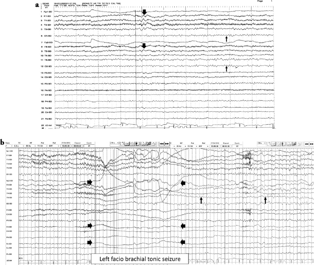
    EEG features a) Interictal EEG showed normal EEG background of 8-9 hertz bilaterally (thin arrows). Intermixed slow transients of 6-7 hertz in the temporal regions bilaterally (thick arrows). No epileptiform discharges seen, b) The ictal EEG showed periods of right hemispheric electro-decremental response with right hemispheric alpha frequency attenuation at FP2, F4, T2, T4, T6, P4 & O2 with some diffusion to the left for 4 seconds time locked with left facio brachial tonic seizure (thick arrows) followed by recovery of the EEG background intermixed with right temporal slow transients of 4-5 hertz at T2, T4 and T6 (thin arrows).

  • Figure 2
    • Download figure
    • Open in new tab
    • Download powerpoint
    Figure 2

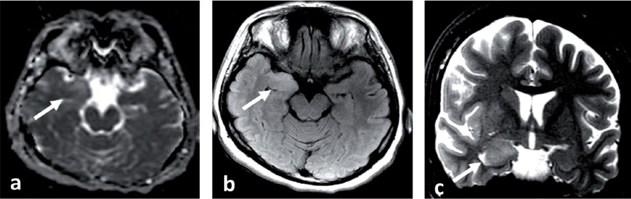
    MRI brain at presentation a) Axial apparent diffusion coefficient (ADC) map, b) Axial fluid attenuated inversion recovery (FLAIR) and c) Coronal T2 spin echo (T2SE) images showing abnormal high T2 and FLAIR signal intensity and swelling with blurring of the margins of right amygdala and medial right temporal lobe cortex and increased diffusivity associated on ADC map (White arrows). No abnormal enhancement (post contrast images not shown).

  • Figure 3
    • Download figure
    • Open in new tab
    • Download powerpoint
    Figure 3

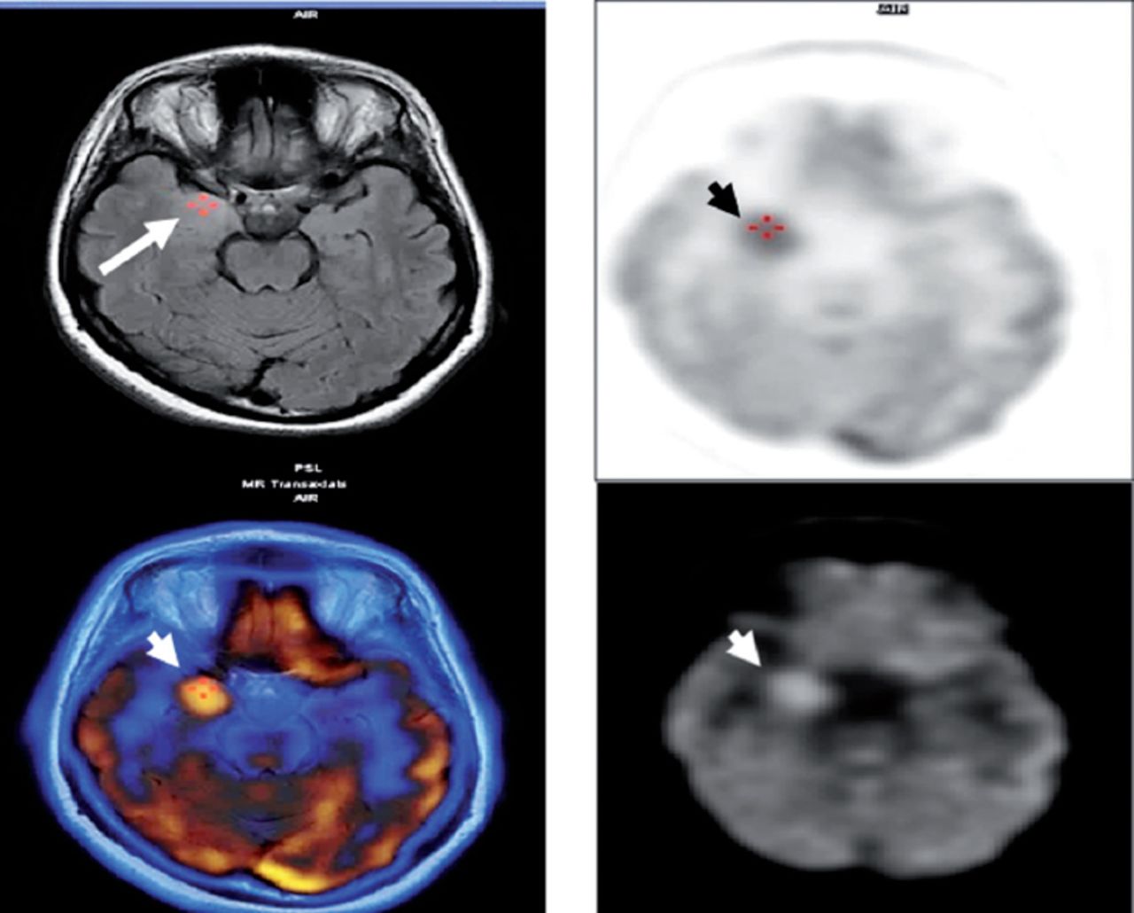
    PET scan of the brain at presentation showed hypermetabolic right mesial temporal area (White & black arrow heads) which matched with MRI abnormality (White arrow).

  • Figure 4
    • Download figure
    • Open in new tab
    • Download powerpoint
    Figure 4

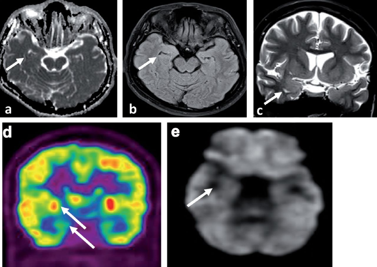
    Neuroimaging at 7 months of treatment a) MRI brain axial ADC map, b) MRI brain axial FLAIR and c) MRI brain coronal T2SE showing almost complete resolution of the signal abnormality with no atrophic changes (white arrows). d-e) The PET scan of the brain showed interval resolution of the focal hypermetabolic activity previously seen in the right temporal lobe with ipsilateral relative mesial temporal and right basal ganglia hypometabolism (white arrows).

PreviousNext
Back to top

In this issue

Neurosciences Journal: 22 (4)
Neurosciences Journal
Vol. 22, Issue 4
1 Oct 2017
  • Table of Contents
  • Cover (PDF)
  • Index by author
Print
Download PDF
Email Article

Thank you for your interest in spreading the word on Neurosciences Journal.

NOTE: We only request your email address so that the person you are recommending the page to knows that you wanted them to see it, and that it is not junk mail. We do not capture any email address.

Enter multiple addresses on separate lines or separate them with commas.
A rare case of autoimmune limbic encephalitis: an uncharted territory!
(Your Name) has sent you a message from Neurosciences Journal
(Your Name) thought you would like to see the Neurosciences Journal web site.
Citation Tools
A rare case of autoimmune limbic encephalitis: an uncharted territory!
Hatim Ibrahim, Abdulelah N. Al Jasser, Sonia A. Khan, Kalthoum G. Tlili
Neurosciences Journal Oct 2017, 22 (4) 292-297; DOI: 10.17712/nsj.2017.4.20170150

Citation Manager Formats

  • BibTeX
  • Bookends
  • EasyBib
  • EndNote (tagged)
  • EndNote 8 (xml)
  • Medlars
  • Mendeley
  • Papers
  • RefWorks Tagged
  • Ref Manager
  • RIS
  • Zotero
Share
A rare case of autoimmune limbic encephalitis: an uncharted territory!
Hatim Ibrahim, Abdulelah N. Al Jasser, Sonia A. Khan, Kalthoum G. Tlili
Neurosciences Journal Oct 2017, 22 (4) 292-297; DOI: 10.17712/nsj.2017.4.20170150
Twitter logo Facebook logo Mendeley logo
  • Tweet Widget
  • Facebook Like
  • Google Plus One
Bookmark this article

Jump to section

  • Article
    • Abstract
    • Case Report
    • Discussion
    • Footnotes
    • References
  • Figures & Data
  • eLetters
  • References
  • Info & Metrics
  • PDF

Related Articles

  • No related articles found.
  • PubMed
  • Google Scholar

Cited By...

  • No citing articles found.
  • Google Scholar

More in this TOC Section

  • Unmasking the mimic: Leprosy neuropathy misdiagnosed as chronic inflammatory demyelinating polyneuropathy: A case report from Saudi Arabia
  • Seminal vesicle schwannoma with chronic hemorrhage
  • Marchiafava-Bignami disease post-bariatric surgery: A case report and review of similar cases
Show more Case Report

Similar Articles

Navigate

  • home

More Information

  • Help

Additional journals

  • All Topics

Other Services

  • About

© 2025 Neurosciences Journal Neurosciences is copyright under the Berne Convention and the International Copyright Convention. All rights reserved. Neurosciences is an Open Access journal and articles published are distributed under the terms of the Creative Commons Attribution-NonCommercial License (CC BY-NC). Readers may copy, distribute, and display the work for non-commercial purposes with the proper citation of the original work. Electronic ISSN 1658-3183. Print ISSN 1319-6138.

Powered by HighWire