
Main menu

  • Home
  • Content
    • Latest
    • Ahead of print
    • Archive
  • Info for
    • Authors
    • Reviewers
    • Subscribers
    • Institutions
    • Advertisers
  • About Us
    • About Us
    • Editorial Office
    • Editorial Board
  • More
    • Alerts
    • Feedback
    • Folders
    • Help
  • Other Publications
    • Saudi Medical Journal

User menu

  • My alerts
  • Log in

Search

  • Advanced search
Neurosciences Journal
  • Other Publications
    • Saudi Medical Journal
  • My alerts
  • Log in
Neurosciences Journal

Advanced Search

  • Home
  • Content
    • Latest
    • Ahead of print
    • Archive
  • Info for
    • Authors
    • Reviewers
    • Subscribers
    • Institutions
    • Advertisers
  • About Us
    • About Us
    • Editorial Office
    • Editorial Board
  • More
    • Alerts
    • Feedback
    • Folders
    • Help
  • Follow psmmc on Twitter
  • Visit psmmc on Facebook
  • RSS
Case ReportCase Report
Open Access

Posterior mediastinal neuroblastoma masked as flaccid paraparesis in a 3 year child

Farah S. Yahya and Hieder A. Al-Shami
Neurosciences Journal October 2019, 24 (4) 320-323; DOI: https://doi.org/10.17712/nsj.2019.4.20190036
Farah S. Yahya
From the Department of Pediatrics (Yahya), College of Medicine, University of Mosul, Mosul, Iraq, and from Department of Neurosurgery (Al-Shami), Al-Ahly Bank Hospital, Cairo, Egypt.
MBCHB, CABP
  • Find this author on Google Scholar
  • Find this author on PubMed
  • Search for this author on this site
Hieder A. Al-Shami
From the Department of Pediatrics (Yahya), College of Medicine, University of Mosul, Mosul, Iraq, and from Department of Neurosurgery (Al-Shami), Al-Ahly Bank Hospital, Cairo, Egypt.
MD, EFNS, MRCS
  • Find this author on Google Scholar
  • Find this author on PubMed
  • Search for this author on this site
  • Article
  • Figures & Data
  • eLetters
  • Info & Metrics
  • References
  • PDF
Loading

Article Figures & Data

Figures

  • Figure 1
    • Download figure
    • Open in new tab
    • Download powerpoint
    Figure 1

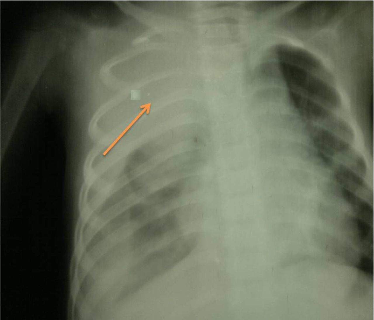
    Chest X-ray posterior-anterior view showing rather a well defined large homogenous soft tissue opacity lesion in the right upper zone area (arrow) that have broad based toward mediastinum and making an obtuse angle with it. There are no any detected calcifications seen within the lesion. The mass have overlapped the upper thoracic spine but not Silhouetting the upper cardiac border suggested to be retro-cardiac. Note the widening of the right sided upper posterior rib interspace highly localizing mass lesion to be in the posterior mediastinum.

  • Figure 2
    • Download figure
    • Open in new tab
    • Download powerpoint
    Figure 2

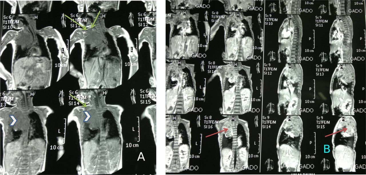
    Chest MRI (multiple sections through the chest and mediastinum). The examination showing A) large well defined soft tissue lesion about 8 * 6 * 5 cm (arrow head), have low signal intensity in T1W image with multiple signal void area in favouring of few spots of calcification, This mass being extra pulmonary lesion at the right side para-vertebral area showing extension upward occupying the right side upper posterio-superior thoracic gutter connecting to another mass at the right side supraclavicular area in favour of multiple discrete & matted involved lymph nodes (arrow) by metastasis that have the same mass criteria in signal intensity enhancement. B) The whole 2 bulk masses showing heterogeneous enhancement post contrast (arrow). There are no any surrounded bony changes. This large mass abutting the descending thoracic aorta displacing trachea to the left side.

  • Figure 3
    • Download figure
    • Open in new tab
    • Download powerpoint
    Figure 3

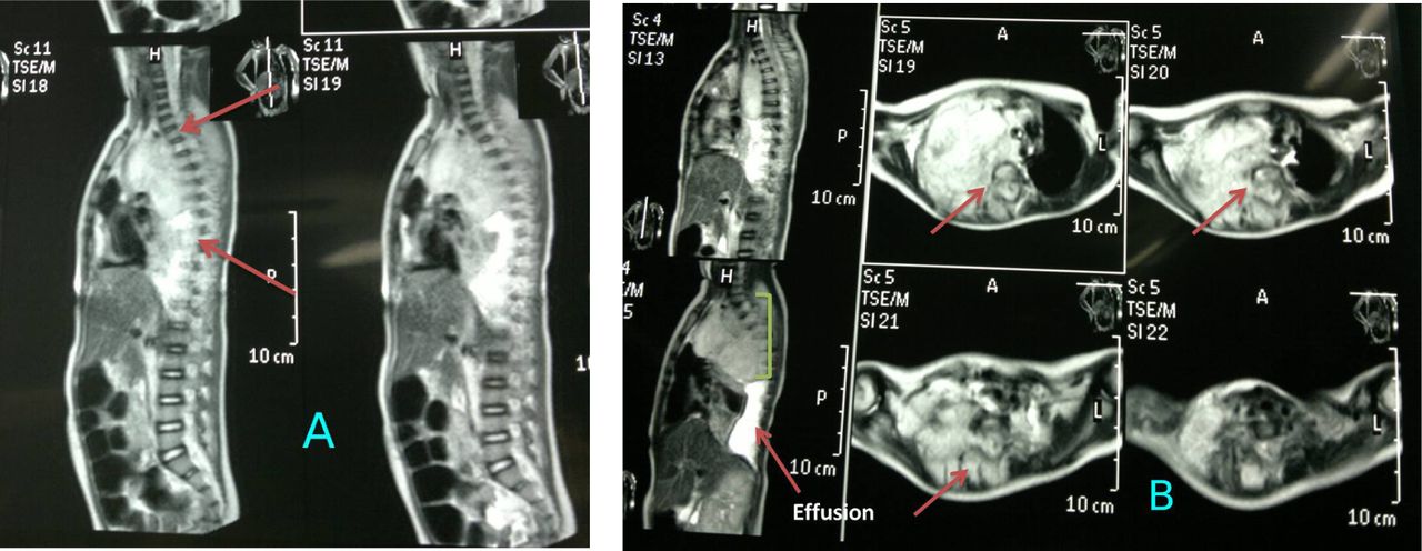
    Spinal MRI (multiple sections through the thoracic spine showing: A) large mass lesion 8 *6* 5 cm enhanced heterogeneously in the right sided posterior mediastinum (arrows) that extended through the neural exit foramen in the right side aspect widened them at multiple levels D1-D8. The mass demonstrated also posterior extra dural component within the thoracic spinal sac deviating & flattening the spinal cord aside from D1 to approximately D8 but not invade spinal cord tissue (arrows). B) Axial view the mass considered to be a (dumbbell) malignant neoplasm (arrow), primarily in the mediastinum with posterior extension through an intervertebral foramen to the spinal canal & causing spinal cord compression (extra dural extension) highly in favour of posterior mediastinal Neuroblastoma. There are a concomitant right sided pleural effusion that indicates involvement of the pleural cavity.

PreviousNext
Back to top

In this issue

Neurosciences Journal: 24 (4)
Neurosciences Journal
Vol. 24, Issue 4
1 Oct 2019
  • Table of Contents
  • Table of Contents (PDF)
  • Cover (PDF)
  • Index by author
Print
Download PDF
Email Article

Thank you for your interest in spreading the word on Neurosciences Journal.

NOTE: We only request your email address so that the person you are recommending the page to knows that you wanted them to see it, and that it is not junk mail. We do not capture any email address.

Enter multiple addresses on separate lines or separate them with commas.
Posterior mediastinal neuroblastoma masked as flaccid paraparesis in a 3 year child
(Your Name) has sent you a message from Neurosciences Journal
(Your Name) thought you would like to see the Neurosciences Journal web site.
Citation Tools
Posterior mediastinal neuroblastoma masked as flaccid paraparesis in a 3 year child
Farah S. Yahya, Hieder A. Al-Shami
Neurosciences Journal Oct 2019, 24 (4) 320-323; DOI: 10.17712/nsj.2019.4.20190036

Citation Manager Formats

  • BibTeX
  • Bookends
  • EasyBib
  • EndNote (tagged)
  • EndNote 8 (xml)
  • Medlars
  • Mendeley
  • Papers
  • RefWorks Tagged
  • Ref Manager
  • RIS
  • Zotero
Share
Posterior mediastinal neuroblastoma masked as flaccid paraparesis in a 3 year child
Farah S. Yahya, Hieder A. Al-Shami
Neurosciences Journal Oct 2019, 24 (4) 320-323; DOI: 10.17712/nsj.2019.4.20190036
Twitter logo Facebook logo Mendeley logo
  • Tweet Widget
  • Facebook Like
  • Google Plus One
Bookmark this article

Jump to section

  • Article
    • Abstract
    • Case Report
    • Discussion
    • Acknowledgments
    • Footnotes
    • References
  • Figures & Data
  • eLetters
  • References
  • Info & Metrics
  • PDF

Related Articles

  • No related articles found.
  • Google Scholar

Cited By...

  • No citing articles found.
  • Google Scholar

More in this TOC Section

  • Unmasking the mimic: Leprosy neuropathy misdiagnosed as chronic inflammatory demyelinating polyneuropathy: A case report from Saudi Arabia
  • Seminal vesicle schwannoma with chronic hemorrhage
  • Marchiafava-Bignami disease post-bariatric surgery: A case report and review of similar cases
Show more Case Report

Similar Articles

Navigate

  • home

More Information

  • Help

Additional journals

  • All Topics

Other Services

  • About

© 2025 Neurosciences Journal Neurosciences is copyright under the Berne Convention and the International Copyright Convention. All rights reserved. Neurosciences is an Open Access journal and articles published are distributed under the terms of the Creative Commons Attribution-NonCommercial License (CC BY-NC). Readers may copy, distribute, and display the work for non-commercial purposes with the proper citation of the original work. Electronic ISSN 1658-3183. Print ISSN 1319-6138.

Powered by HighWire