


Article Figures & Data
Figures
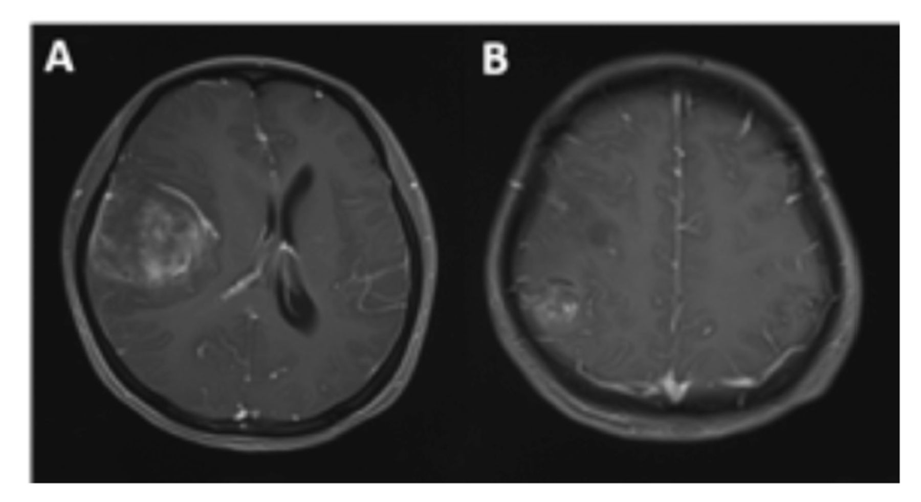
- Figure 1
- Radiographic features of case 1. Contrast-enhanced MRI shows 2 lesions in the right frontal-temporal (A) and right parietal lobes (B).
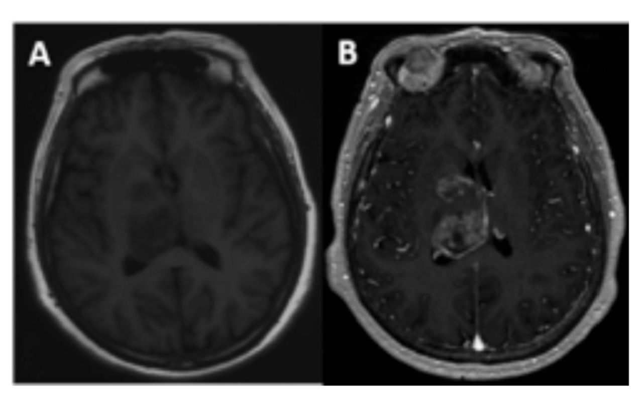
- Figure 2
- Radiographic features of case 2. T1-weighted image shows 2 lesions located in the right basal ganglia and right thalamus (A), with obvious inhomogeneous enhancement on enhanced scans (B).
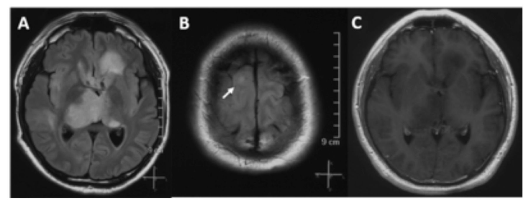
- Figure 3
- Radiographic features of case 3. The lesion was unevenly enhanced on contrast-enhanced MRI. The FLAIR image shows multiple lesions in the left frontal lobe, bilateral thalamus (A), and right frontal lobe (B), and the lesions are not enhanced (C).
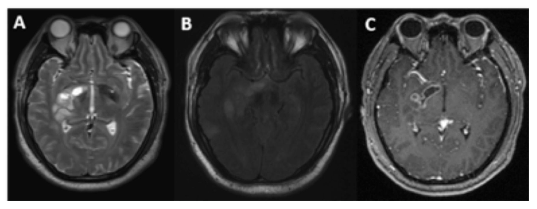
- Figure 4
- Radiographic features of case 4. The T2-weighted image shows that the lesion was located in the right basal ganglia, involving the thalamus (A), and the FLAIR image shows that another lesion was located in the right temporal lobe (B). Contrast-enhanced MRI shows that the lesion was enhanced in the right basal ganglia (C).
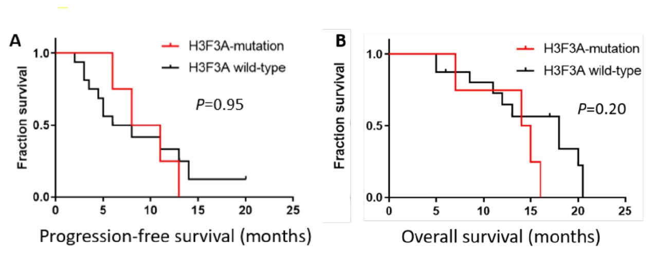
- Figure 5
- Kaplan–Meier survival curve. Kaplan–Meier survival curves and log-rank tests for PFS (A) and OS (B). The median PFS and OS were 9.5 months and 14.5 months, respectively, in M-HGGs patients with H3F3A mutations. For non-H3F3A-mutated M-HGGs patients, the median PFS and OS were 7.0 months and 18.0 months, respectively. Comparisons between the 2 groups using the log-rank test indicated no significant difference.


Tables
No. Age (y) Sex Histo-diagnosis No. of lesions Location H3F3A IDH TERT MGMT Treatment PFS (mo.) OS (mo.) 1 27 F GBM 2 FL+TL+PL, R H3G34R wt wt met STR+RT(66Gy)+TMZ 13 16 2 65 F HGG 2 BG+TH, R H3K27 M wt wt met Biopsy+RT(60Gy)+TMZ 11 14 3 37 M AA 3 FL+TH, B H3K27 M wt wt unmet Biopsy+RT(54Gy) 8 15 4 35 M HGG 2 TL+BG+TH, R H3K27 M wt wt unmet Biopsy+RT(60Gy)+TTF 6 7 M - male; F - female; GBM - glioblastoma; HGG - high-grade glioma; AA - anaplastic astrocytoma; FL - frontal lobe; PL - parietal lobe; TH - thalamus; TL - temporal lobe; BG - basal ganglia; R - right; L - left; B - bilateral; wt - wild-type; met - methylated; unme t- unmethylated; STR - subtotal resection; RT - radiotherapy; TMZ - temozolomide; TTF - tumor-treating fields; mo - month(s)
In this issue


Jump to section
Related Articles
Cited By...
- No citing articles found.